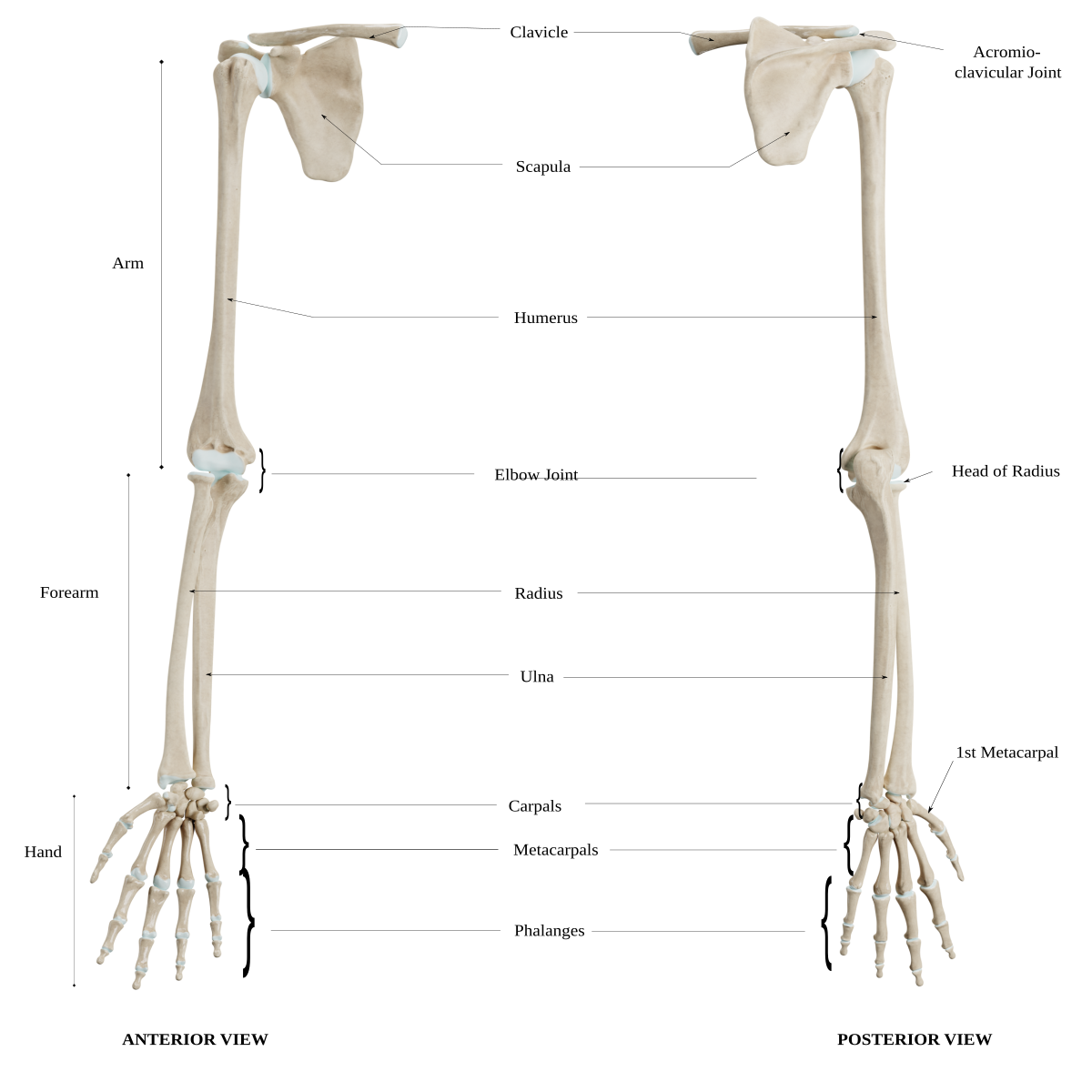
Functional anatomy of the upper extremity
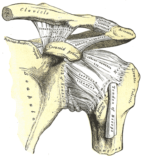
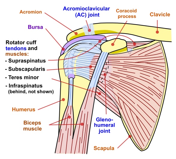
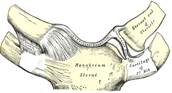
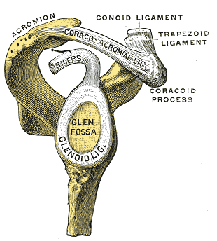
Shoulder region
The primary movements of the shoulder are aided by the three shoulder joints and the scapulo thoracic articulation are:
- Shoulder flexion/extension
- Shoulder elevation/depression
- Shoulder external rotation/internal rotation
- Shoulder abduction/adduction
- Scapular elevation/depression
- Scapular upward rotation/downward rotation
- Scapular abduction/adduction
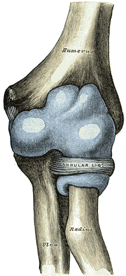
Elbow region
The primary movements of the elbow that are aided by the four joints of the elbow are:
- Elbow flexion/extension
- Forearm supination/pronation
The elbow joint has other important anatomical features:
- Capsule- a thin structure that surrounds the anterior and posterior components of the joint
- Bursae- act as fluid-filled sacs that provide cushioning and friction reduction between tendons, joints, muscles, and bone
The ligaments that aid in the movement of the elbow region are:
- Ulnar collateral ligament
- Ligament is triangular in shape, running anteriorly, posteriorly, and obliquely to reinforce the medial humeroradial joint
- Radial collateral ligament
- Fan-shaped ligament that runs from the lateral epicondyle to the annular ligament, to support the humeroradial joint laterally
- Annular ligament
- Cone-shaped ligament that envelopes the radial head and attaches to the medial ulna; protects the radial head
- Quadrate ligament
- Extends from the radial notch on the ulna surface to the neck of the radius; reinforces the inferior portion of the joint capsule
- Distal radioulnar ligament
- Composed of the anterior and posterior radioulnar ligaments to provide strength to the capsule
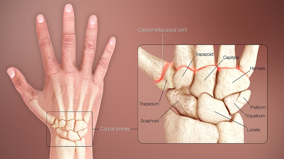
Wrist and hand region
The primary movements of the wrist and hand region are:
- Wrist flexion/extension
- Wrist radial deviation/ulnar deviation
- Finger flexion/extension
- Finger abduction/adduction
- Intrinsic finger movement
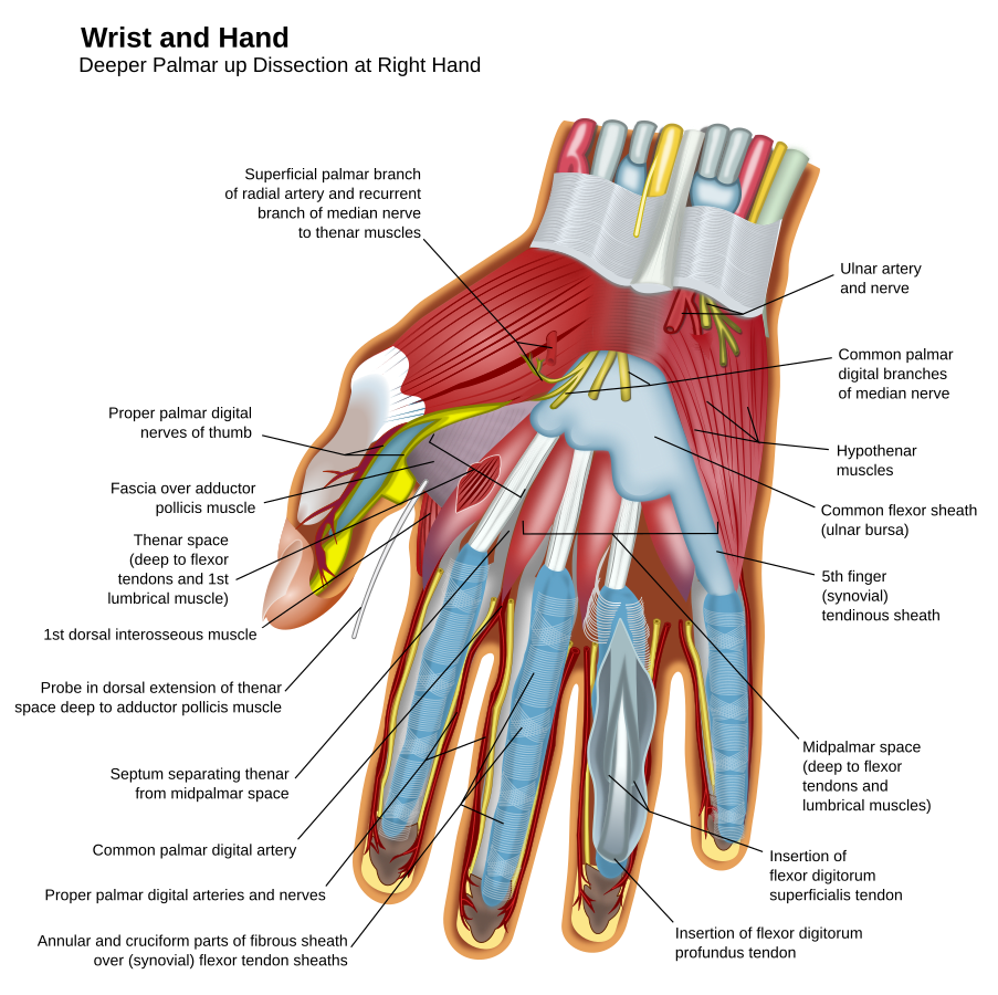
The wrist and hand joint has other important anatomical features:
- Capsule of wrist and hand — provide support to internal structures
- Volar plate— present on the palmar aspect of the MCP, PIP, and DIP to protect joints
- Palmar aponeurosis — sheet of fibrous tissue in the palm that protects underlying tendons and nerves.
- Extensor hood — fibrous connection on the dorsum of the finger that aids in extension of the PIP and DIP
- Nerves— radial, median, and ulnar are the primary nerves of innervation for the wrist and hand
Ligaments of the wrist:
- Dorsal radiocarpal
- Limits flexion, pronation
- Radiate
- Radial collateral ligament
- Ulnar collateral ligament
- Palmar ulnocarpal
- Limits extension and supination
- Palmar radiocarpal
- Limits extension and supination through the knuckles
Ligaments of fingers:
- Collateral ligaments
- Oriented from the lateral condyle to the distal phalanx and on the lateral volar plate to each metacarpal, PIP, and DIP
- All fibers tighten during flexion, but only volar fibers tighten during extension
- Accessory
- Oriented from the condylar head to the volar plate
- Transverse
- Provide stability linking the MCP joints and reinforcing the anterior capsule
Upper extremity range of motion normals
Shoulder range
Elbow range
Wrist range
MCP range
PIP range
DIP range
1st CMC range
1st MCP range
1st IP range