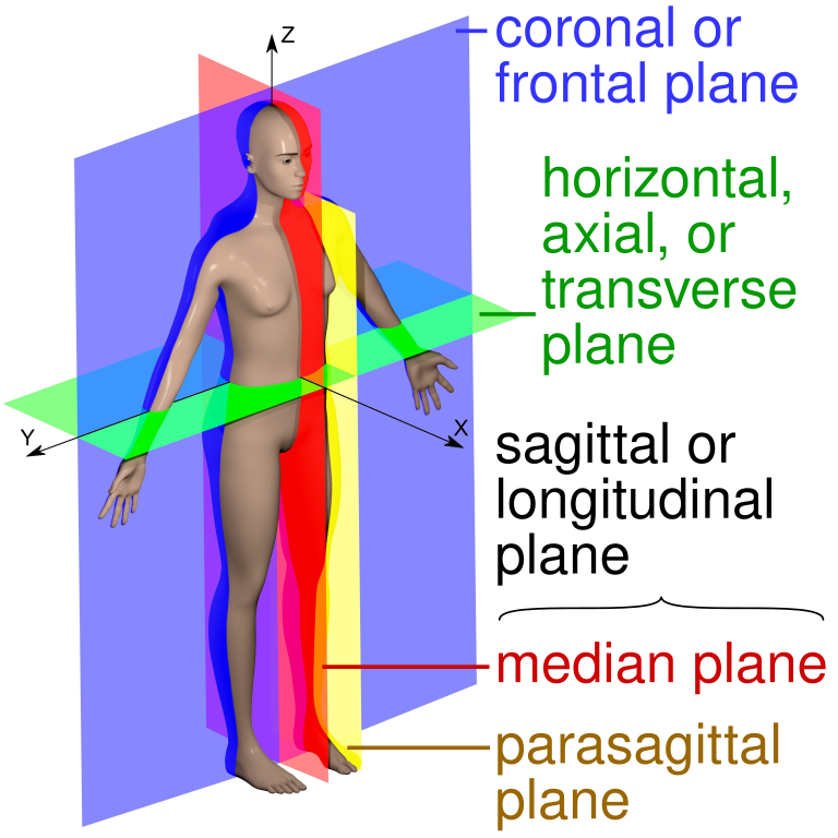
Anatomical position
Anatomical position refers to a standardized posture of the human body used as a reference point for describing anatomical structures and movements.
Specifications:
The body is upright and facing forward.
Feet are flat and parallel, with toes pointing straight ahead.
Arms are extended at the sides, with palms facing forward.
Head is held erect, with eyes looking straight ahead.
Definitions
Anterior
Front of the body, or the direction toward the front
Posterior
Back of the body, or the direction toward the back
Median
Towards the middle of the body
Lateral
Towards the side of the body
Superior
Above or higher than another body part
Inferior
Below or lower than another body part
Distal
Further away from the center of the body
Proximal
Closer to the trunk of the body
Ventral
Close to the anterior of a structure
Dorsal
Close to the back of a structure
Sagittal plane
Runs vertically through the body, dividing it into left and right halves
Frontal (coronal) plane
Runs vertically through the body, dividing it into front (anterior) and back (posterior) halves
Axial (transverse) plane
Runs horizontally through the body, dividing it into top (superior) and bottom (inferior) halves
Basics of strength training
Strength training refers to a type of exercise program designed to increase muscle strength and mass. It involves performing exercises that challenge the muscles to work against resistance, such as lifting weights, using resistance bands, or performing bodyweight exercises. There are multiple factors contributing to the overall success of an appropriate strength training program.
Muscle fiber types
Muscle types are defined by how they produce energy and how quickly they fatigue.
.
Slow Twitch (Type I)
Slow contraction speed
Low force production
Highly resistant to fatigue
Example: Postural muscles
Fast Twitch (Type IIA)
Fast contraction speed
Fatigue resistant
Can be influenced by training
Examples: Muscles used for strength and movement
Fast Twitch (Type IIB)
Fast contraction speed
High force production
Susceptible to quick fatigue
Examples: Muscles for quick movements and eye muscles
Guidelines for strength training
The foundational concepts of strength training are:
Overload principle
Increasing the amount of resistance added to the muscle over a period of time
Specificity of training
Train the muscle or muscle groups necessary to perform the activity
Resistance needs to be added differently to each muscle and/or muscle group to exhibit hypertrophy
Training effects are reversible
If training ceases, the amount of resistance that can be applied to a muscle will be lost
Changes to the muscle fibers and the amount of motor units recruited with initiation of strength training take 6-8 weeks to occur with a consistent training program.
.
Contraindications to strength training
Active inflammation or acute condition
Severe pain during or greater than 24 hours after initiation of exercise
Types of exercise for strength training
Isometric
Muscle contraction without change in muscle lengthExample: Quad sets
Typically used during the acute phase of healing or when learning muscle control
Isotonic
A muscle produces force and changes length, with tension remaining relatively constant, allowing movement at a joint.
Example: Bicep curls with a 20-pound weight
Used during sub-acute and chronic phases of healing when pain and inflammation are reduced. Requires adequate motor planning with movement control have been established
Concentric: The muscle shortens, for example, standing from a seated position with shortening of the hip and knee extensors.
Eccentric: The muscle lengthens while holding tension, for example, controlled sitting from a standing position, controlled with lengthening of the knee and hip extensors.
Isokinetic
Muscle changes length (shortening or lengthening) while moving at a constant, controlled velocity.
Example: Isokinetic machines using electric or hydraulic dynamometers
Provides accommodating resistance that matches the force the muscle generates, it loads the muscle maximally throughout the entire range of motion
Used during the sub-acute phase of healing when the concern of re-injuring muscle is a concern due to control of movement and resistance throughout the movement
Endurance training
Endurance training refers to a type of physical exercise that aims to improve the body’s ability to sustain physical activity for prolonged periods. It involves engaging in activities that require moderate-to-high intensity effort over extended timeframes, such as running, swimming, cycling, or hiking. Endurance training helps enhance cardiovascular health, pulmonary ventilation, muscle strength, and overall fitness by increasing the body’s oxygen consumption and utilization capacity.
Effects of endurance training:
Capillary growth : More capillaries in muscles improve blood flow and oxygen delivery.Cardiac adaptations : Stronger heart muscle, increased stroke volume, and improved cardiac output.Metabolic benefits: Enhanced ability to utilize fat as fuel during exercise.Improved VO2 max: The maximum amount of oxygen your body can use during exercise increases significantly with endurance training
Guidelines for endurance training
At least 150 minutes of moderate-intensity aerobic activity or 75 minutes of vigorous aerobic activity per week (per American Heart Association)
Spread exercise out throughout the week
Begin with low duration and intensity while progressing to longer duration and intensity over a period of time
Contraindications for endurance training
Unstable angina
Acute coronary syndrome
Uncontrolled arrhythmias
Acute heart failure
High-degree atrioventricular block
Severe aortic stenosis
Coronary artery stenosis
Recent stroke or transient ischemic attack
Uncontrolled diabetes mellitus
Uncontrolled hypertension
Hyperthyroidism
Severe COPD
Cerebrovascular or musculoskeletal disease
Significant anemia
Important electrolyte imbalances
Coordination and balance training
Coordination and balance training are ways to improve your ability to control your body’s movement and maintain stability.
Effects of coordination and balance training
Reduced fall risk
Improving ability to react quickly and maintain stability, overall reducing fall risk
Improved coordination
Allows for smoother movements, better agility, and quicker reaction times
Improved proprioception:
Balance exercises stimulate the nervous system to better sense your body’s position in space, enhancing proprioception.
Better posture:
Regular balance training can help maintain proper alignment and posture by strengthening the muscles that support the axial skeleton
Muscle strength and endurance:
Improved muscle activation, leading to increased muscle strength and endurance throughout the body
Guidelines for coordination and balance training
The typical sequence of coordination and balance training interventions begins with axial/postural stability activities, progressing to the peripheral system. The activities will also begin as static and progress to more dynamic activities in nature.
Interventions that can be used are as follows:
Therapeutic exercises
Postural training
Weight shifting activities
Sit-to-stands
Gait training
Dual tasking
Changing surfaces
Sensory training
Contraindications for coordination and balance training
Acute injuries
Severe pain
Unstable joints
Recent surgery
Significant neurological impairments
Severe dizziness or vertigo
Poor vision
Cardiovascular instability
Uncontrolled medical conditions,
Aquatic Therapy
Aquatic therapy, also known as hydrotherapy, is a form of physical therapy that involves performing exercises and movements in water. It utilizes the buoyancy, resistance, and warmth of the water to improve physical function, reduce pain, and enhance rehabilitation.
Related Physics
Buoyancy: Force of water on immersed body segment, decreasing body weight and joint off-loadingCohesion: Water molecules adhere to each other, creating resistanceDensity: Proportional to water depth, providing additional resistance
The deeper the individual is submerged, the harder the activity will be for the individual
Water Temperatures
Cooler water: Used for high-intensity exercises- seen more in athletes
Multiple sclerosis patients: 80—84°F (26—29°C) to reduce spasticity and fatigue
Warmer water: Used to improve mobility and flexibility while decreasing pain -common rehab population
83—86°F (28—30°C) is a common range for fitness
88—92°F (31—33°C is common range of osteoarthritis
Precautions for aquatic therapy
Fear of water
Patients with heat intolerance
Contraindications for aquatic therapy
Bowel/bladder incontinence
Severe kidney disease
Seizures
Uncontrolled cardiac or respiratory disorders
Peripheral vascular disease (PVD)
Open wounds
Active bleeding
Active infections
Ways to progress therapeutic activities
Exercise progression is the process of making an exercise more challenging over time. It’s a key part of any training routine that helps you maintain and improve your fitness level. Below are ways in which a therapist can progress exercise over a period of time.
Progressions examples
Small Motion → Large Motion
Low Center of Gravity → High Center of Gravity
Low Resistance → High Resistance
Slow Movements → Fast Movements
Stable Surface → Unstable Surface
Large Base of Support → Small Base of Support
Closed Environment → Open Environment
All Sensory Input → Limited Sensory Input
Extrinsic Feedback → Intrinsic Feedback
Eccentric Exercises → Concentric Exercises
Osteokinematics and arthrokinematics
Osteokinematics is the study of bone movement, while arthrokinematics is the study of joint surface movement. Both are branches of biomechanics that describe how the body moves.
Definitions
Osteokinematics
Movement between two bones (flexion/extension, IR/ER)
Arthrokinematics
Movement of joint surfaces, such as roll, glide, spin- such movements are used to improve the range of motion
Rule of convex surface moving on a fixed concave surface
Roll and glide occur in opposite directions to allow for motion to occur- movement of distal and proximal segments are in opposite directions
Rule of the concave surface moving on a fixed convex surface
Roll and glide occur in the same direction to allow for motion to occur- movement of distal and proximal segments is in the same direction
Arthrokinematics rules
Shoulder (Convex on concave rule)
Flexion: Roll anterior, slide posterior
Horizontal Adduction: Roll anterior, slide posterior
Internal Rotation: Roll anterior, slide posterior
Extension: Roll posterior, slide anterior
Horizontal Abduction: Roll posterior, slide anterior
External Rotation: Roll posterior, slide anterior
Abduction: Roll superior, slide inferior
Elbow (Concave on convex rule)
Flexion: Roll anterior, slide anterior
Extension: Roll posterior, slide posterior
Wrist (Convex on concave rule)
Flexion: Roll anterior, slide posterior
Extension: Roll posterior, slide anterior
Radial Deviation: Roll radial, slide ulnar
Ulnar Deviation: Roll ulnar, slide radial
Hip (Convex on concave rule)
Flexion: Roll anterior, slide posterior
Extension: Roll posterior, slide anterior
Adduction: Roll medial, slide superior
Abduction: Roll lateral, slide** inferior**
Internal Rotation: Roll medial, slide posterior
External Rotation: Roll lateral, slide anterior
Knee (Concave on convex rule)
Flexion: Roll posterior, slide anterior
Extension: Roll anterior, slide posterior
Ankle (Convex on concave rule)
Dorsiflexion: Roll anterior, slide posterior
Plantarflexion: Roll posterior, slide anterior
Supination/Inversion: Roll medial, slide lateral
Pronation/Eversion: Roll lateral, slide medial
Joint mobilization
Joint mobilization is a manual therapy technique that involves moving a joint passively to improve its range of motion and reduce pain. The above arthrokinematics chart is how the therapist will perform joint mobilizations at each joint.
Indications for joint mobilization
Pain
Muscle spasm
Joint hypomobility
Functional limitation at joint ROM
Precautions for joint mobilization
Joint hypermobility
Joint effusion
Inflammation
Contraindications for mobilization
Malignancy
Fracture
Bone disease
Rheumatoid arthritis (RA)
Individuals on anticoagulants
The grades of joint mobilization (Maitland approach)
Grade I - small amplitude movement at the beginning of the joint’s range of motion
Typically used in acute phases for pain management
Grade II - large amplitude movement within the joint’s range
Typically used in sub-acute phases for return of range of motion
Grade III - large amplitude movement reaching the limit of the joint’s range
Typically used in sub-acute phases for return of range of motion
Grade IV - - small amplitude movement at the end of the joint’s range
Used to increase tissue extensibility, reduce stiffness, and improve range of motion
Joint positions
Joint position or mechanics are ways to define the joint’s level of stability. An open position (also called loose-packed position or resting position) refers to a joint position where the articulating surfaces have minimal contact, ligaments are relaxed, and the joint has the least stability. A closed position (also called a close-packed position) is when the joint surfaces are fully congruent, ligaments are maximally taut, and the joint is at its most stable position. The open positions are those in which joint mobilizations will occur.
Resting and closed positions
Sternoclavicular
Resting position: arm resting at the side
Closed position: arm maximally elevated
Acromioclavicular
Resting position: arm resting at the side
Closed position: arm abducted to 90 degrees
Glenohumeral
Resting position: 40-55 degrees abduction; 30 degrees of horizontal adduction
Closed position: maximum abduction and external rotation
Humeroulnar (elbow)
Resting position: 70 degrees flexion, 10 degrees supination
Closed position: full extension and supination
Humeroradial (elbow)
Resting position: full extension and supination
Closed position: 90 degrees flexion and 5 degrees supination
Proximal radioulnar (forearm)
Resting position: 70 degrees flexion and 35 degrees supination
Closed position: 5 degrees of supination
Proximal radioulnar (forearm)
Resting position: 10 degrees of supination
Closed position: 5 degrees of supination
Radio/ulnarcarpal
Resting position: neutral with slight ulnar deviation
Closed position: full extension with radial deviation
Hip
Resting position: 30 degrees flexion, 30 degrees abduction, and slight lateral rotation
Closed position: full extension, abduction, and internal rotation
Knee
Resting position: 25-degree flexion
Closed position: full extension and external rotation
Talocrural (ankle/foot)
Resting position: mid inversion/eversion and 10 degrees plantar flexion
Closed position: full dorsiflexion
Subtalar (ankle/foot)
Resting position: midway between inversion and eversion
Closed position: full inversion
Midtarsal (ankle/foot)
Resting position: midway between inversion and eversion
Closed position: full supination
Tarsometatarsal (ankle/foot)
Resting position: midway between supination/pronation
Closed position: full supination
Capsular patterns
A capsular pattern is a specific limitation in the range of motion that indicates joint tightness or inflammation. It’s a combination of pain and limited movement that can occur in any joint that’s controlled by muscles. Certain pathologies, as denoted in later chapters, will be defined specifically from the capsular pattern that exists within the joint.
Below are capsular patterns of joints
Glenohumeral
External rotation, abduction, internal rotation
Sternoclavicular
Acromioclavicular
Humeroulnar
Humeroradial
Proximal radioulnar
Distal radioulnar
Wrist
Hip
Flexion, internal rotation, abduction
Knee
Tibiofibular (proximal and distal)
Equal limitations of flexion and extension
End feels of joints
End feel of a joint refers to the sensation a clinician feels when they reach the limit of a joint’s passive range of motion during an assessment, essentially describing the quality of tissue resistance at the end of movement. Tendons can be characterized as soft, firm, or hard depending on the tissues that limit the motion. By evaluating the end feel, a clinician can determine if a joint is moving within its normal range and identify potential abnormalities like inflammation, ligamentous damage, or joint stiffness.
Below are the characteristics of normal end feels:
Soft end feel:
Occurs when soft tissue, es as muscles, meet, often felt as a cushioned sensation (example: knee flexion)
Firm end feel:
A more defined resistance, usually due to the tension of ligaments or joint capsule at the end of range (example: wrist flexion)
Hard end feel: (Bony)
A sudden, abrupt stop to movement, typically caused by bone-on-bone contact (example: elbow extension)
Pathological end feels are those indicating there has been injury to the joint, tendon, or muscle.
Pathological end feels that may be present
Springy block
A rebounding sensation is often associated with internal joint derangements , like a torn meniscus
Empty end feel:
Significant pain experienced before reaching the end of the range of motion, usually indicating acute inflammation
Boggy or soft end feel
A “mushy” sensation due to joint effusion or edema, typically seen in acute injuries
Hard end feel
Excessive bony resistance beyond the normal endpoint, potentially from osteoarthritis or bone spurs
Muscle spasm end feel
Sudden, sharp resistance due to muscle guarding, causing pain and limiting movement
Phases of healing and types of musculoskeletal interventions
The musculoskeletal system has three stages in which disease processes can be classified: 1. Acute or inflammatory phase, 2. Subacute or proliferative phase, and 3. Chronic phase.
The acute phase refers to the initial inflammatory stage following an injury, characterized by pain, swelling, and redness. The acute phase typically lasts for 3-7 days.
The subacute phase is the subsequent repair stage where new tissue begins to grow, and the body starts to rebuild damaged structures, usually occurring a few days after the initial injury and lasting several weeks.
The chronic phase is not a natural part of healing but occurs when healing has stopped in either the acute or subacute phase. Injuries are classified as chronic if no progression past the acute or subacute phases for greater than 3 months.
Interventions in each phase
Acute phase
Pain management Maitland mobilizations — grade I or IV
Joint protection to prevent further injury
Edema management Therapeutic Exercise: 40%-60% of 1 rep max in pain-free ROM Stretching is contraindicated
Subacute phase
Avoid overuse pain, as resting pain should be at a minimum
Stretching is initiated to aid in restoring the full range of motion
Endurance training will begin
Resistance training will begin
Postural and biomechanical education
Chronic phase
Identify healing phase (acute vs subacute) and make intervention selections based on phase of healing
Emphasize postural and biomechanical strengthening Improve flexibility and joint alignment