Medications, imaging, and fractures
Imaging
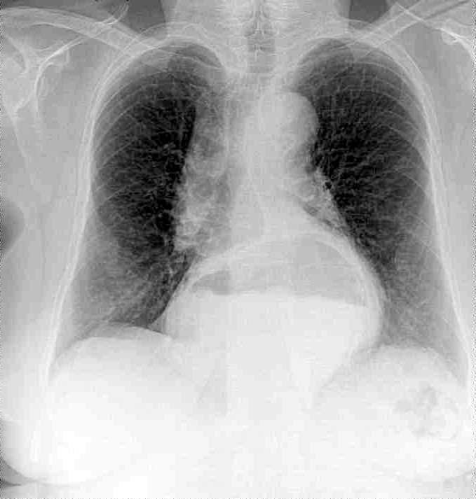
X-ray (radiography)
- Best for: Bones, fractures, joint alignment, lung conditions
- How it works: Uses ionizing radiation to create images of dense structures
- Common uses:
- Fractures & dislocations
- Arthritis
- Lung infections (e.g., pneumonia)
- Foreign objects
- Limitations: Limited soft tissue visibility, radiation exposure
- Interpreting an X-Ray
- High-density tissue (e.g., bone) – absorbs x-rays to a greater degree, and appears white on the film. Low-density tissue (e.g., the lungs) –absorbs X-rays to a lesser degree, and appears black on the film
- Intermediate density tissue (e.g., muscle and fat) – appears as shades of grey on the x-ray film
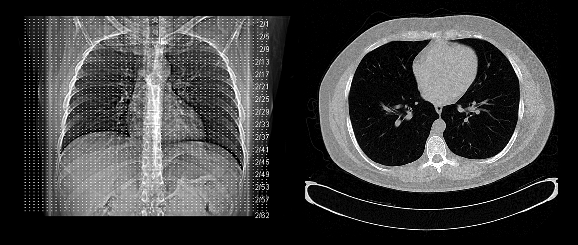
Computed tomography (CT Scan)
- Best for: Bones, soft tissues, internal bleeding, internal organs, and the brain.
- How it works: Combines multiple X-rays to create cross-sectional images
- Common uses:
- Bone fractures and complex injuries
- Internal bleeding (trauma)
- Stroke
- Tumors
- Lung and abdominal conditions
- Limitations: Higher radiation exposure than X-rays
- Interpreting CT scan
- Dense structures (like bone and calcifications) appear lighter (white).
- Lucent structures (like air and fat) appear darker (black).
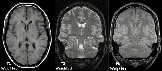
Magnetic resonance imaging (MRI)
- Best for: Soft tissues, brain, muscles, ligaments, and nerves
- How it works: Uses strong magnets and radio waves to generate detailed images
- Common uses:
- Ligament and tendon injuries
- Brain and spinal cord conditions
- Tumors
- Disc herniations
- Limitations: Expensive, time-consuming, cannot be used with metal implants
- Interpreting MRI
- T1 MRI highlights anatomy, provides crisp images, and shows fluids as dark
- T2 MRI focuses on pathology, making fluids bright, which is ideal for visualizing inflammation, edema
Ultrasound
- Best for: Soft tissues, pregnancy, blood flow
- How it works: Uses high-frequency sound waves to create real-time images.
- Common uses:
- Pregnancy monitoring
- Soft tissue injuries (e.g., muscle tears)
- Blood clots (Doppler ultrasound)
- Organ imaging (e.g., liver, kidneys)
- Limitations: Poor image quality for bones and deep structures
Nuclear medicine imaging (e.g., PET Scan, Bone Scan)
- Best for: Organ function, cancer detection, metabolic activity
- How it works: Uses radioactive tracers to highlight metabolic activity
- Common uses:
- Cancer detection (PET scan)
- Bone metastases (Bone scan)
- Stress fracture/microfracture (Bone scan)
- Thyroid and kidney function
- Limitations: Radiation exposure, high cost
Fluoroscopy
- Best for: Real-time imaging of movement (e.g., swallowing, joint motion)
- How it works: Continuous X-ray imaging allows real-time assessment
- Common uses:
- Barium swallow for digestive tract
- Cardiac catheterization
- Joint injections
- Limitations: Higher radiation exposure than standard X-rays
Medications
-
Non-steroidal anti-inflammatory drugs (NSAIDs)
- Mechanism of Action: Decrease inflammation, fever, and pain
- System Interactions:
-
Cardiac: High blood pressure (hypertension)
-
Circulatory: increases bleeding risk
-
Gastrointestinal: Indigestion, diarrhea, vomiting, GI ulcers, GERD
-
Neuro: Dizziness, headache
-
-
Opioids
- Mechanism of Action: Decreases pain in the musculoskeletal system
- System Interactions:
- Cardiac: Decrease heart rate, arrhythmia
- Pulmonary: Decrease respiration rate
- Gastrointestinal: Delayed gastric emptying (causes constipation)
- Musculoskeletal: Muscle rigidity, muscle jerks
- Integumentary: Itchy skin
- General: Dry mouth, addiction
-
Corticosteroids
- Mechanism of Action: Decrease inflammation in the musculoskeletal system
- System Interactions:
- Cardiac: High blood pressure (hypertension)
- Gastrointestinal: Indigestion, diarrhea
- Musculoskeletal: Osteoporosis (high risk for fractures if taken for a long period of time)
- General: Weight gain, diabetes
- Integumentary: Acne
-
Baclofen
- Mechanism of action: decrease spasticity
- Systems interactions
- Musculoskeletal: muscle stiffness, abnormal posturing, bone/joint stiffness/pain, muscle weakness
-
Muscle relaxants
- Mechanism of action: acting on the central nervous system (CNS) to interfere with the transmission of nerve impulses to muscles, effectively reducing muscle spasms and tension by depressing neuronal activity
- Systems interactions
- Neuro: drowsiness, fatigue, dizziness, headache
- Musculoskeletal: weakness
- Gastrointestinal: nausea, constipation
- General: dry mouth, blurred vision
Fractures
Types of fractures
- Open (compound): The bone breaks through the skin, exposing it to the environment
- Closed (simple): The bone breaks but does not penetrate the skin.
- Transverse: A straight break across the bone
- Oblique: A diagonal break at an angle to the bone
- Spiral: A twisting break that spirals around the bone
- Greenstick: A partial break that occurs in children’s flexible bones
- Comminuted: The bone breaks into multiple fragments
- Stress fracture: A small, hairline crack caused by repetitive stress
- Impacted fracture: The broken ends of the bone are driven into each other
- Avulsion fracture: A small piece of bone is pulled away by a tendon or ligament
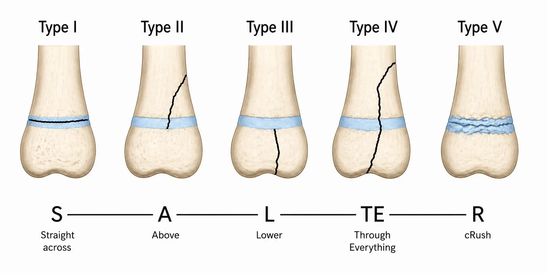
Salter-Harris fracture
Salter-Harris fractures are classified into five types based on the location and extent of the fracture:
-
S – Straight across (Type I) Fracture through the growth plate (physis) only → No bone involvement
-
A – Above (Type II) Fracture through the physis and metaphysis (above) → Most common type
-
L – Lower (Type III) Fracture through the physis and epiphysis (below) → Involves joint surface
-
TE – Through Everything (Type IV) Fracture through metaphysis, physis, and epiphysis → Crosses entire bone
-
R – Crush (Type V) Crush injury to the growth plate → Often not visible initially, worst prognosis