The primary movements of the shoulder are that are aided by the four joints of the shoulder are:
Shoulder flexion/ extension
Shoulder elevation/ depression
Shoulder external rotation/ internal rotation
Shoulder abduction/adduction
Scapular elevation/depression
Scapular upward rotation/ downward rotation
Scapular abduction/ adduction
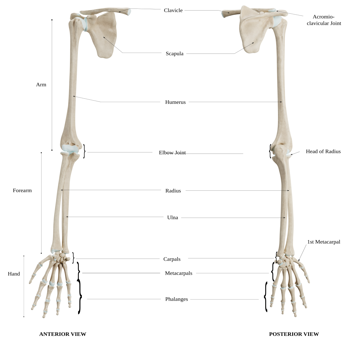
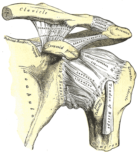
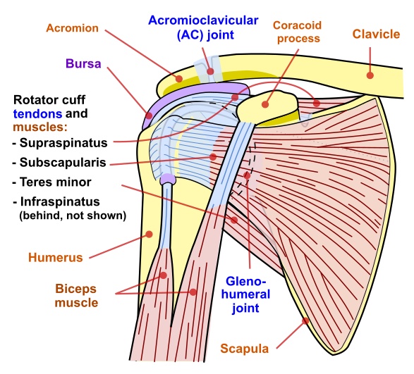
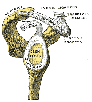
Glenohumeral joint
Rotator cuff
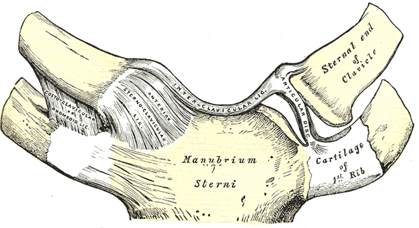
Sternoclavicular joint
Coracoclavicular joint
Elbow region
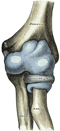
Elbow joint
The primary movements of the elbow that are aided by the four joints of the elbow are:
Elbow flexion/extension
Forearm supination/pronation
The elbow joint has other important anatomical features:
Capsule- thin, structure that surrounds the anterior and posterior components of the joint
Bursae- act as fluid-filled sac that provides cushioning and friction reduction between tendons, joints, muscles and bone
The ligaments that aid in movement of the elbow region are:
Ulnar collateral ligament
Ligament is triangle in shape running anteriorly, posteriorly, and obliquely to reinforce the medial humeroradial joint
Radial collateral ligament
Fan shaped ligament that runs from lateral epicondyle to annular ligament to support the humeroradial joint laterally
Annular ligament
Cone shaped ligament that envelopes the radial head and attaches to the medial ulna; provides protection to radial head
Quadrate ligament
Extends from radial notch on ulna surface to the neck of the radius; reinforces the inferior portion of the joint capsule
Distal radioulnar ligament
Comprised of anterior and posterior radioulnar ligament to provide strength to the capsule
Wrist and hand region
The primary movements of the wrist and hand region are:
Wrist flexion/extension
Wrist radial deviation/ulnar deviation
Finger flexion/extension
Finger abduction/adduction
Intrinsic finger movement
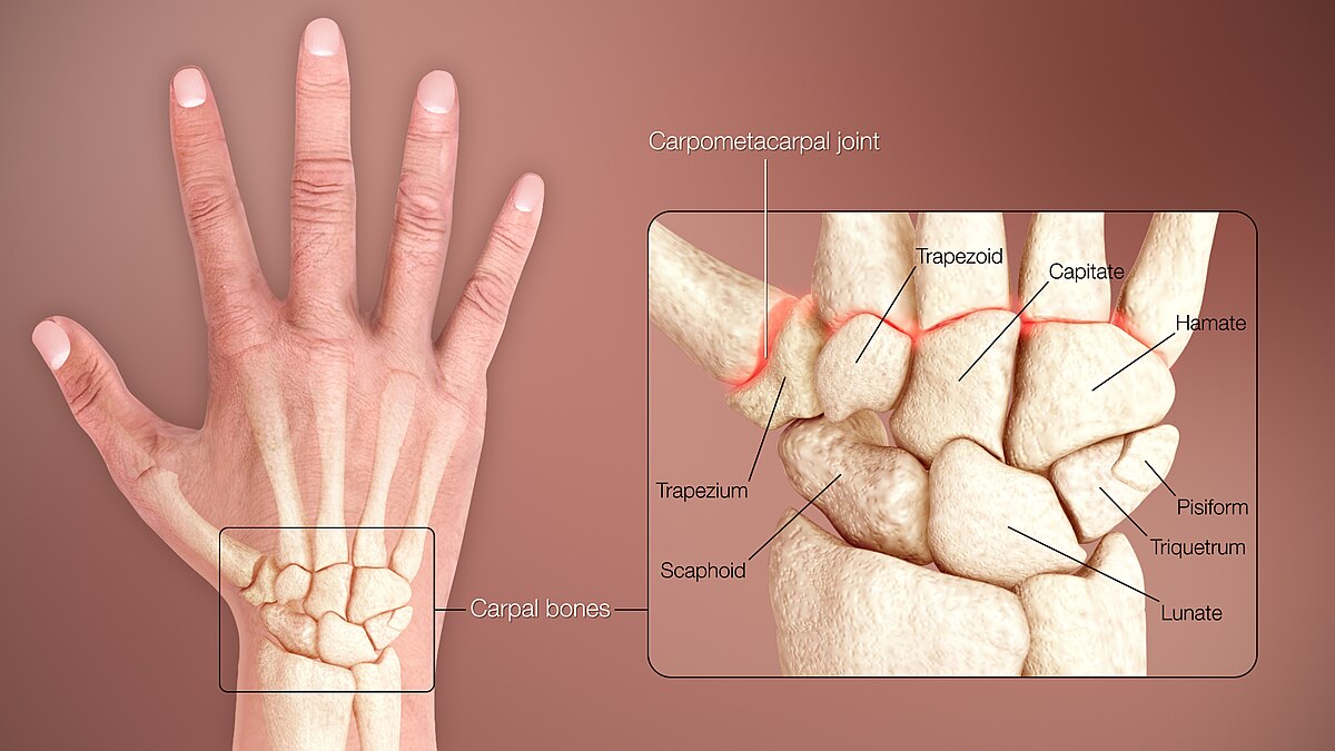
Carpal bones
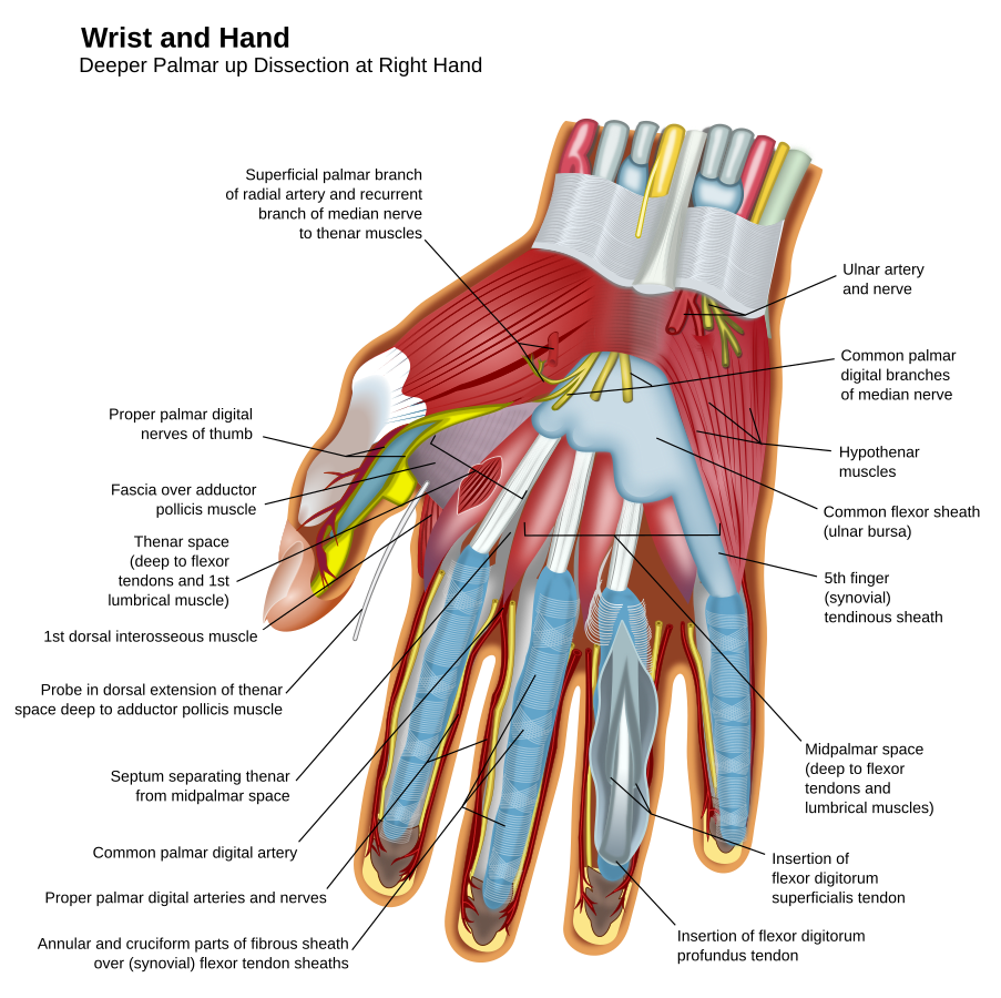
Anatomy of hand
The wrist and hand joint has other important anatomical features:
Capsule of wrist and hand- provide support to internal structures
Volar plate- present on palmar aspect of MCP, PIP, and DIP to protect joints
Extensor hood- fibrous connection on the dorsum of finger that aids in extension of the PIP and DIP
Nerves- radial, medial, and ulnar are the primary nerves of innervation for the wrist and hand
Ligaments of the wrist:
Dorsal radiocarpal
Limits flexion, pronation
Radiate
Stabilizes hand
Radial collateral ligament
Limits ulnar deviation
Ulnar collateral ligament
Limits radial deviation
Palmar ulnocarpal
Limits extension and supination
Palmar radiocarpal
Limits extension and supination through knuckles
Ligaments of fingers:
Collateral ligaments
Oriented from lateral condyle to distal phalanx and lateral volar plate to each metacarpal, PIP, and DIP
All fibers tighten during flexion but only volar fibers tighten during extension
Accessory
Oriented from condylar head to volar plate
Transverse
Provide stability linking MCP joints and reinforcing the anterior capsule
Upper extremity range of motion normals
Shoulder range
Elbow range
Wrist range
MCP range
PIP range
DIP range
1st CMC range
1st MCP range
1st IP range
Special tests of upper extremity
Shoulder special tests
Apprehension test (tests anterior instability)
Patient is supine with shoulder in 90 degrees abduction; therapist attempts to externally rotate
Positive: patient seems apprehensive about performing movement and resists motion
Sulcus sign (tests posterior and inferior instability)
Patient stands with arm relaxed at side; therapist pulls arm distally
Positive: presence of sulcus inferior to the acromion with symptom reproduction
Drop arm test
Patient seated with shoulder passively abducted to 120 degrees; patient instructed to slowly lower arm back to sign
Positive: patient is unable to lower arm down slowly and suddenly drops to side without control
Infraspinatus/supraspinatus muscle test
Patient is seated or standing and therapist resists external rotation with arm in neutral position and adducted to trunk
Positive: patient is unable to sustain external rotation
External rotation lag sign
Patient is seated or standing with shoulder passively abducted to 90 degrees and externally rotated
Positive: patient is unable to maintain external rotation
Internal rotation lag sign
Patient is seated with arm held behind back in internal rotation passively
Positive: patient is unable to maintain internal rotation
Empty can test
The patient stands or sits with their arms at their sides. The patient abducts their arm to 90 degrees, with their elbow extended. The patient internally rotates their shoulder, so that their thumbs point towards the floor. The examiner applies downward pressure on the patient’s wrist or forearm.
Positive: Pain in the shoulder, Weakness in the arm, and The patient’s arm dropping involuntarily.
Tests supraspinatus muscle
Neer’s
The patient sits comfortably, and the examiner stands behind them. The examiner stabilizes the patient’s scapula (shoulder blade) with one hand to prevent scapular movement during the test. The examiner passively flexes the patient’s arm forward while internally rotating it, bringing the greater tuberosity of the humerus (the bony bump on the upper arm) under the acromion.
Positive: The patient reports pain or tenderness during the arm movement, particularly in the anterior or lateral aspect of the shoulder.
Tests for impingement
Hawkins- Kennedy
The patient sits with their arm flexed at the shoulder and elbow to 90 degrees. The examiner stabilizes the patient’s shoulder with one hand and internally rotates the arm with the other hand.
Positive: The test is considered positive if the patient experiences pain in the anterior shoulder during internal rotation.
Tests for impingement
Acromioclavicular (AC) joint
Horizontal adduction test
Patient standing with shoulder flexed to 90 degrees and adducted across chest
Positive: localized pain over AC joint
SLAP (superior labrum anterior to posterior) lesions
O’Brien’s test
The patient stands or sits with their affected arm flexed at 90° and adducted 10–15°; the patient internally rotates their shoulder; the examiner applies downward pressure on the patient’s arm while the patient resists; repeated with upper extremity in external rotation
Positive: pain or clicking noise found when performing internal rotation and symptoms relieved when performing external rotation
Differential diagnosis needs to be made to determine if AC joint vs glenoidhumeral joint dysfunction
Bicep tendonitis tests
Bicep load II test
Patient in supine with shoulder abducted to 120 degrees elbow flexed to 90 degrees, forearm supinated; shoulder fully externally rotated; if the patient demonstrates apprehension when performing then asked patient to flex the elbow against resistance
Positive: if apprehension remains the same or shoulder becomes more painful
Yergasons test
Patient sitting with shoulder in neutral position against trunk, elbow at 90 degrees, and forearm pronated, resist supination of forearm and external rotation of shoulder
Tests for transverse ligament, bicipital tendonitis, and SLAP lesions
Positive: bicep tendon of long head will be palpable outside of bicipital groove or a reproduction of pain
Speed’s test
Patient sitting or standing with upper limb in full extension and forearm supination, resist shoulder flexion
Can also place shoulder in 90 degrees of flexion and push upper limb into extension (causing eccentric contraction)
Tests bicipital tendonitis and SLAP lesions
Positive: pain in anterior shoulder
Neurological dysfunction
Upper limb tension tests- assists with identifying peripheral nerve injury by placing the upper limb in positions of that will stress nerve
Upper limb tension test 1 (ULTT1)- median and anterior interosseous nerve
Cervical spine: contralateral lateral flexion
Shoulder: depression and abduction to 110 degrees
Elbow: extension
Forearm: supination
Wrist: extension
Fingers and thumb: extension
Upper limb tension test 2 (ULTT 2)- median, axillary, and musculocutaneous nerve
Cervical spine: contralateral lateral flexion
Shoulder: depression and abduction to 10 degrees, lateral rotation
Elbow: extension
Forearm: supination
Wrist: extension
Upper limb tension test 3 (ULTT 3)- radial nerve
Cervical spine: contralateral lateral flexion
Shoulder: depression and abduction to 10 degrees; internal rotation
Elbow: extension
Forearm: pronation
Wrist: flexion with ulnar deviation
Fingers and thumb: flexion
Upper limb tension test 4 (ULTT 4)- ulnar nerve
Cervical spine: contralateral lateral flexion
Shoulder: depression and abduction (10 - 90 degrees) with hand to ear
Elbow: flexion
Forearm: pronation
Wrist: extension and radial deviation
Fingers and thumb: extension
Thoracic outlet syndrome- assess for structural damage of nerves and arteries that pass through the thoracic inlet
Adson’s test (compression of the subclavian artery and/or nerves as it passes through the interscalene space)
Patient sitting with radial nerve palpated; head rotated toward extremity being tested with shoulder extended and externally rotated; extend head
Positive: reproduction neurological (pain, weakness, numbness, and loss of hand coordination) and vascular symptoms (loss of radial pulse)
Roos elevated arm test (nerves and/or blood vessels in the space between the collarbone and the first rib are compressed)
Patient standing with shoulders fully externally rotated, 90 degrees abducted, elbows flexed to 90 degrees- patient then rapidly opens and closes hand for 3 minutes
Positive: reproduction neurological (pain, weakness, numbness, and loss of hand coordination) and vascular symptoms (loss of radial pulse)
Wright test (compression at the space behind the pectoralis minor muscle)
Patient seated with passive movement of arm into abduction and external rotation
Positive: reproduction neurological (pain, weakness, numbness, and loss of hand coordination) and vascular symptoms (loss of radial pulse)
Costoclavicular test (compression of the neurovascular bundle between the clavicle and first rib)
To perform the test the patient sitting, the therapist assists the patient in performing the following 4 movements: scapula retraction, scapula depression, elevation, and protraction- the patient holds each position for up to 30 seconds, while the patient rests his or her forearms on his thighs
Positive: reproduction neurological (pain, weakness, numbness, and loss of hand coordination) and vascular symptoms (loss of radial pulse)
Elbow special tests
Elbow extension test
Patient in seated position attempts to fully extend elbow
Positive: patient unable to extend due to possible fracture- imaging will be needed to confirm
Varus/valgus test
Patient siting or supine with elbow flexed to 20 degrees; valgus force applied to test ulnar collateral ligament and then varus force applied to test for radial collateral ligament
Positive; joint laxity and possible pain- needs to be performed bilaterally to determine joint laxity
Bicep rupture sign
Observation of distal bunching of bicep muscle along with complete loss of function (unable to perform elbow flexion)
Positive: Indicates rupture of proximal long head of biceps tendon
Cozen’s test
Patient can be seated or standing. Position the patient with their elbow extended, forearm in pronation, wrist in slight radial deviation, and then ask them to make a fist and resist wrist extension while the examiner palpates the lateral epicondyle.
Positive: Pain indicates lateral epicondylitis
Mills test
Patient can be seated or standing. The patient extends their arm straight out in front of them with their palm facing down. The examiner then flexes the patient’s wrist and supinates their forearm, causing a stretch in the flexor tendons.
Positive: Pain with this maneuver suggests medial epicondylitis.
Neurological dysfunction
Elbow flexion test
Patient supine with shoulder in full external rotation and elbow held in maximal flexion with wrist extended for one minute
Positive: pain present at medial elbow with hypoanesthesia n ulnar distribution of involved side
Entrapment of ulnar nerve at cubital tunnel
Wrist and hand special tests
Ligamentous, capsule, and joint instability
Watson (scaphoid shift)
Patient seated with elbow rested on table, forearm pronated, wrist palace in full ulnar deviation, with slight extension while stabilization of metacarpals by therapist. Pressure placed on side of scaphoid while radially deviating and flexing wrist
Positive: painful shift of scaphoid with a “clunk” sound when pressure is removed (wrist placed into resting position) indicates carpal instability
Interphalangeal joint varus/valgus tests
Patient in seated position with fingers supported and stabilized; valgus/varus force applied to PIP and DIP joints of all digits
Positive: joint laxity and possible pain - needs to be performed bilaterally to determine extent of laxity
Tendon and muscle
Wrist hyperabduction and abduction of thumb test (WHAT)
Patient in seated position with wrist hyperflexed and thumb abducted in full MCP and IP extension with resistance applied against therapist’s index finger
Positive: reproduction of pain in wrist- needs to be performed bilaterally
Indicates de Quervain’s tenosynovitis
Most preferred test due to sensitivity for de Quervain’s tenosynovitis
Eichoff’s test
Patient seated makes fist with thumb flexed within fingers while examiner passively moves wrist into ulnar deviation
Positive: reproduction of pain in wrist- needs to be performed bilaterally
Indicates de Quervain’s tenosynovitis
2nd preferred test due to sensitivity for de Quervain’s tenosynovitis
Finkelstein’s test
Patient seated while therapist passively pulls the thumb and wrist into ulnar deviation and pulses in longitudinal direction
Positive: reproduction of pain in wrist- needs to be performed bilaterally
Indicates de Quervain’s tenosynovitis
3rd preferred test due to sensitivity for de Quervain’s tenosynovitis
Neurological dysfunction
Phalen’s test (wrist flexion test)
Patient in seated position maximally flexes both wrists while holding them together for one minute
Positive: reproduces tingling sensation or paresthesia in median nerve distribution
Indicates carpal tunnel syndrome
Tinel’s test
Patient in seated position and therapists taps peripheral nerve (can be any nerve palpable)
Positive: reproduces tingling sensation or paresthesia nerve distribution
Vascular dysfunction
Modified Allen’s test
Patient in seated position has therapist palpate radial and ulnar nerve followed by patient quickly opening and closing hand several times; patient then makes a fist
Therapist then compresses radial artery, has patient open hand, observes palm of hand, releases compressed radial artery, and observes for radial filling time; the same procedure will be done with ulnar artery
Positive: abnormal re-filling time- needs to be performed bilaterally
Sign up for free to take 15 quiz questions on this topic