Spine, pelvis, and temporomandibular joint anatomy
Spine and pelvis region
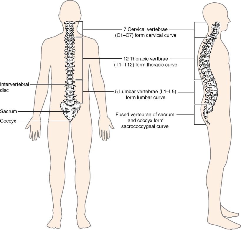
Components of spine (vertebral column)
- 24 vertebrae
- 2 fused bones: the sacrum and coccyx
- Divided into 5 areas — cervical, thoracic, lumbar, sacral, and coccygeal
Function of the spine
- Provides a strong and flexible framework that supports the body’s weight, allowing for standing, walking, and moving freely.
- Encases and protects the spinal cord
- Connecting discs allow for a wide range of motion, including flexion, extension, rotation, and lateral flexion.
- The thoracic spine and ribs protect vital internal organs such as the heart and lungs.
- Maintains proper balance and posture by distributing the body’s weight evenly.
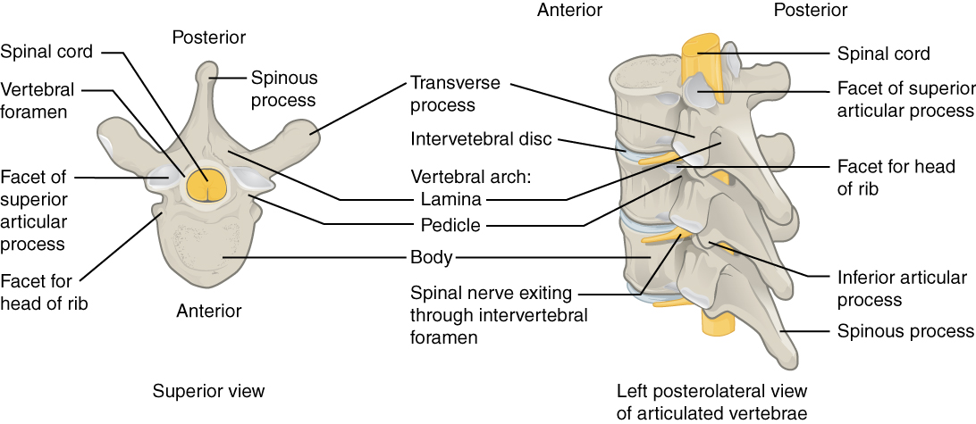
Bony structures
- Typical components in definitions below
Special bony components of each segment
-
Cervical: supports the head and neck; muscles are attached to allow a large freedom of movement
- C1 and C2 allow for rotation without compression of the spinal cord
-
Costotransverse and costovertebral
- Articulation between ribs and transverse processes of the spine
- Prominent (easily palpable) spinous processes
-
Thoracic: supports axial loading, attached to ribs to form the most rigid section of the spine.
- Prominent (easily palpable) spinous processes
-
Lumbar: the largest vertebrae transfer weight. Bearing loads and increasing mobility of the trunk
-
Sacrum
- Composed of 5 fused bones
Special joints of the spine
- Atlanto-occipital joint- articulation between the occipital bone and C1, which allows head nodding to occur
- Atlanto-axial joint- articulation between C1 and C2 which allows for head rotation
- Facet joints
- Aid in the movement of the spine as a whole
- Intervertebral joints
- Allow for movement at a single vertebral body and assist with transferring the load from one vertebral body to another
- Sacroiliac joints
- Aid in movements of the lower axial skeleton and lower extremities
Other important features of the spine
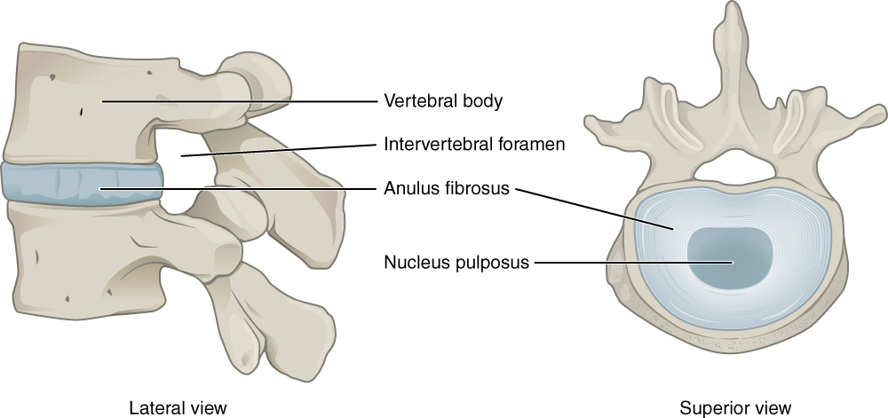
- Intervertebral discs
- Annulus fibrosus
- Composed of collagen fibers, fibrocartilage, and water
- Vascularized and neural connections
- Functions to maintain the integrity of the vertebral column during compression, torsion, shearing, and distracting forces
- Nucleus pulposus
- Composed of proteoglycans and water with minimal collagen
- Avascular and has no neural connections
- Functions to maintain the integrity of the vertebral column during compression, torsion, shearing, and distracting forces
- Vertebral endplate
- Composed of proteoglycans, collagen, fibrocartilage, and water
- Functions to diffuse nutrients to the annulus fibrosus and the nucleus pulposus
- Annulus fibrosus
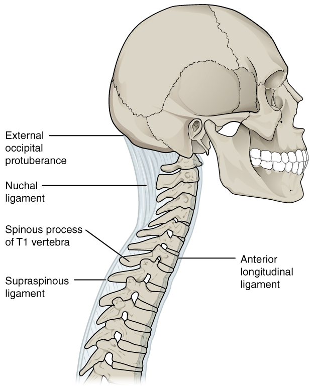
Ligaments
- Alar
- Connects dens to the occipital condyle
- Functions to limit flexion, contralateral lateral flexion, and contralateral rotation
- Anterior longitudinal ligament
- Connects the anterior and lateral surfaces of vertebral bodies from C2 to the sacrum
- Functions to reinforce the anterolateral portion of the intervertebral discs
- Posterior longitudinal ligament
- Located on the posterior surface of vertebral bodies from C2 to the sacrum
- Functions to prevent hyperflexion of the vertebral column and to protect the spinal cord from foreign objects and displaced disc material.
- Ligamentum flavum
- Connects vertebra to the lamina above it; runs from C2 to the sacrum
- Functions to limit flexion (greatest in lumbar spine)
- Interspinous ligament
- Runs between spinous processes
- Functions to limit flexion
- Iliolumbar ligament
- Connects L5 vertebrae to the ilium
- Functions to limit motion between L5 and S1
Capsule
- Facet joints
- Role is to reinforce ligaments by limiting motion and stabilizing the spine
- Sacroiliac joints
- Role is to stabilize the sacroiliac joint, limiting its movement, and protecting the joint by distributing biomechanical loads.
Nerves
- Spinal nerves are mixed nerves that connect the spinal cord to the rest of the body. They carry both sensory (dorsal rami) and motor (ventral rami) information between the central nervous system (brain and spinal cord) and the peripheral nervous system (body and muscles).
- Location of spinal nerves
- Cervical: exit at the level above the associated vertebrae
- Thoracic/lumbar: exit below the level of associated vertebrae
Spinal and pelvis movements
General rules regarding facet joint opening and closing
- Flexion (bending forward):
- When you bend forward, the facet joints on the back of the spine open up, allowing for more space between the vertebrae.
- Extension (bending backward):
- Conversely, when you bend backward, the facet joints close together, creating a more stable and compressed position.
- Side bending:
- During side bending, the facet joints on the side you are bending towards close, while the facet joints on the opposite side open.
- Rotation:
- During rotation, the facet joints on the ipsilateral side compress together, while the facet joints on the opposite side tend to open.
Joint movements (arthrokinematics)
- Flexion
- The inferior facet of the vertebra above slides upward and forward relative to the superior facet of the vertebra below
- Extension
- The superior facets glide downward and backward, while the inferior facets, the lower articular processes, move upward and forward.
Lateral bending
- Right lateral bending The left (contralateral) facet joint moves upward and anteriorly to open, while the right (ipsilateral) facet joint moves down and posteriorly to close. * Causing facet closure on the right side and opening on the left side
- Left lateral bending The left (contralateral) facet joint moves upward and anteriorly to open, while the right (ipsilateral) facet joint moves down and posteriorly to close. * Causing facet closure on the left side and opening on the right side
Cervical rotation
-
Right rotation The left facet joint of the upper vertebra moves anteriorly (forward) while the right facet joint moves posteriorly (backward). At the same time, the intervertebral foramen on the right side (ipsilateral side of the rotation) closes, and the foramen on the left side (contralateral side) opens.
-
Left rotation The right facet joint of the upper vertebra moves anteriorly (forward) while the left facet joint moves posteriorly (backward).
-
At the same time, the intervertebral foramen on the left side (ipsilateral side of the rotation) closes, and the foramen on the right side (contralateral side) opens.
Coupled movements
- Cervical spine
- C1: lateral bending and rotation occur in different directions (occurs if the spine is in neutral, flexion, and extension)
- C2-C7: lateral bending and rotation occurring in the same direction (occurs if the spine is in neutral, flexion, and extension)
- Lumbar/thoracic spine
- If the spine is in flexion, side bending and rotation will occur in the same direction
- If the spine is in extension/neutral, side bending and rotation will occur in opposite directions
- Lumbopelvic joint
- When in flexion: lumbar spine flexes followed by pelvis rotation anteriorly, ending with hip flexion
- When in extension (when coming from a flexed position): the hips extend, followed by the pelvis rotating posteriorly, and the spine extending
- Sacroiliac joint
- Nutation: flexion of the sacrum causes posterior tilt of the ilium (in the frontal plane)
- Counternutation: extension of the sacrum causes an anterior tilt of the ilium (in the frontal plane)
- Anterior innominate: anterior superior iliac spine (ASIS) moves downward (in the sagittal plane)
- Posterior innominate: anterior superior iliac spine (ASIS) moves upward (in the sagittal plane)