Lower extremity anatomy
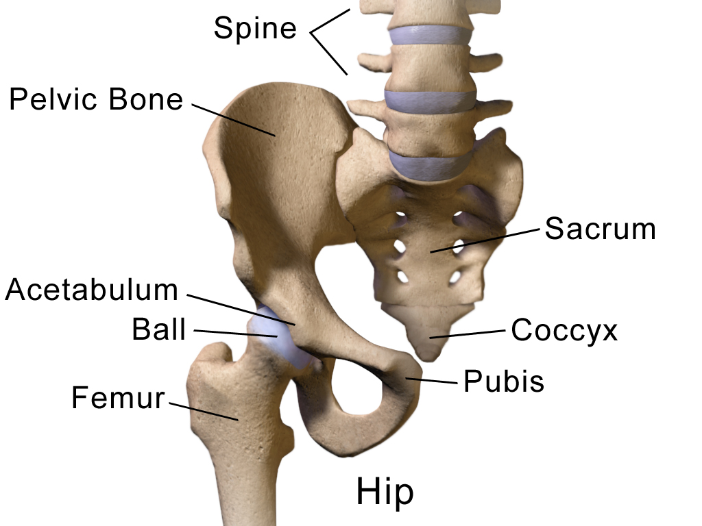
Hip region
The hip region comprises two (2) bony structures- the acetabulum and femur.
The normal angle of inclination is 115-125 degrees (angle of inclination between femur and acetabulum)- if the angle is >125 degrees, then referred to as coxa valga; if the angle is <115 degrees, then referred to as coxa varus. Femoral neck angle is positioned anteriorly at a 10-15 degree angle; excessive anterior rotation >25 degrees is anteversion, and excessive posterior rotation <10 degrees is retroversion.
The primary movements of the hip are:
- Hip flexion/extension
- Hip external rotation/internal rotation
- Hip abduction/adduction
The hip joint is a stable synovial joint due to the bony anatomy and strength of ligaments, capsule, and labrum.
- Capsule encloses the entire joint
- Labrum
- Attached to the acetabulum and serves to deepen the structure to allow for greater articulation
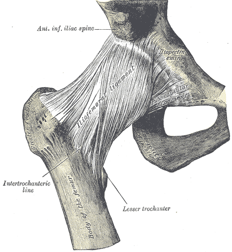
- Ligaments
- Iliofemoral
- Two (2) bands that originate at the anterior iliac spine (ASIS), run medially to the distal intertrochanteric line, and laterally to the proximal aspect of the intertrochanteric line
- Both bands tighten with extension and external rotation; the superior band tightens with adduction; the inferior band tightens with abduction
- Pubofemoral
- Band tightens with extension, external rotation, and abduction
- Ischiofemoral
- Band tightens with medial rotation, abduction, and extension
- Iliofemoral
Other pertinent structures of the hip joint are:
- Inguinal ligament — forms a tunnel for vital arteries, veins, and nerves in the lower extremity
- Bursae — act as fluid-filled sacs that provide cushioning and friction reduction between tendons, joints, muscles, and bone
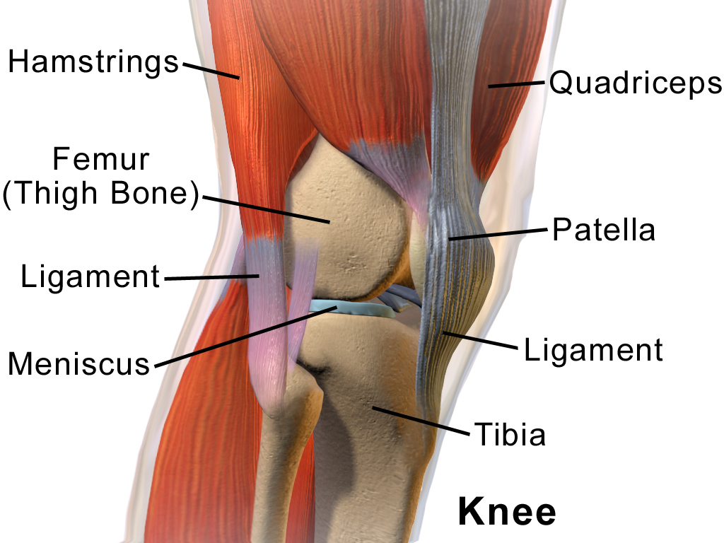
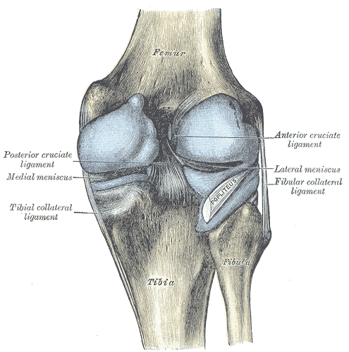
Knee region
The knee region is composed of four (4 bony the femur, tibia, fibula, and patella.
These bony structures then form three (3) joints: tibiofemoral, patellafemoral, and proximal tibiofibular joint.
The primary movements of the knee that are aided by the three joints of the knee are:
- Knee flexion/extension
Other pertinent structures of the knee joint are:
- Capsule
- Tibiofemoral capsule covers the distal femur and proximal tibia- posteriorly divided into medial and lateral sections, anterior cut-out for the patella
- Proximal tibiofibular capsule is continuous with the knee 10% of the time
- Ligaments
- Tibiofemoral and patellofemoral joints
- Medial collateral ligament
- Tightened in extension; slackened in flexion
- Prevents internal rotation and provides stability against valgus forces
- Lateral collateral ligament
- Tightened in extension; slackened in flexion
- Prevents external rotation and provides stability against varus forces
- Anterior cruciate ligament
- Prevents anterior displacement of the tibia on the femur and provides rotational stability
- Posterior cruciate ligament
- Prevents posterior displacement of the tibia on the femur
- Medial collateral ligament
- Proximal tibiofibular joint ligaments
- Anterior tibiofibular ligament
- Reinforces anterior capsule
- Posterior tibiofibular
- Reinforces the posterior capsule
- Anterior tibiofibular ligament
- Tibiofemoral and patellofemoral joints
- Menisci
- Function
- Deepen the fossa of the tibia
- Increased congruency of the tibia and femur
- Reduces friction between joints during movement
- Improves weight distribution
- Provides shock absorption and lubrication to the knee
- Provide stability to the tibiofemoral joint
- Lateral meniscus
- Outer side of joint
- Attached to the popliteus and the joint capsule
- Stabilizes the knee against lateral rotation and tibial rotation
- Medial meniscus
- Inner side of joint
- Attached to the medial collateral ligament and joint capsule
- Stabilizes the knee against medial rotation and tibial translation
- Function
- Bursae- act as fluid-filled sacs that provide cushioning and friction reduction between tendons, joints, muscles, and bone
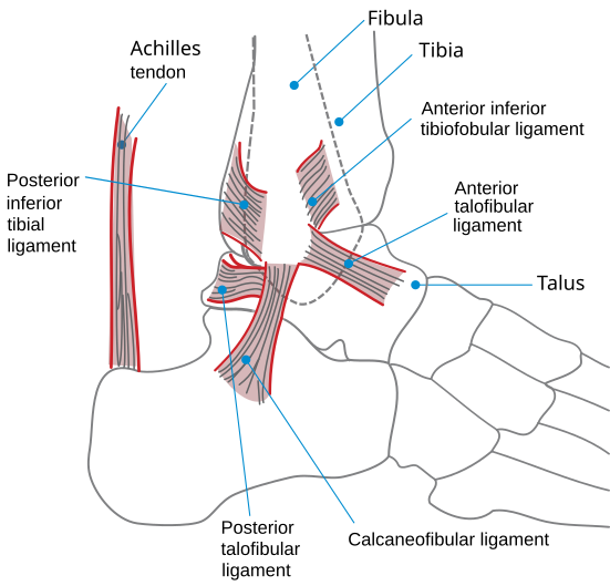
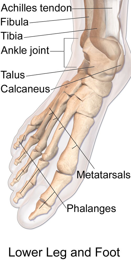
Foot and ankle region
The ankle/foot joint is composed of eight (4) major articulations, which include the talocrural, subtalar, metatarsophalangeal, and interphalangeal joints.
The primary movements of the foot/ankle joint are:
- Ankle plantarflexion/dorsiflexion
- Ankle inversion/eversion
Other pertinent structures of the ankle/foot joint are:
- Capsule
- Provides stability to the joints of the ankle/foot
- Ligaments
-
Talocrural joint
- The medial collateral ligament (Deltoid ligament) has deep and superficial fibers
- Lateral collateral ligament: Anterior talofibular, calcaneofibular, posterior talofibular
-
Syndesmosis tibiofibular joint (high ankle ligaments) * Anterior inferior tibiofibular ligament * Posterior inferior tibiofibular ligament
-
Metatarsophalangeal joint
- Plantar ligaments
- Collateral ligaments
-
Interphalangeal joint
- Plantar ligament
- Collateral ligament
-
- Plantar fascia
- Collagen fibers connecting from the medial calcaneus to the phalanges
- Fascia tightens during dorsiflexion- leads to supination and inversion
- Bursae - act as fluid-filled sacs that provide cushioning and friction reduction between tendons, joints, muscles, and bone