Comparing clinical presentation and interventions for upper extremity
Contrasting shoulder conditions
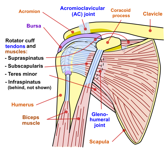
- Glenohumeral dislocations and instability
- Dislocations of the glenohumeral joint caused by traumatic or atraumatic reasons
- Trauma is due to direct injury, most commonly a fall on an outstretched hand (FOOSH) mechanism of injury
- Atraumatic can be due to repetitive injury, causing hypermobility
- Types of dislocations:
- Anterior-inferior dislocations: Most common type (95%). Mechanism is a combination of motions including: excessive horizontal abduction, abduction, external (lateral) rotation, and extension/hyperextension of the upper extremity.
- If traumatic can lead to: Disruption anterior glenohumeral/capsular ligament, subscapularis, and anterior/inferior glenoid labrum
- Sulcus sign: a depression or groove appears between the acromion and the humeral head.
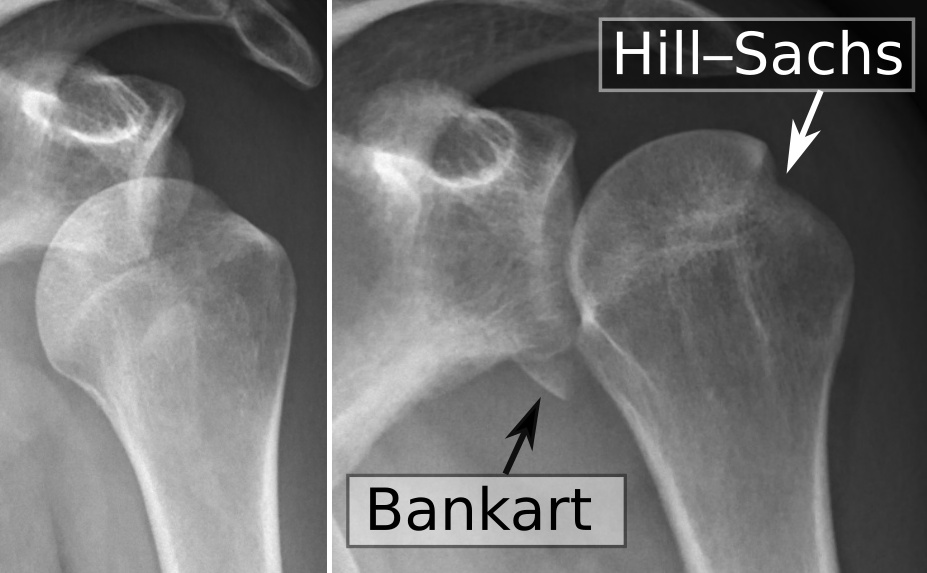
- Hills-Sachs lesion: compression fracture of the posterior humeral head
- Bankart lesion: avulsion of the anterior-inferior glenoid labrum
- Axillary nerve injury: numbness, tingling, and weakness in the deltoid
- If traumatic can lead to: Disruption anterior glenohumeral/capsular ligament, subscapularis, and anterior/inferior glenoid labrum
- Anterior-inferior dislocations: Most common type (95%). Mechanism is a combination of motions including: excessive horizontal abduction, abduction, external (lateral) rotation, and extension/hyperextension of the upper extremity.
- Dislocations of the glenohumeral joint caused by traumatic or atraumatic reasons
- Posterior dislocations: rare, caused by horizontal adduction and internal rotation
- Labral tears
-
Tear in the cartilage ring that surrounds the shoulder joint; divided into above the middle of the socket and below the middle of the socket
- Above the middle of the socket is called a SLAP (superior labral anterior-posterior) tear; can also involve the biceps tendon
- Below the middle of the socket is called a Bankart lesion; it can also involve an avulsion fracture of the anterior/inferior lip of the glenoid (bony Bankart)
-
Labral tears are associated with traumatic injury or repetitive shoulder dislocations
-
- Rotator cuff tendonitis
- Caused by mechanical impingement of the distal attachment of the rotator cuff, causing inflammation of the tendons
- Increased risk for development of tendonitis due to poor vascularity at attachment sites
- Caused by mechanical impingement of the distal attachment of the rotator cuff, causing inflammation of the tendons
- Impingement syndrome
- Impingement (entrapment) of the rotator cuff tendon against the acromion due to mechanical repetition
- Rotator cuff tear/damage
- Causes acute tear from a fall on an outstretched hand (FOOSH), sudden heavy lifting, or the shoulder in an awkward position
- Partial and degenerative damage from an increased risk for the development of tendonitis due to poor vascularity at attachment sites
- Subacromial/subdeltoid bursitis
- Subacromial and subdeltoid bursae become inflamed (close relationship with rotator cuff tendonitis )
- The bursa becomes trapped (impinged) beneath the acromion arch
- Subacromial and subdeltoid bursae become inflamed (close relationship with rotator cuff tendonitis )
- Bicipital tendonitis
- Inflammation of the long head of the biceps
- Cases can be mechanical trapping (impingement) of the long head of the biceps between the acromion and the bicipital groove of the humerus
- Inflammation of the long head of the biceps
- Adhesive capsulitis
- Restriction in shoulder motion due to inflammation of the joint capsule
- Restrictions are in external rotation (greatest), abduction, and flexion (capsular pattern of the shoulder)
- Reason for diagnosis can be repetitive motion, diabetes, cardiovascular disease, or thyroid disease
- Restriction in shoulder motion due to inflammation of the joint capsule
- Acromioclavicular and sternoclavicular disorders
- Occurs when falling on the adducted shoulder or when in collision with another individual, particularly during a sporting event
- Grades of injury
- Type I
- A minor sprain of the acromioclavicular ligament
- No radiographic displacement
- No tear of the acromioclavicular or coracoclavicular ligament
- Type II
- A tear of the acromioclavicular ligament, but not the coracoclavicular ligaments
- Less than 25% increase in the coracoclavicular interspace
- Type III
- Tears of both the acromioclavicular and coracoclavicular ligaments
- 25% to 100% displacement of the clavicle
- Type IV
- Tears of both the acromioclavicular and coracoclavicular ligaments
- Posterior displacement of the distal clavicle into the trapezius fascia
- Type I
- Grades of injury
- Occurs when falling on the adducted shoulder or when in collision with another individual, particularly during a sporting event
- Proximal humeral fracture
- Occurs due to a fall on an outstretched arm and motor vehicle accident
- Stable fractures that do not require surgery
- Occurs due to a fall on an outstretched arm and motor vehicle accident
- Distal humeral fracture
- Trauma causes a fracture at the distal humerus
- Immediate attention must be given to whether a supracondylar fracture is due to the increased likelihood of neurovascular involvement
-
Radial nerve involvement associated with posterior type injuries may lead to damage of vascular structures, pulselessness, and/or paralysis
-
Ulnar nerve involvement associated with flexion type injuries may lead to paralysis and a loss of fine motor control in the hand
-
In children, it can cause malunion due to growth plate involvement
-
- Lateral epicondyle fractures will require internal fixation (rod and screws implanted in arm) for adults, percutaneous removal pins for non-skeletally mature fractures, for proper alignment
- Immediate attention must be given to whether a supracondylar fracture is due to the increased likelihood of neurovascular involvement
- Trauma causes a fracture at the distal humerus
- Thoracic outlet syndrome
- Compression of the neurovascular bundle that includes the brachial plexus, sympathetic trunk, subclavian artery and vein, and phrenic and vagus nerves due to alteration in thoracic outlet size
- Common areas of compression are:
- Superior thoracic outlet
- Scalene triangle
- Between the clavicle and the first rib
- Between the pectoralis minor and the thoracic wall
Contrasting elbow conditions
- Medial epicondylitis
- Inflammation of the pronator teres and the flexor carpi radialis tendons at the attachment of the medial epicondyle
- Typically due to overuse in activities that require excessive pronation at the forearm
- Commonly referred to as golfer’s elbow
- Typically due to overuse in activities that require excessive pronation at the forearm
- Inflammation of the pronator teres and the flexor carpi radialis tendons at the attachment of the medial epicondyle
- Lateral epicondylitis
- Inflammation of the extensor carpi radialis brevis tendon at its attachment to the lateral epicondyle
- Gradual onset occurring with repetitive wrist extension, resulting in overloading of the extensor carpi radialis
- Inflammation of the extensor carpi radialis brevis tendon at its attachment to the lateral epicondyle
- Ulnar collateral ligament injuries
- Due to repetitive valgus stress to the medial elbow, causing stress to the ulnar collateral ligament
- Elbow dislocation
- Caused by trauma to the elbow, causing misalignment from the anatomical position
- Posterior dislocation is the most common
- Posterolateral dislocation occurs as a result of hyperextension from a fall on an outstretched arm
- Posterior dislocations commonly cause an avulsion fracture of the medial epicondyle
- Complete dislocation will impact all of the following structures
- Lateral collateral ligament, anterior capsule, brachialis muscle, wrist flexor muscles, and wrist extensor muscles
- Posterior dislocation is the most common
- Caused by trauma to the elbow, causing misalignment from the anatomical position
- Nerve entrapments
- Medial nerve entrapment
- Tightness of the pronator teres muscle and under the superficial head of the flexor digitorum superficialis secondary to repetitive gripping activities
- Symptoms
- Pain, numbness, tingling, and weakness in the median nerve distribution in the forearm and below
- Diagnosis
- Clinical presentation
- Manual muscle test of forearm muscles
- Positive Tinel’s test in the median nerve distribution
- Radial nerve entrapment
- Entrapment of the posterior interosseous nerve within the radial tunnel as a result of overhead activities and throwing
- Symptoms
- Lateral elbow pain
- Pain, numbness, tingling, and weakness in the radial nerve distribution in the forearm and below
- Diagnosis
- Clinical presentation
- Manual muscle test of forearm muscles
- Positive Tinel’s test in radial nerve distribution
- Ulnar nerve entrapment
- Compression or trauma at the cubital tunnel, thickened retinaculum, or hypertrophy of the flexor carpi ulnaris muscle
- Symptoms
- Medial elbow pain
- Pain, numbness, tingling, and weakness in the ulnar nerve distribution in the forearm and below
- Diagnosis
- Clinical presentation
- Manual muscle test of forearm muscles
- Positive Tinel’s test in the ulnar nerve distribution
- Medical management for all nerve entrapments
- Acetaminophen or non-steroidal anti-inflammatory (NSAIDs)
- Physical therapy management for all nerve entrapments
- Early interventions — rest, modalities to reduce inflammation/pain
- Medial nerve entrapment
Contrasting conditions of wrist and hand
- Carpal tunnel syndrome
- Compression of the median nerve at the carpal tunnel at the wrist due to inflammation of the wrist flexor tendon or inflammation of the median nerve
- Caused by repetitive wrist motions; other causes may be pregnancy, diabetes, or rheumatoid arthritis
- Compression of the median nerve at the carpal tunnel at the wrist due to inflammation of the wrist flexor tendon or inflammation of the median nerve
- De Quervain’s tenosynovitis
- Inflammation of the extensor pollicis brevis and abductor pollicis longus
- Due to repetitive microtrauma or occur during pregnancy
- Inflammation of the extensor pollicis brevis and abductor pollicis longus
- Colles fracture
- Fracture causing posterior displacement of the distal radius with radial shift of the wrist and hand
- Most common fracture from falling on an outstretched hand with the wrist in extension and radial deviation.
- Can cause median nerve damage if edema is unmanaged
- Fracture causing posterior displacement of the distal radius with radial shift of the wrist and hand
- Scaphoid fracture
- Due to falling onto an outstretched hand
- This is the most common fractured carpal bone
- Due to falling onto an outstretched hand
- Dupuytren’s contracture
- Contracture of the palmar fascia leading to flexion of the digits towards the palm
- Common in the metacarpophalangeal (MCP) and proximal interphalangeal (PIP) joints of the fourth and fifth digits in non-diabetic and the third and fourth in diabetic
- Contracture of the palmar fascia leading to flexion of the digits towards the palm
- Boutonnière deformity
- Rupture of the central tendon slip of the extensor hood
- Commonly occurs after trauma to the hand or with the diagnosis of rheumatoid arthritis
- Deformity noted is extension of MCP and DIP with flexion of PIP
- Rupture of the central tendon slip of the extensor hood
- Swan neck deformity
- Contracture of intrinsic muscles with dorsal subluxation of lateral extensor tendons
- Commonly occurs after trauma to the hand or with the diagnosis of rheumatoid arthritis
- Deformity noted is flexion of the MCP and DIP with hyperextension of the PIP
- Contracture of intrinsic muscles with dorsal subluxation of lateral extensor tendons
- Mallet finger
- Rupture or avulsion of the extensor tendon at its insertion into the distal phalanx digit
- Commonly occurs after trauma, forcing the distal phalanx into a flexed position
- Deformity noted is flexion of the DIP
- Rupture or avulsion of the extensor tendon at its insertion into the distal phalanx digit
- Ape hand deformity
- Median nerve dysfunction causes thenar muscle weakness with the first digit moving dorsally until it becomes aligned with the second digit