



Elbow, wrist, and hand tests
Each test below follows the same pattern: a technique the clinician performs and a positive finding that points to a specific structure or condition. Laxity and vascular tests are compared bilaterally so a finding can be judged as a side-to-side difference.
Elbow special tests
Fracture and ligamentous instability tests
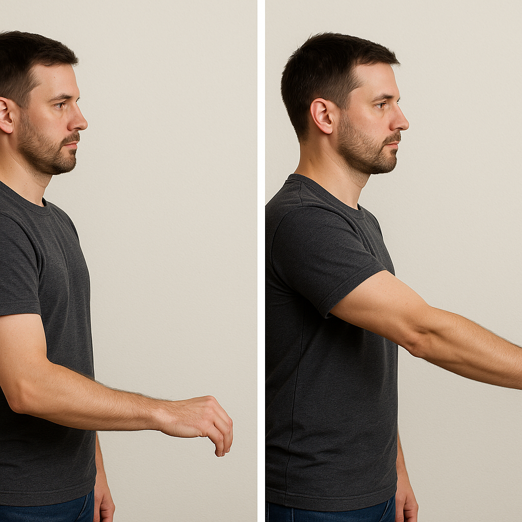
- Elbow extension test
- Patient in seated position attempts to fully extend the elbow
- Positive: patient unable to extend due to possible fracture - stop and report to the supervising PT; imaging may be needed
- Patient in seated position attempts to fully extend the elbow
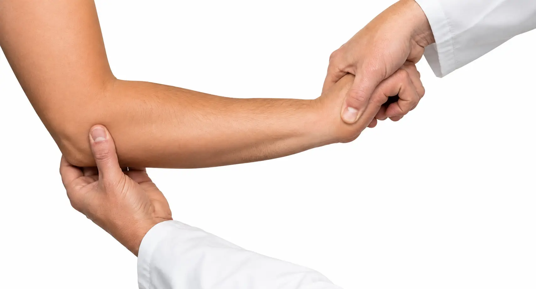
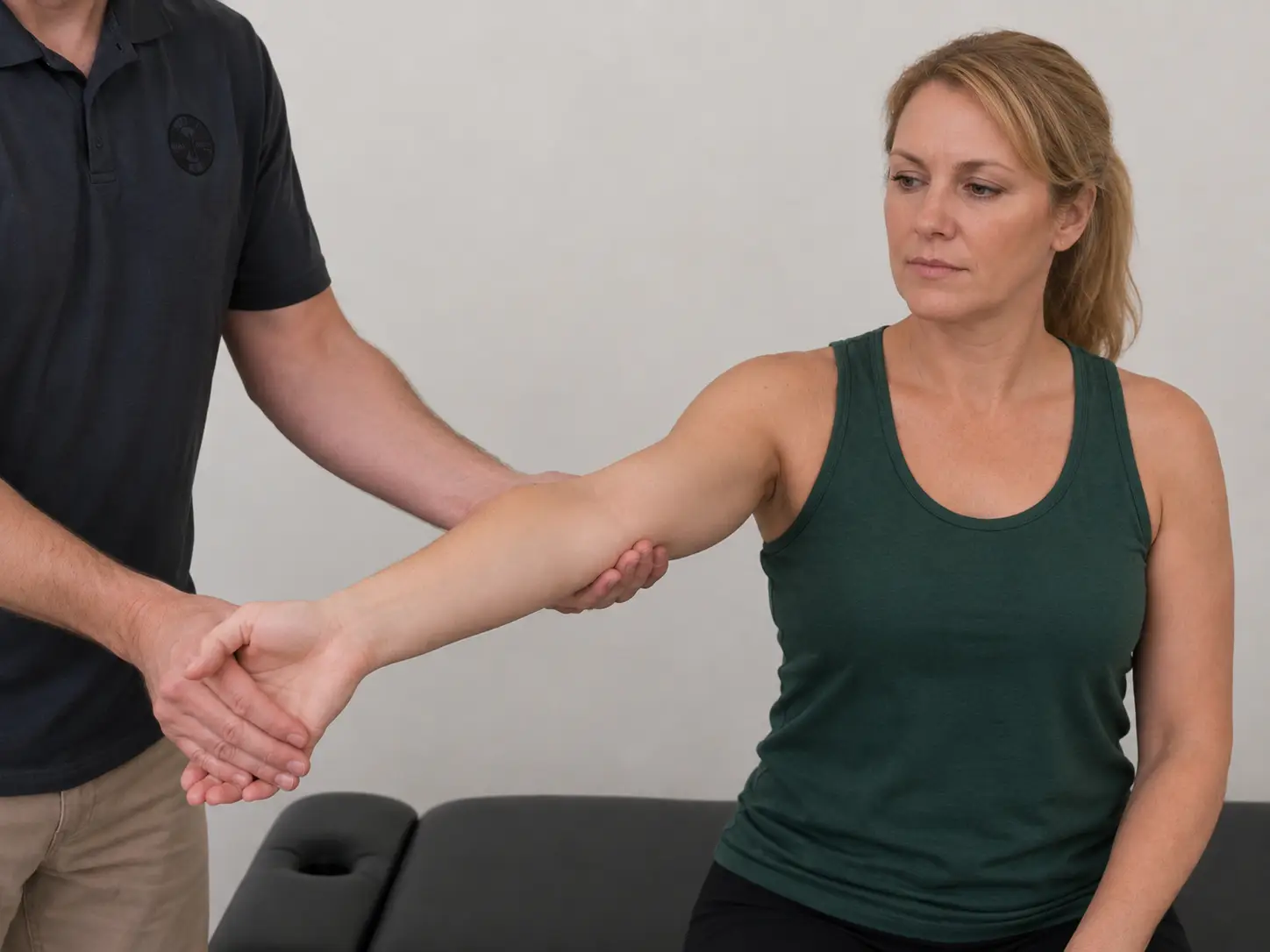
- Varus/valgus test
- Patient sitting or supine with elbow flexed to 20 degrees; valgus force applied to test the ulnar collateral ligament, then varus force applied to test the radial collateral ligament
- Positive: joint laxity and possible pain
- Patient sitting or supine with elbow flexed to 20 degrees; valgus force applied to test the ulnar collateral ligament, then varus force applied to test the radial collateral ligament
Muscle and tendon rupture tests
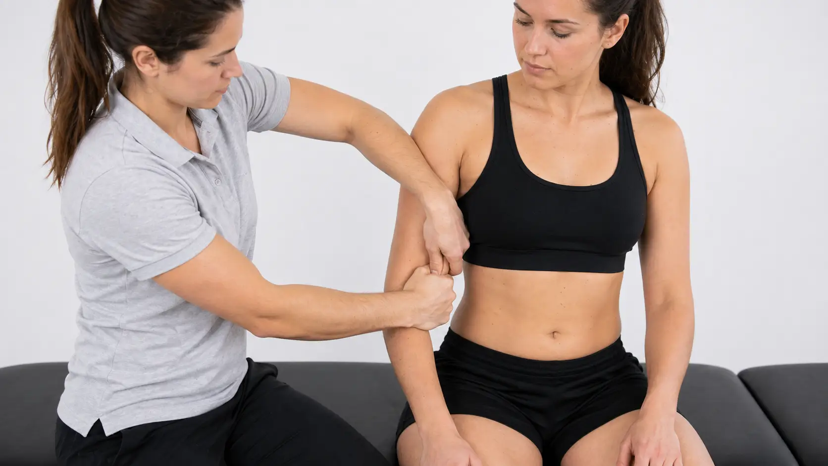
- Biceps squeeze test (for distal biceps tendon rupture)
- The clinician squeezes the biceps muscle belly with the patient’s elbow flexed to about 60-80 degrees, similar to the Thompson test for the Achilles tendon
- Positive: absence of forearm supination when the muscle is squeezed, indicating rupture of the distal biceps tendon at the elbow
- The clinician squeezes the biceps muscle belly with the patient’s elbow flexed to about 60-80 degrees, similar to the Thompson test for the Achilles tendon
Lateral epicondylitis tests
- Cozen’s test
- Patient seated or standing with the elbow extended, forearm pronated, and wrist in slight radial deviation; the patient makes a fist and extends the wrist against the clinician’s resistance while the clinician palpates the lateral epicondyle
- Positive: pain indicates lateral epicondylitis
- Patient seated or standing with the elbow extended, forearm pronated, and wrist in slight radial deviation; the patient makes a fist and extends the wrist against the clinician’s resistance while the clinician palpates the lateral epicondyle
- Mills test
- Patient seated; the clinician palpates the lateral epicondyle with one hand while pronating the patient’s forearm and fully flexing the wrist, elbow extended
- Positive: pain with this maneuver suggests lateral epicondylitis
- Patient seated; the clinician palpates the lateral epicondyle with one hand while pronating the patient’s forearm and fully flexing the wrist, elbow extended
Medial epicondylitis tests
- Reverse Mills test (also called the golfer’s elbow test)
- Patient seated or standing; the clinician stabilizes the patient’s humerus, palpates the medial epicondyle, then passively supinates the forearm, extends the wrist, and fully extends the elbow, stretching the flexor tendons
- Positive: pain with this maneuver suggests medial epicondylitis
- Patient seated or standing; the clinician stabilizes the patient’s humerus, palpates the medial epicondyle, then passively supinates the forearm, extends the wrist, and fully extends the elbow, stretching the flexor tendons
Neurological dysfunction
- Elbow flexion test (Wadsworth test)
- Patient supine with the shoulder in full external rotation and the elbow held in maximal flexion, wrist extended, for one minute
- Tests for ulnar nerve entrapment at the cubital tunnel
- Positive: tingling or paresthesia in the ulnar nerve distribution of the hand (4th and 5th digits), suggesting cubital tunnel syndrome
- Patient supine with the shoulder in full external rotation and the elbow held in maximal flexion, wrist extended, for one minute
Wrist and hand special tests
Ligamentous, capsule, and joint instability
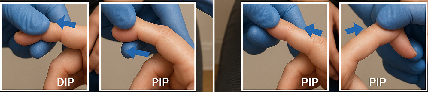
- Interphalangeal joint varus/valgus tests
- Patient in a seated position with fingers supported and stabilized; valgus/varus force applied to the PIP and DIP joints of all digits
- Positive: joint laxity and possible pain
- Patient in a seated position with fingers supported and stabilized; valgus/varus force applied to the PIP and DIP joints of all digits
Tendon and muscle
Finkelstein’s test and the WHAT test both stress the first dorsal compartment tendons (abductor pollicis longus and extensor pollicis brevis) to provoke de Quervain’s tenosynovitis. Strictly, the thumb-in-fist version below is Eichhoff’s maneuver (in Finkelstein’s original test, the clinician pulls the thumb while the hand moves into ulnar deviation, with no fist), but most PT texts call it Finkelstein’s test.
-
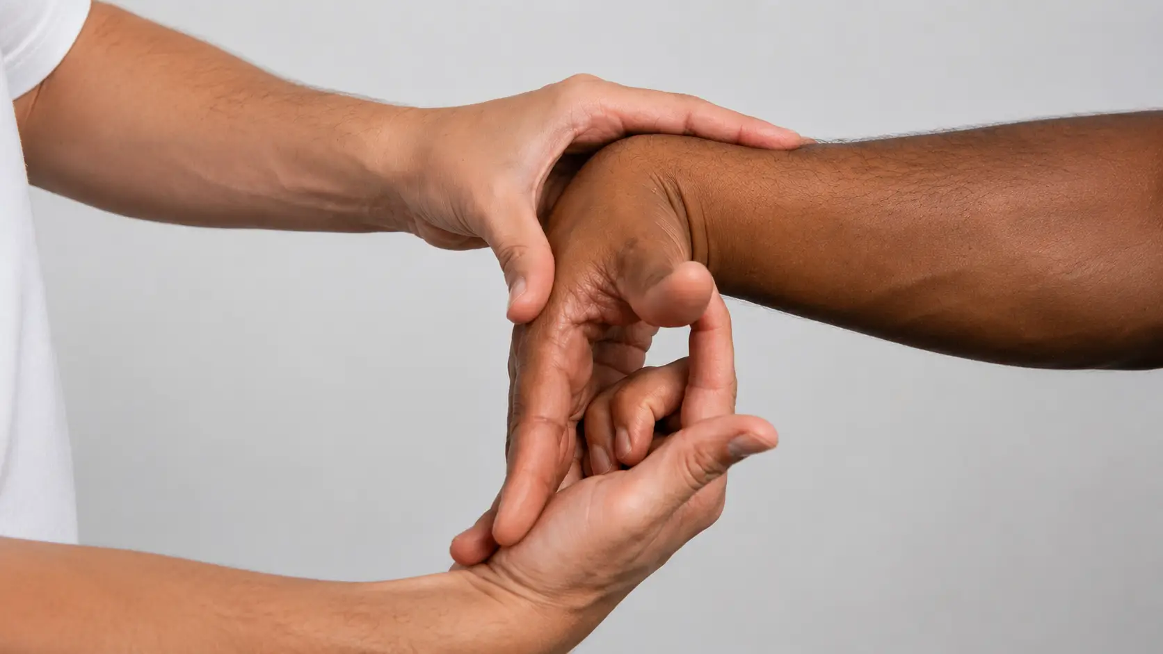
Finkelstein’s test (thumb-in-fist version)
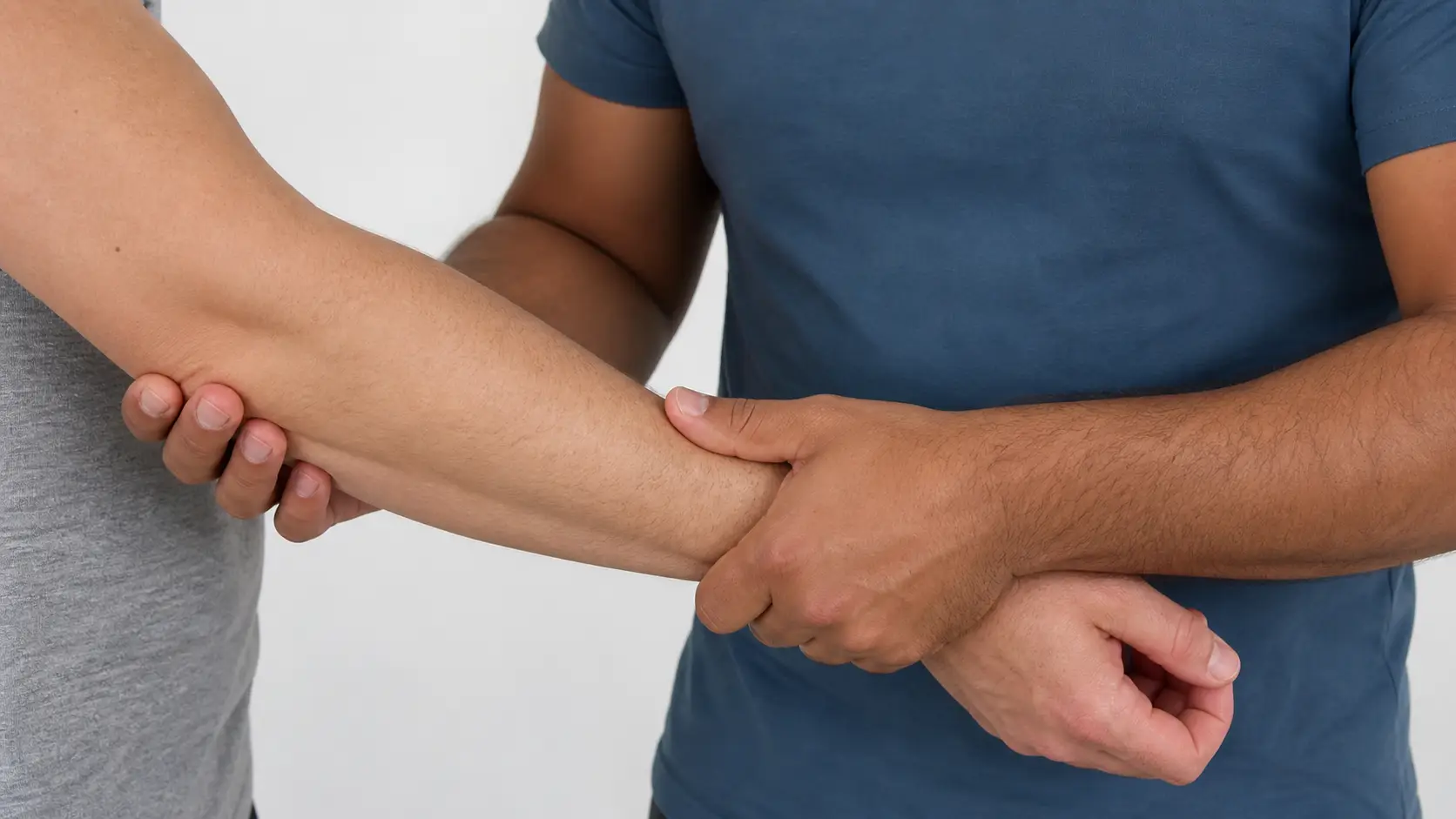
- Patient makes a fist with the thumb tucked inside the fingers; the clinician passively moves the wrist into ulnar deviation
- Positive: pain over the radial wrist suggests de Quervain’s tenosynovitis
- Patient makes a fist with the thumb tucked inside the fingers; the clinician passively moves the wrist into ulnar deviation
-
Wrist hyperflexion and abduction of the thumb test (WHAT)
- Patient in seated position with the wrist hyperflexed and the thumb abducted in full MCP and IP extension. The clinician applies gradually increasing abduction resistance to the thumb.
- Positive: reproduction of pain in the wrist
- Indicates de Quervain’s tenosynovitis in the 1st dorsal compartment (abductor pollicis longus and extensor pollicis brevis), the same structure implicated by a positive Finkelstein’s or Eichhoff’s test
- Patient in seated position with the wrist hyperflexed and the thumb abducted in full MCP and IP extension. The clinician applies gradually increasing abduction resistance to the thumb.
Neurological dysfunction
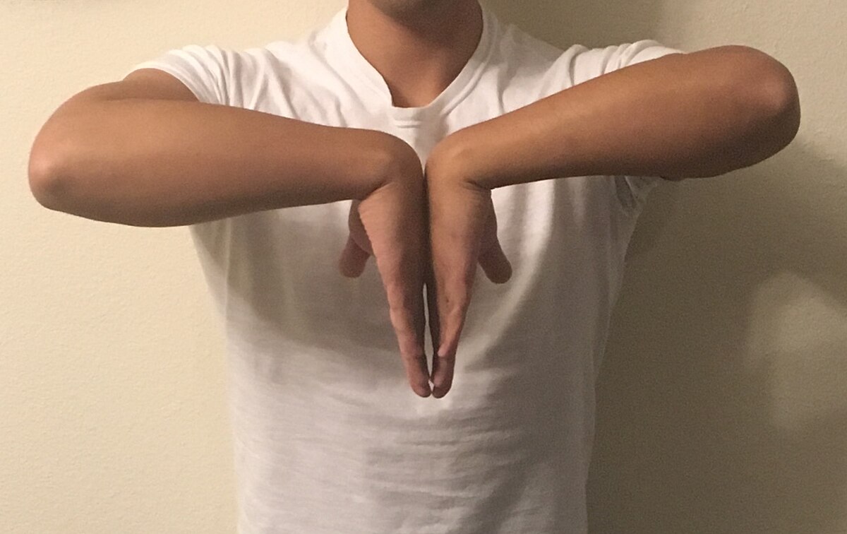
- Phalen’s test (wrist flexion test)
- Patient in seated position maximally flexes both wrists while holding them together for one minute
- Positive: reproduces tingling sensation or paresthesia in the median nerve distribution
- Indicates carpal tunnel syndrome
- Positive: reproduces tingling sensation or paresthesia in the median nerve distribution
- Patient in seated position maximally flexes both wrists while holding them together for one minute
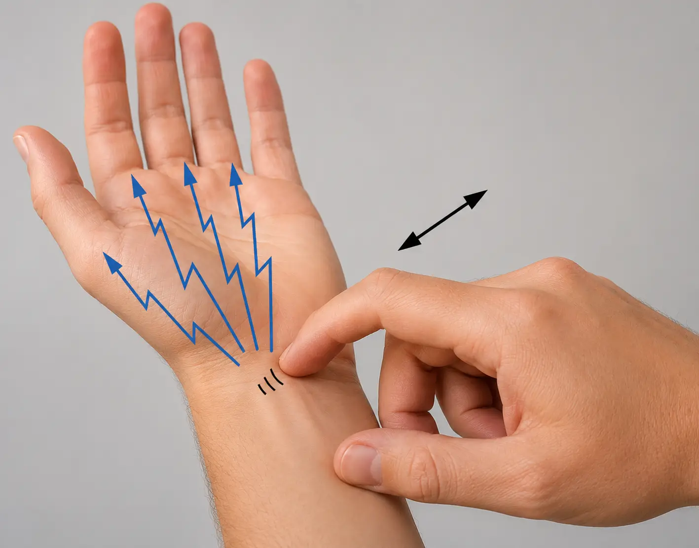
- Tinel’s test
- Patient in seated position; the clinician taps a palpable peripheral nerve - commonly the median nerve at the carpal tunnel or the ulnar nerve at the cubital tunnel
- Positive: reproduces tingling sensation or paresthesia in the nerve distribution
- Patient in seated position; the clinician taps a palpable peripheral nerve - commonly the median nerve at the carpal tunnel or the ulnar nerve at the cubital tunnel
Triangular fibrocartilage complex (TFCC)
The TFCC is the cartilage-and-ligament complex on the ulnar side of the wrist that stabilizes the distal radioulnar joint.
- Press test
- The seated patient pushes the body up off the chair with both hands, axially loading the ulnar side of the wrist
- Positive: focal ulnar-sided wrist pain is reproduced, consistent with a TFCC tear
- The seated patient pushes the body up off the chair with both hands, axially loading the ulnar side of the wrist
A positive press test does not confirm a tear (imaging or arthroscopy does). Manual wheelchair users are prone to TFCC injury because pressure-relief push-ups, transfers, and propulsion load the wrist in ulnar deviation; report ulnar-sided wrist pain reproduced by these tasks to the supervising PT.
Vascular dysfunction
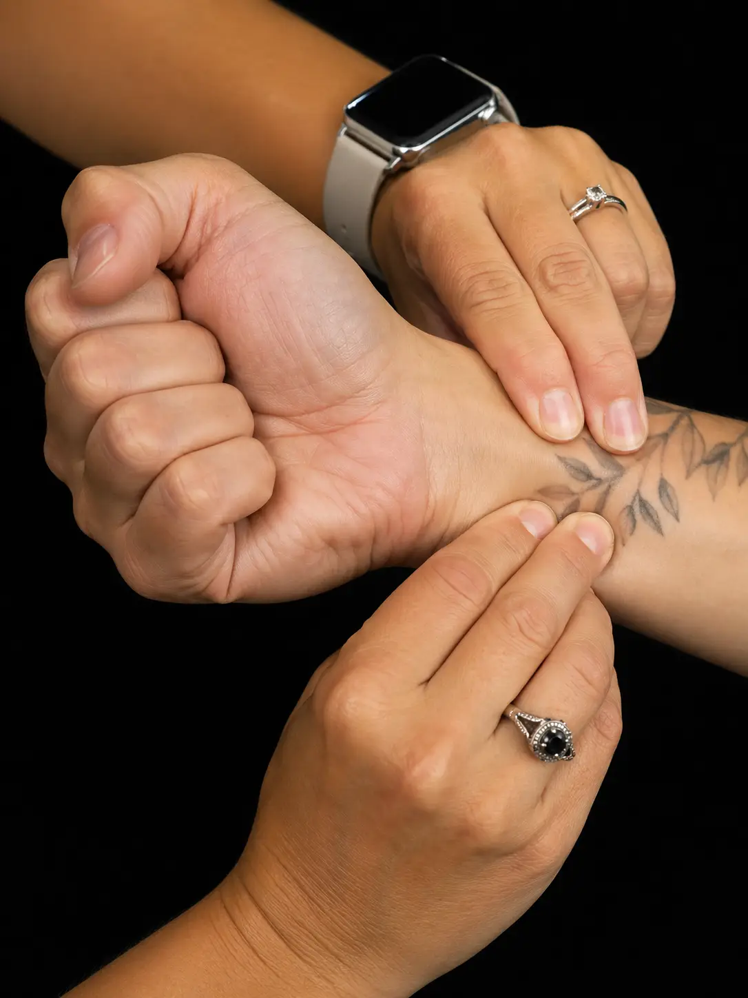
- Modified Allen’s test
- With the patient seated, the clinician palpates the radial and ulnar arteries, then has the patient quickly open and close their hand several times before making a tight fist
- The clinician compresses both the radial and ulnar arteries while the patient makes a tight fist; the patient opens the now-blanched hand, the clinician releases one artery and observes how quickly the palm flushes, then repeats the test releasing the other artery
- Positive: delayed or absent flushing after an artery is released suggests that artery is not supplying the hand adequately. Compare both sides.
Outcome measures complement these special tests: the DASH (Disabilities of the Arm, Shoulder and Hand) or its shorter QuickDASH version, and the PRWE (Patient-Rated Wrist Evaluation), are common self-report measures of upper-extremity function that a PTA may administer as data collection, reporting score changes to the supervising PT.
Comparing epicondylitis and nerve entrapment tests
Several of the tests above look alike because they stress similar structures from different angles. This table lines up the lateral vs. medial epicondylitis tests and the median vs. ulnar nerve entrapment tests side by side.
The following table compares seven special tests by position or stress applied, positive finding, and the structure implicated: Cozen’s test, Mills test, reverse Mills test, Phalen’s test, Tinel’s test at the wrist and at the elbow, and the elbow flexion test.
| Test | Position or stress | Positive finding | Structure implicated |
|---|---|---|---|
| Cozen’s test | Elbow extended, forearm pronated, wrist extended against resistance | Pain at the lateral epicondyle | Lateral epicondylitis |
| Mills test | Forearm pronated, wrist passively fully flexed, elbow extended | Pain at the lateral epicondyle | Lateral epicondylitis |
| Reverse Mills test | Forearm supinated, wrist and elbow passively extended | Pain at the medial epicondyle | Medial epicondylitis |
| Phalen’s test | Both wrists maximally flexed together for 1 minute | Tingling or paresthesia in the median nerve distribution | Carpal tunnel syndrome |
| Tinel’s test (wrist) | Clinician taps over the carpal tunnel | Tingling or paresthesia in the median nerve distribution | Carpal tunnel syndrome |
| Tinel’s test (elbow) | Clinician taps over the cubital tunnel | Tingling or paresthesia in the ulnar nerve distribution | Cubital tunnel syndrome |
| Elbow flexion test | Elbow held in maximal flexion, wrist extended, for 1 minute | Tingling or paresthesia in the ulnar nerve distribution | Cubital tunnel syndrome |