


This browser is too old to run Achievable.

Update it, or open this page in a current version of Chrome, Safari, Firefox or Edge. On an iPhone or iPad, update iOS: every browser there runs on the version of Safari that comes with it.

Achievable logoAchievable logo
NPTE-PTA
Sign in
Sign up
Purchase
Textbook
Practice exams
Support
How it works
Resources
Exam catalog
Mountain with a flag at the peak
Textbook
Introduction
1. Cardiovascular system
2. Pulmonary system
3. Neuromuscular system
4. Pediatrics
5. Musculoskeletal system
5.1 Anatomy of musculoskeletal system
5.2 Anatomical terminology and exercise training principles
5.3 Joint mechanics and phases of healing
5.4 Upper extremity anatomy
5.5 Special tests of upper extremity
5.6 Comparing clinical presentation and interventions for upper extremity
5.7 Lower extremity anatomy
5.8 Special tests of lower extremity
5.9 Comparing clinical presentation and interventions for lower extremity
5.10 Spine and pelvis anatomy
5.11 Special tests of the spine, pelvis, and temporomandibular joint
5.12 Comparing clinical presentation and interventions for the spine, pelvis, and temporomandibular joint
5.13 Other musculoskeletal conditions
5.14 Gait
5.15 Prosthetics
5.16 Orthotics
5.17 Medications, imaging, and fractures
5.18 Surgical protocols
6. Other systems
7. Non-systems
Wrapping up
Achievable logoAchievable logo
5.10 Spine and pelvis anatomy
Achievable NPTE-PTA
5. Musculoskeletal system
Our NPTE-PTA course is now in "early access" - get 50% off for a limited time.

Spine and pelvis anatomy

11 min read
Font
Discuss
Share
Feedback

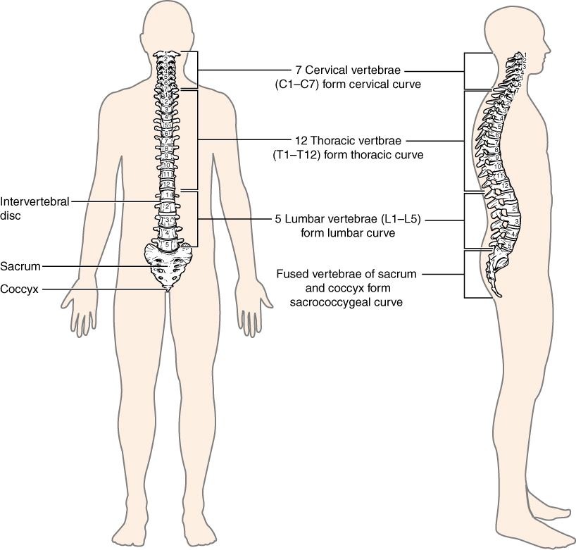
Spine and pelvis region

Components of spine (vertebral column)

  • 24 vertebrae
  • 2 fused bones: the sacrum and coccyx
  • The column is divided into 5 areas - cervical, thoracic, lumbar, sacral, and coccygeal

Function of the spine

  • Provides a strong and flexible framework that supports the body’s weight, allowing for standing, walking, and moving freely.
  • Encases and protects the spinal cord
  • Angled facet joints and intervertebral discs allow for a wide range of motion, including flexion, extension, rotation, and lateral flexion.
  • The thoracic spine and ribs protect vital internal organs such as the heart and lungs.
  • Maintains proper balance and posture by distributing the body’s weight evenly.
Vertebral column
Vertebral column
Anatomy & Physiology by Lindsay M. Biga, Sierra Dawson, Amy Harwell, Robin Hopkins, Joel Kaufmann, Mike LeMaster, Philip Matern, Katie Morrison-Graham, Devon Quick & Jon Runyeon
/
Oregon State University
/
CC BY 4.0

Components of the pelvis

  • Two innominate (hip) bones, each formed by the fused ilium, ischium, and pubis
  • The innominates meet anteriorly at the pubic symphysis and join the sacrum posteriorly at the sacroiliac joints

Bony structures

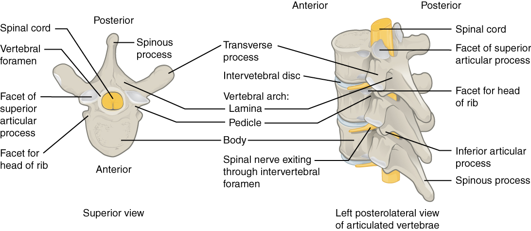
Vertebrae of spine
Vertebrae of spine
Anatomy & Physiology by Lindsay M. Biga, Sierra Dawson, Amy Harwell, Robin Hopkins, Joel Kaufmann, Mike LeMaster, Philip Matern, Katie Morrison-Graham, Devon Quick & Jon Runyeon
/
Oregon State University
/
CC BY 4.0
Definitions
Vertebral body
The cylindrical, weight-bearing anterior part of every vertebra from C2 to L5 (C1 has no body): cancellous bone inside a thin shell of compact bone.
Pedicles
Either of two short cylindrical bony processes lying on either side of a vertebra that project posteriorly from the vertebral body and fuse with the laminae to form a neural arch
Lamina
Flat, arched bone that forms the posterior (back) part of each vertebra, joining the pedicles to complete the neural arch.
Articular facets (facet joints)
The superior and inferior articular processes that project from each vertebra and meet the processes of the vertebrae above and below to form the paired facet (zygapophyseal) joints, which guide and limit spinal motion.
Transverse processes
Bony projections on the sides of the vertebrae that help support the spine and attach muscles via muscular and ligamentous attachments.
Spinous process
A bony projection located at the back of each vertebra in the spinal column. The only exceptions are the atlas (C1), which lacks one entirely, and the sacrum and coccyx (fused segments)

Special bony components of each segment

  • Cervical: supports the head and neck; muscles are attached to allow a large freedom of movement
    • The head on C1 the atlanto-occipital (condyloid joint) allows the motion of flexion-extension of head, nodding motion.
    • C1 and C2, the atlanto-axial joint allows for rotation without compression of the spinal cord via a special projection from C2 called the dens.
  • Thoracic: costotransverse and costovertebral
    • Costovertebral joints: the rib head articulates with the vertebral bodies; costotransverse joints: the rib tubercle articulates with the transverse process.
    • Prominent (easily palpable) spinous processes
    • Supports axial loading, attached to ribs to form the most rigid section of the spine.
  • Lumbar: the largest vertebrae transfer weight bearing loads and increase mobility of the trunk
  • Sacrum
    • Composed of 5 fused bones
  • Coccyx (tailbone)
    • Final spinal segment composed of 3-5 small fused bones

Special joints of the spine

  • Facet joints
    • Aid in the movement of the spine as a whole
  • Intervertebral (disc) joints
    • Allow a small amount of motion between two adjacent vertebral bodies and transfer load from one body to the next
  • Sacroiliac joints
    • Aid in movements of the lower axial skeleton and lower extremities

Other important features of the spine

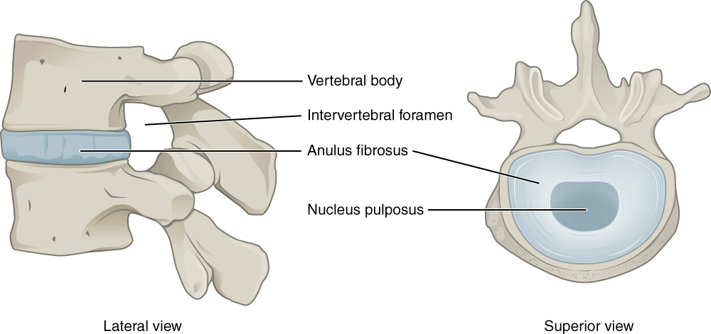
  • Intervertebral discs: assist in transferring loads between vertebral bodies.
    • Annulus fibrosus
      • Only the outer annulus fibrosus is vascularized and innervated; the inner annulus is avascular and aneural.
      • Functions to maintain the integrity of the vertebral column during compression, torsion, shearing, and distracting forces
    • Nucleus pulposus
      • Avascular and has no neural connections
      • Gel-like core that resists compression by building hydrostatic pressure, which the surrounding annulus fibrosus contains through tension in its fibers
    • Vertebral endplate
      • Functions to diffuse nutrients to the annulus fibrosus and the nucleus pulposus
Intervertebral disk of spine
Anatomy & Physiology by Lindsay M. Biga, Sierra Dawson, Amy Harwell, Robin Hopkins, Joel Kaufmann, Mike LeMaster, Philip Matern, Katie Morrison-Graham, Devon Quick & Jon Runyeon
/
Oregon State University
/
CC BY 4.0

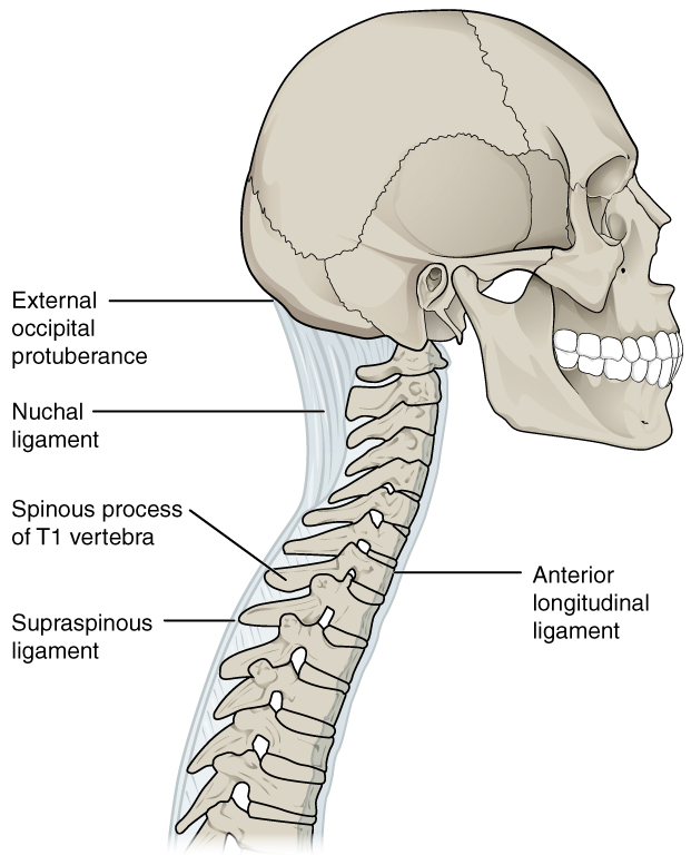
Ligaments

  • Alar
    • Connects dens to the occipital condyle
    • Functions to limit flexion, contralateral lateral flexion, and contralateral rotation
  • Anterior longitudinal ligament
    • Runs along the anterior surfaces of the vertebral bodies and discs from the occiput and anterior tubercle of C1 to the sacrum
    • Functions to limit extension and reinforce the anterior intervertebral discs
  • Posterior longitudinal ligament
    • Located on the posterior surface of vertebral bodies from C2 to the sacrum
    • Functions to prevent hyperflexion of the vertebral column and to protect the spinal cord from foreign objects and displaced disc material.
  • Ligamentum flavum
    • Connects vertebra to the lamina above it; runs from C2 to the sacrum
    • Functions to limit flexion (greatest in lumbar spine)
  • Interspinous ligament
    • Runs between spinous processes
    • Functions to limit flexion
  • Iliolumbar ligament
    • Connects L5 vertebrae to the ilium
    • Functions to limit motion between L5 and S1
Ligaments of spine
Ligaments of spine
Anatomy & Physiology by Lindsay M. Biga, Sierra Dawson, Amy Harwell, Robin Hopkins, Joel Kaufmann, Mike LeMaster, Philip Matern, Katie Morrison-Graham, Devon Quick & Jon Runyeon
/
Oregon State University
/
CC BY 4.0

Capsule

  • Facet joints
    • Role is to reinforce ligaments by limiting motion and stabilizing the spine
  • Sacroiliac joints
    • Role is to stabilize the sacroiliac joint, limiting its movement, and protecting the joint by distributing biomechanical loads.

Nerves

  • Spinal nerves are mixed nerves that connect the spinal cord to the rest of the body. Sensory (afferent) fibers enter the spinal cord via the dorsal root, and motor (efferent) fibers exit via the ventral root. Once these roots combine to form the spinal nerve, each dorsal and ventral ramus that branches off carries both sensory and motor fibers between the central nervous system (brain and spinal cord) and the peripheral nervous system (body and muscles).
  • Location of spinal nerves
    • Cervical: C1-C7 exit above their same-numbered vertebra; C8 (there’s no C8 vertebra) exits below C7
    • Thoracic/lumbar: exit below the level of the associated vertebrae, continuing the pattern C8 started

PTA insight: During data collection under an established plan of care, you won’t diagnose spine or pelvic pathology, but you will observe and measure findings like asymmetrical facet motion, restricted coupled movement, or sacroiliac joint asymmetry, and report them to the supervising PT. Watch for red flags during any spine intervention, such as a sudden increase in radicular pain, saddle anesthesia, or loss of bowel or bladder control, and stop and notify the PT immediately, since these can signal a medical emergency. The next chapter, special tests of the spine, pelvis, and temporomandibular joint, covers how these findings are formally assessed.

Spinal and pelvis movements

Facet joint mechanics (arthrokinematics)

As a general rule, facet joints open (increasing space between vertebrae) during flexion and on the side opposite a side-bend, and they close (compressing together) during extension and on the side you’re bending toward. Rotation follows a different pattern by region: in the cervical spine, rotation closes the facet and foramen on the side you rotate toward (because rotation couples with same-side side-bending), while in the lumbar spine, pure axial rotation compresses the facet on the side opposite the rotation.

  • Flexion
    • The facet joints open. The inferior facet of the vertebra above slides upward and forward relative to the superior facet of the vertebra below.
  • Extension
    • The facet joints close. The inferior facets of the vertebra above glide downward and backward relative to the superior facets of the vertebra below.

Lateral bending

  • Right lateral bending The left (contralateral) facet joint moves upward and anteriorly to open, while the right (ipsilateral) facet joint moves down and posteriorly to close.
    • Left-sided motion is the mirror image.

Cervical rotation

  • Right rotation The left facet joint of the upper vertebra moves anteriorly (forward) while the right facet joint moves posteriorly (backward). At the same time, the intervertebral foramen on the right side (ipsilateral side of the rotation) closes, and the foramen on the left side (contralateral side) opens.
    • Left-sided motion is the mirror image.
    • Clinically, closing the foramen on the side of rotation can reproduce radicular symptoms on that side, which you’d observe and report to the supervising PT.

Coupled movements

Coupled movements are secondary movements that consistently accompany a primary motion, most importantly lateral flexion paired with rotation.

  • Cervical spine
    • C1: lateral bending and rotation occur in different directions (occurs if the spine is in neutral, flexion, and extension)
    • C2-C7: lateral bending and rotation occurring in the same direction (occurs if the spine is in neutral, flexion, and extension)
  • Lumbar/thoracic spine (Kaltenborn’s manual therapy model; research on lumbar coupling is mixed, so treat these as tendencies)
    • If the spine is in flexion, side bending and rotation will occur in the same direction
    • If the spine is in extension/neutral, side bending and rotation will occur in opposite directions

Lumbopelvic rhythm and sacroiliac motion

  • Lumbopelvic joint
    • When in flexion: lumbar spine flexes followed by pelvis rotation anteriorly, ending with hip flexion
    • When in extension (when coming from a flexed position): the hips extend, followed by the pelvis rotating posteriorly, and the spine extending
  • Sacroiliac joint
    • Nutation: flexion of the sacrum causes posterior tilt of the ilium (in the sagittal plane)
    • Counternutation: extension of the sacrum causes an anterior tilt of the ilium (in the sagittal plane)
    • Anterior innominate: anterior superior iliac spine (ASIS) moves downward (in the sagittal plane)
    • Posterior innominate: anterior superior iliac spine (ASIS) moves upward (in the sagittal plane)

Spine structure & function

  • 24 vertebrae + sacrum/coccyx; 5 regions: cervical, thoracic, lumbar, sacral, coccygeal
  • Supports body weight, protects spinal cord, enables flexion/extension/rotation/lateral flexion
  • Thoracic spine/ribs protect organs; maintains posture and balance
  • Pelvis: two innominates (each fused ilium, ischium, pubis) meet at the pubic symphysis anteriorly and join the sacrum at the SI joints posteriorly

Bony anatomy of vertebrae

  • Vertebral body: weight-bearing anterior portion (absent in C1)
  • Pedicles + lamina form neural arch; articular facets guide/limit motion
  • Transverse processes (muscle attachment) and spinous process (absent in atlas)
  • Regional specializations:
    • Cervical: atlanto-occipital (nodding), atlanto-axial (rotation via dens)
    • Thoracic: costovertebral/costotransverse joints, rigid due to rib attachment
    • Lumbar: largest vertebrae, weight transfer/mobility
    • Sacrum (5 fused bones), coccyx (3-5 fused bones)

Key joints

  • Atlanto-occipital (nodding) vs atlanto-axial (rotation)
  • Facet joints: overall spinal movement
  • Intervertebral (disc) joints: small motion between two adjacent vertebral bodies, load transfer
  • Sacroiliac joints: lower axial skeleton/extremity movement

Intervertebral disc

  • Annulus fibrosus: outer vascularized/innervated, inner avascular/aneural; resists compression/torsion/shear
  • Nucleus pulposus: avascular, gel-like, builds hydrostatic pressure
  • Vertebral endplate: diffuses nutrients to disc

Ligaments (function = limit specific motions)

  • Alar: limits flexion, contralateral side-bend/rotation
  • Anterior longitudinal: limits extension
  • Posterior longitudinal: limits hyperflexion, protects cord
  • Ligamentum flavum: limits flexion (strongest in lumbar)
  • Interspinous: limits flexion
  • Iliolumbar: limits L5-S1 motion

Nerves & clinical flags

  • Spinal nerves mixed: sensory via dorsal root, motor via ventral root
  • Cervical nerves C1-C7 exit above same-numbered vertebra; C8 and below exit below vertebra
  • PTA role: observe/report asymmetries, not diagnose
  • Red flags: sudden radicular pain increase, saddle anesthesia, bowel/bladder loss → stop, notify PT

Facet joint mechanics

  • Flexion: facets open (upward/forward glide); Extension: facets close (downward/backward glide)
  • Lateral bending: contralateral facet opens, ipsilateral facet closes
  • Cervical rotation: ipsilateral foramen closes (can cause radicular symptoms), contralateral opens

Coupled movements

  • C1: lateral flexion & rotation occur in opposite directions
  • C2-C7: lateral flexion & rotation occur in same direction
  • Thoracic/lumbar (general tendency, not fixed): flexion = same-direction coupling; extension/neutral = opposite-direction coupling

Lumbopelvic rhythm & SI joint motion

  • Flexion sequence: lumbar flexion → anterior pelvic rotation → hip flexion
  • Extension sequence (from flexion): hip extension → posterior pelvic rotation → spinal extension
  • SI joint: nutation (sacral flexion/posterior ilium tilt) vs counternutation (sacral extension/anterior ilium tilt)
  • Innominate motion: anterior innominate = ASIS moves down; posterior innominate = ASIS moves up

Sign up for free to take 22 quiz questions on this topic

Previous
Next  | 5.11.1 Cervical spine tests
All rights reserved ©2016 - 2026 Achievable, Inc.

Spine and pelvis anatomy

Spine and pelvis region

Components of spine (vertebral column)

  • 24 vertebrae
  • 2 fused bones: the sacrum and coccyx
  • The column is divided into 5 areas - cervical, thoracic, lumbar, sacral, and coccygeal

Function of the spine

  • Provides a strong and flexible framework that supports the body’s weight, allowing for standing, walking, and moving freely.
  • Encases and protects the spinal cord
  • Angled facet joints and intervertebral discs allow for a wide range of motion, including flexion, extension, rotation, and lateral flexion.
  • The thoracic spine and ribs protect vital internal organs such as the heart and lungs.
  • Maintains proper balance and posture by distributing the body’s weight evenly.

Components of the pelvis

  • Two innominate (hip) bones, each formed by the fused ilium, ischium, and pubis
  • The innominates meet anteriorly at the pubic symphysis and join the sacrum posteriorly at the sacroiliac joints

Bony structures

Definitions
Vertebral body
The cylindrical, weight-bearing anterior part of every vertebra from C2 to L5 (C1 has no body): cancellous bone inside a thin shell of compact bone.
Pedicles
Either of two short cylindrical bony processes lying on either side of a vertebra that project posteriorly from the vertebral body and fuse with the laminae to form a neural arch
Lamina
Flat, arched bone that forms the posterior (back) part of each vertebra, joining the pedicles to complete the neural arch.
Articular facets (facet joints)
The superior and inferior articular processes that project from each vertebra and meet the processes of the vertebrae above and below to form the paired facet (zygapophyseal) joints, which guide and limit spinal motion.
Transverse processes
Bony projections on the sides of the vertebrae that help support the spine and attach muscles via muscular and ligamentous attachments.
Spinous process
A bony projection located at the back of each vertebra in the spinal column. The only exceptions are the atlas (C1), which lacks one entirely, and the sacrum and coccyx (fused segments)

Special bony components of each segment

  • Cervical: supports the head and neck; muscles are attached to allow a large freedom of movement
    • The head on C1 the atlanto-occipital (condyloid joint) allows the motion of flexion-extension of head, nodding motion.
    • C1 and C2, the atlanto-axial joint allows for rotation without compression of the spinal cord via a special projection from C2 called the dens.
  • Thoracic: costotransverse and costovertebral
    • Costovertebral joints: the rib head articulates with the vertebral bodies; costotransverse joints: the rib tubercle articulates with the transverse process.
    • Prominent (easily palpable) spinous processes
    • Supports axial loading, attached to ribs to form the most rigid section of the spine.
  • Lumbar: the largest vertebrae transfer weight bearing loads and increase mobility of the trunk
  • Sacrum
    • Composed of 5 fused bones
  • Coccyx (tailbone)
    • Final spinal segment composed of 3-5 small fused bones

Special joints of the spine

  • Facet joints
    • Aid in the movement of the spine as a whole
  • Intervertebral (disc) joints
    • Allow a small amount of motion between two adjacent vertebral bodies and transfer load from one body to the next
  • Sacroiliac joints
    • Aid in movements of the lower axial skeleton and lower extremities

Other important features of the spine

  • Intervertebral discs: assist in transferring loads between vertebral bodies.
    • Annulus fibrosus
      • Only the outer annulus fibrosus is vascularized and innervated; the inner annulus is avascular and aneural.
      • Functions to maintain the integrity of the vertebral column during compression, torsion, shearing, and distracting forces
    • Nucleus pulposus
      • Avascular and has no neural connections
      • Gel-like core that resists compression by building hydrostatic pressure, which the surrounding annulus fibrosus contains through tension in its fibers
    • Vertebral endplate
      • Functions to diffuse nutrients to the annulus fibrosus and the nucleus pulposus

Ligaments

  • Alar
    • Connects dens to the occipital condyle
    • Functions to limit flexion, contralateral lateral flexion, and contralateral rotation
  • Anterior longitudinal ligament
    • Runs along the anterior surfaces of the vertebral bodies and discs from the occiput and anterior tubercle of C1 to the sacrum
    • Functions to limit extension and reinforce the anterior intervertebral discs
  • Posterior longitudinal ligament
    • Located on the posterior surface of vertebral bodies from C2 to the sacrum
    • Functions to prevent hyperflexion of the vertebral column and to protect the spinal cord from foreign objects and displaced disc material.
  • Ligamentum flavum
    • Connects vertebra to the lamina above it; runs from C2 to the sacrum
    • Functions to limit flexion (greatest in lumbar spine)
  • Interspinous ligament
    • Runs between spinous processes
    • Functions to limit flexion
  • Iliolumbar ligament
    • Connects L5 vertebrae to the ilium
    • Functions to limit motion between L5 and S1

Capsule

  • Facet joints
    • Role is to reinforce ligaments by limiting motion and stabilizing the spine
  • Sacroiliac joints
    • Role is to stabilize the sacroiliac joint, limiting its movement, and protecting the joint by distributing biomechanical loads.

Nerves

  • Spinal nerves are mixed nerves that connect the spinal cord to the rest of the body. Sensory (afferent) fibers enter the spinal cord via the dorsal root, and motor (efferent) fibers exit via the ventral root. Once these roots combine to form the spinal nerve, each dorsal and ventral ramus that branches off carries both sensory and motor fibers between the central nervous system (brain and spinal cord) and the peripheral nervous system (body and muscles).
  • Location of spinal nerves
    • Cervical: C1-C7 exit above their same-numbered vertebra; C8 (there’s no C8 vertebra) exits below C7
    • Thoracic/lumbar: exit below the level of the associated vertebrae, continuing the pattern C8 started

PTA insight: During data collection under an established plan of care, you won’t diagnose spine or pelvic pathology, but you will observe and measure findings like asymmetrical facet motion, restricted coupled movement, or sacroiliac joint asymmetry, and report them to the supervising PT. Watch for red flags during any spine intervention, such as a sudden increase in radicular pain, saddle anesthesia, or loss of bowel or bladder control, and stop and notify the PT immediately, since these can signal a medical emergency. The next chapter, special tests of the spine, pelvis, and temporomandibular joint, covers how these findings are formally assessed.

Spinal and pelvis movements

Facet joint mechanics (arthrokinematics)

As a general rule, facet joints open (increasing space between vertebrae) during flexion and on the side opposite a side-bend, and they close (compressing together) during extension and on the side you’re bending toward. Rotation follows a different pattern by region: in the cervical spine, rotation closes the facet and foramen on the side you rotate toward (because rotation couples with same-side side-bending), while in the lumbar spine, pure axial rotation compresses the facet on the side opposite the rotation.

  • Flexion
    • The facet joints open. The inferior facet of the vertebra above slides upward and forward relative to the superior facet of the vertebra below.
  • Extension
    • The facet joints close. The inferior facets of the vertebra above glide downward and backward relative to the superior facets of the vertebra below.

Lateral bending

  • Right lateral bending The left (contralateral) facet joint moves upward and anteriorly to open, while the right (ipsilateral) facet joint moves down and posteriorly to close.
    • Left-sided motion is the mirror image.

Cervical rotation

  • Right rotation The left facet joint of the upper vertebra moves anteriorly (forward) while the right facet joint moves posteriorly (backward). At the same time, the intervertebral foramen on the right side (ipsilateral side of the rotation) closes, and the foramen on the left side (contralateral side) opens.
    • Left-sided motion is the mirror image.
    • Clinically, closing the foramen on the side of rotation can reproduce radicular symptoms on that side, which you’d observe and report to the supervising PT.

Coupled movements

Coupled movements are secondary movements that consistently accompany a primary motion, most importantly lateral flexion paired with rotation.

  • Cervical spine
    • C1: lateral bending and rotation occur in different directions (occurs if the spine is in neutral, flexion, and extension)
    • C2-C7: lateral bending and rotation occurring in the same direction (occurs if the spine is in neutral, flexion, and extension)
  • Lumbar/thoracic spine (Kaltenborn’s manual therapy model; research on lumbar coupling is mixed, so treat these as tendencies)
    • If the spine is in flexion, side bending and rotation will occur in the same direction
    • If the spine is in extension/neutral, side bending and rotation will occur in opposite directions

Lumbopelvic rhythm and sacroiliac motion

  • Lumbopelvic joint
    • When in flexion: lumbar spine flexes followed by pelvis rotation anteriorly, ending with hip flexion
    • When in extension (when coming from a flexed position): the hips extend, followed by the pelvis rotating posteriorly, and the spine extending
  • Sacroiliac joint
    • Nutation: flexion of the sacrum causes posterior tilt of the ilium (in the sagittal plane)
    • Counternutation: extension of the sacrum causes an anterior tilt of the ilium (in the sagittal plane)
    • Anterior innominate: anterior superior iliac spine (ASIS) moves downward (in the sagittal plane)
    • Posterior innominate: anterior superior iliac spine (ASIS) moves upward (in the sagittal plane)
Key points

Spine structure & function

  • 24 vertebrae + sacrum/coccyx; 5 regions: cervical, thoracic, lumbar, sacral, coccygeal
  • Supports body weight, protects spinal cord, enables flexion/extension/rotation/lateral flexion
  • Thoracic spine/ribs protect organs; maintains posture and balance
  • Pelvis: two innominates (each fused ilium, ischium, pubis) meet at the pubic symphysis anteriorly and join the sacrum at the SI joints posteriorly

Bony anatomy of vertebrae

  • Vertebral body: weight-bearing anterior portion (absent in C1)
  • Pedicles + lamina form neural arch; articular facets guide/limit motion
  • Transverse processes (muscle attachment) and spinous process (absent in atlas)
  • Regional specializations:
    • Cervical: atlanto-occipital (nodding), atlanto-axial (rotation via dens)
    • Thoracic: costovertebral/costotransverse joints, rigid due to rib attachment
    • Lumbar: largest vertebrae, weight transfer/mobility
    • Sacrum (5 fused bones), coccyx (3-5 fused bones)

Key joints

  • Atlanto-occipital (nodding) vs atlanto-axial (rotation)
  • Facet joints: overall spinal movement
  • Intervertebral (disc) joints: small motion between two adjacent vertebral bodies, load transfer
  • Sacroiliac joints: lower axial skeleton/extremity movement

Intervertebral disc

  • Annulus fibrosus: outer vascularized/innervated, inner avascular/aneural; resists compression/torsion/shear
  • Nucleus pulposus: avascular, gel-like, builds hydrostatic pressure
  • Vertebral endplate: diffuses nutrients to disc

Ligaments (function = limit specific motions)

  • Alar: limits flexion, contralateral side-bend/rotation
  • Anterior longitudinal: limits extension
  • Posterior longitudinal: limits hyperflexion, protects cord
  • Ligamentum flavum: limits flexion (strongest in lumbar)
  • Interspinous: limits flexion
  • Iliolumbar: limits L5-S1 motion

Nerves & clinical flags

  • Spinal nerves mixed: sensory via dorsal root, motor via ventral root
  • Cervical nerves C1-C7 exit above same-numbered vertebra; C8 and below exit below vertebra
  • PTA role: observe/report asymmetries, not diagnose
  • Red flags: sudden radicular pain increase, saddle anesthesia, bowel/bladder loss → stop, notify PT

Facet joint mechanics

  • Flexion: facets open (upward/forward glide); Extension: facets close (downward/backward glide)
  • Lateral bending: contralateral facet opens, ipsilateral facet closes
  • Cervical rotation: ipsilateral foramen closes (can cause radicular symptoms), contralateral opens

Coupled movements

  • C1: lateral flexion & rotation occur in opposite directions
  • C2-C7: lateral flexion & rotation occur in same direction
  • Thoracic/lumbar (general tendency, not fixed): flexion = same-direction coupling; extension/neutral = opposite-direction coupling

Lumbopelvic rhythm & SI joint motion

  • Flexion sequence: lumbar flexion → anterior pelvic rotation → hip flexion
  • Extension sequence (from flexion): hip extension → posterior pelvic rotation → spinal extension
  • SI joint: nutation (sacral flexion/posterior ilium tilt) vs counternutation (sacral extension/anterior ilium tilt)
  • Innominate motion: anterior innominate = ASIS moves down; posterior innominate = ASIS moves up

More from Musculoskeletal system

  • Anatomy of musculoskeletal system
  • Anatomical terminology and exercise training principles
  • Joint mechanics and phases of healing
  • Upper extremity anatomy
  • Lower extremity anatomy