



Cervical spine tests
The PTA performs these cervical special tests as part of data collection within the PT’s plan of care - interpreting findings, diagnosing, and changing the plan of care remain the supervising PT’s responsibility. If a test produces a red-flag sign, stop immediately, keep monitoring the patient, and notify the supervising PT right away.
Safety and stability screens (perform first)
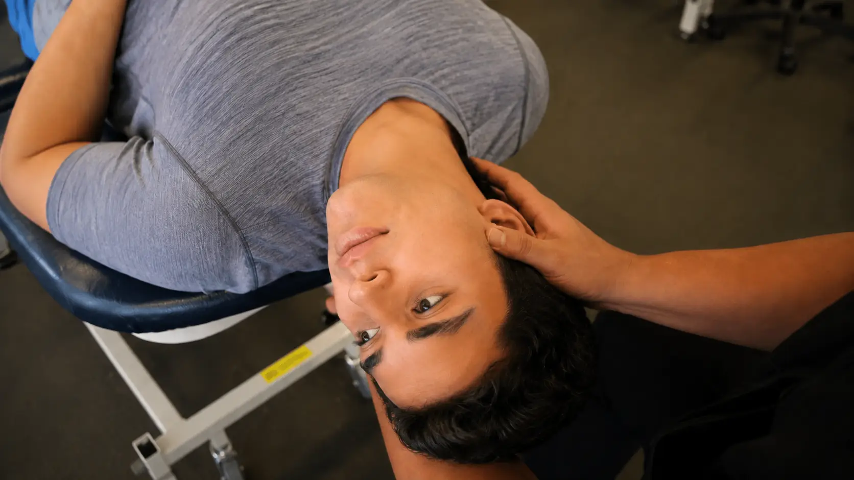
- Vertebral artery test
- The examiner carefully extends the patient’s neck, then rotates the head to one side and holds it there for up to 30 seconds. The patient’s eyes are kept open and monitored, and they are usually asked to count backward or speak to check for changes in their voice.
- Tests the integrity of the vertebrobasilar vascular system
- Positive: dizziness, diplopia, dysarthria, dysphagia, drop attacks, nystagmus, nausea, or numbness. This is a red flag - stop the test and any cervical spine intervention immediately, continue monitoring the patient, and notify the supervising PT right away.
- Current evidence questions the validity and reliability of this positional test on its own. Rather than relying on the positional test alone as the standard of care, current frameworks call for a broader cervical arterial dysfunction screen before cervical spine mobilization or manipulation - including patient history, risk factors, blood pressure, and a cranial nerve exam - to identify patients at risk before proceeding
- The examiner carefully extends the patient’s neck, then rotates the head to one side and holds it there for up to 30 seconds. The patient’s eyes are kept open and monitored, and they are usually asked to count backward or speak to check for changes in their voice.
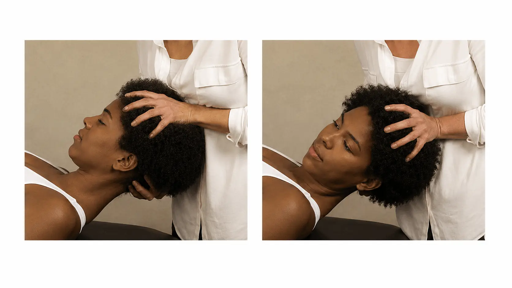
- Flexion rotation test
- Patient in supine with maximal flexion of neck performed with rotation to left and right
- Provocative test for atlantoaxial dysfunction or cervicogenic headache
- Positive: headache reproduced, or rotation to one side reduced by more than 10 degrees compared with the other side
- Patient in supine with maximal flexion of neck performed with rotation to left and right
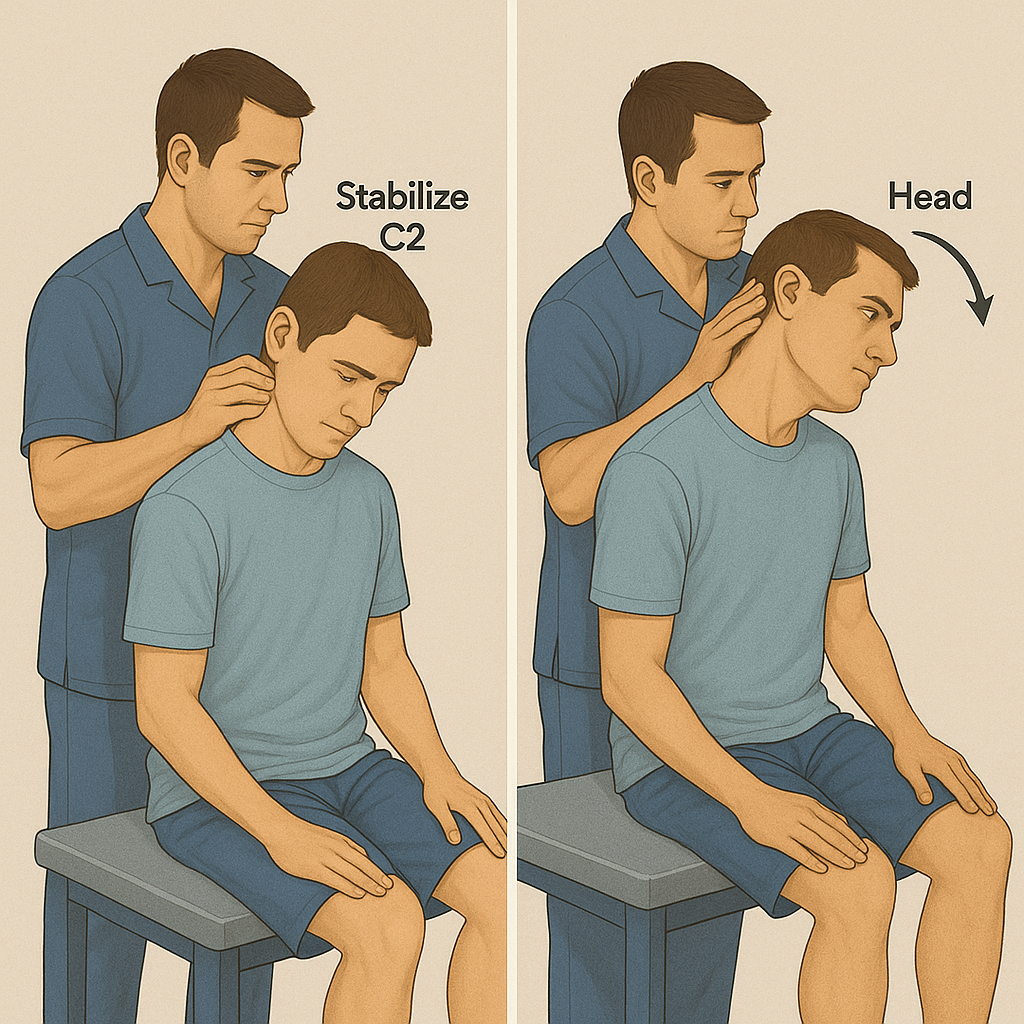
- Alar ligament test
- Patient seated (or supine) with the neck in slight flexion. The therapist stabilizes the C2 spinous process and passively side-bends the head to each side.
- Tests the integrity of the alar ligament
- Positive: excessive side-bending motion compared with the other side, or a soft end-feel (with an intact alar ligament, little to no side-bending occurs and the end-feel is firm). This suggests alar ligament laxity or upper cervical instability - stop and report the finding to the supervising PT.
- Patient seated (or supine) with the neck in slight flexion. The therapist stabilizes the C2 spinous process and passively side-bends the head to each side.
Nerve root and cord tests (Spurling’s and compression narrow the foramen to provoke radicular symptoms; distraction opens it to relieve them)
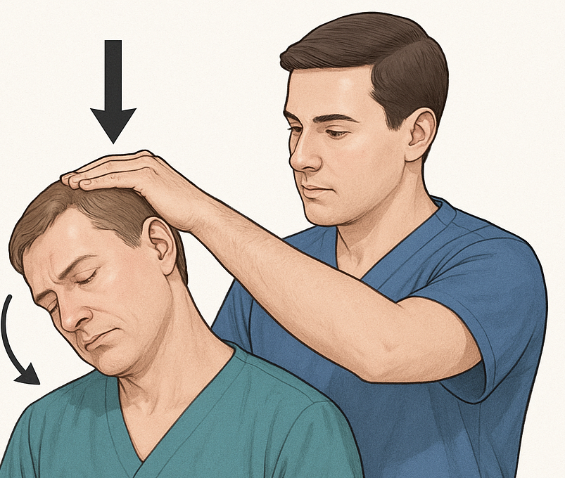
- Spurling’s test (foraminal compression)
- Patient sitting with head laterally bending towards the involved side while the therapist applies pressure straight down
- Provokes/suggests cervical radiculopathy
- Cervical radiculopathy is compression of a cervical nerve root, and the pain travels down the arm in the area served by that root; extending the neck or turning the head toward the side narrows the foramen and increases symptoms, so extension positions are avoided, while traction in slight flexion opens the foramen
- Positive: pain and paresthesia in a dermatomal pattern of the cervical root
- Patient sitting with head laterally bending towards the involved side while the therapist applies pressure straight down
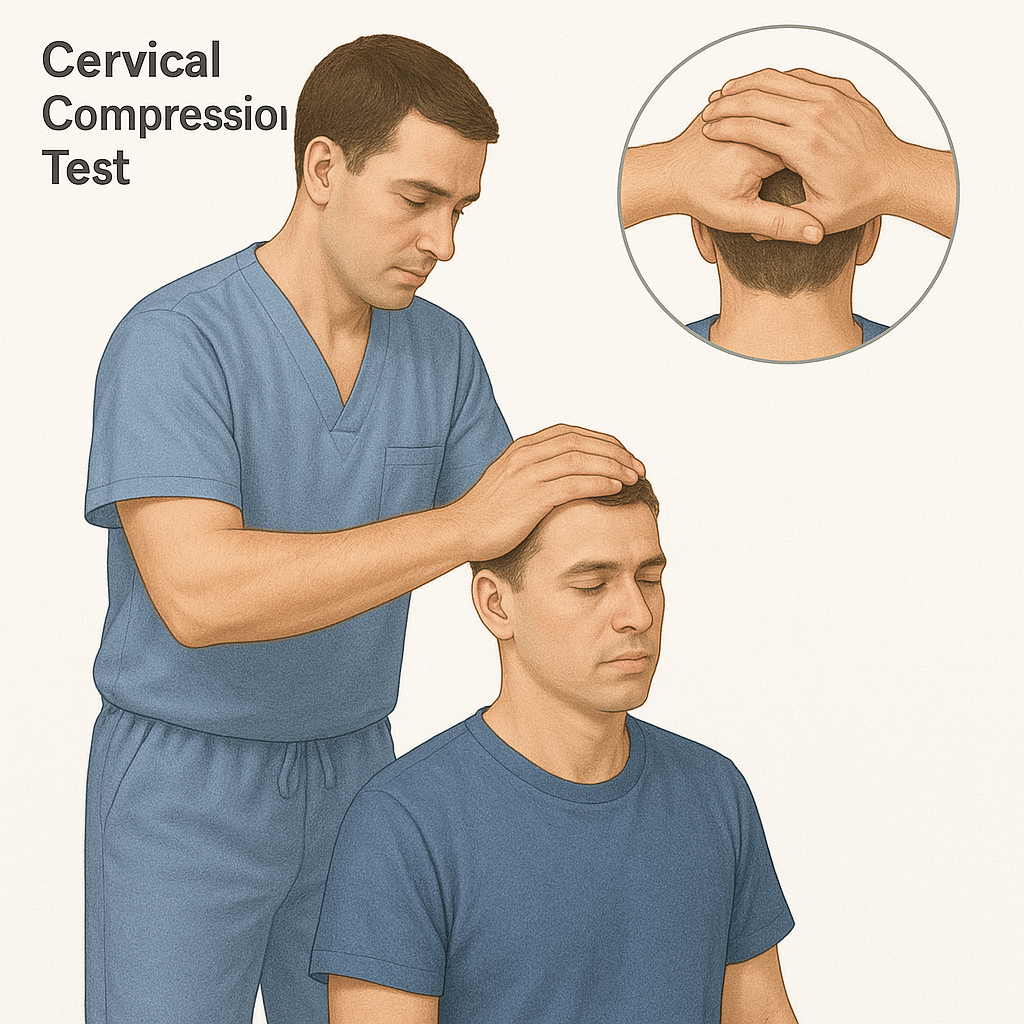
- Cervical compression test
- Patient sitting and neck passively moved into lateral flexion and rotation to the non-painful side, followed by extension. Repeat the painful side.
- Identifies intervertebral foramen and/or facet dysfunction
- Positive: pain and paresthesia in a dermatomal pattern of cervical root or localized pain in the neck if facet dysfunction
- Patient sitting and neck passively moved into lateral flexion and rotation to the non-painful side, followed by extension. Repeat the painful side.
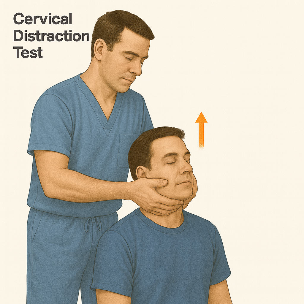
- Distraction test
- Patient sitting with head passively distracted
- Identifies intervertebral foramen and/or facet dysfunction
- Positive: decrease in symptoms or decrease in upper limb pain
- Patient sitting with head passively distracted
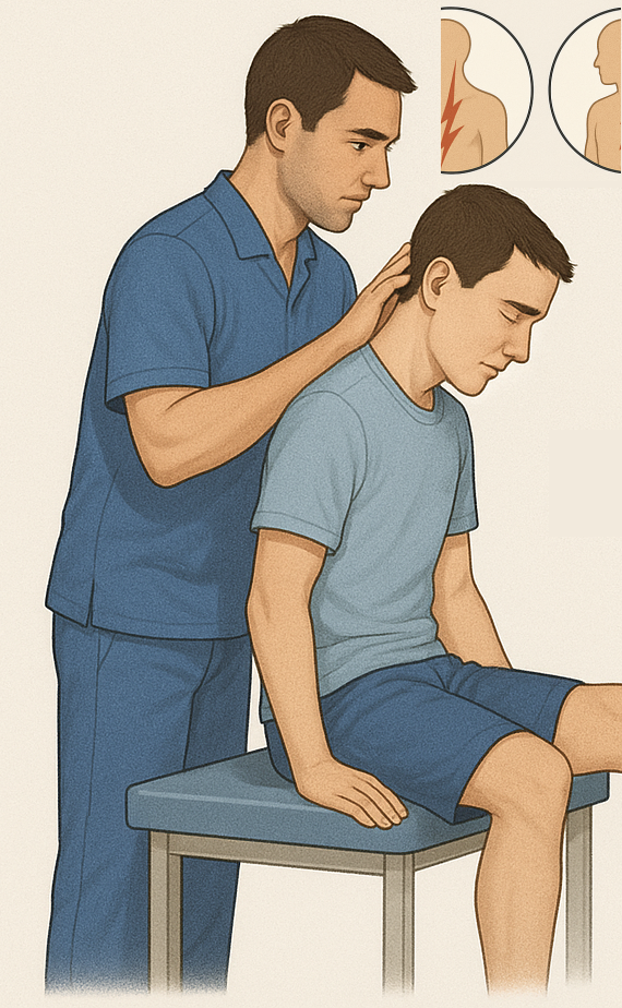
- Lhermitte’s sign
- Patient sitting upright; the therapist passively flexes the patient’s neck (chin toward chest), or the patient actively flexes their own neck
- Identifies upper motor neuron lesions (often associated with multiple sclerosis)
- Positive: a brief, electric shock-like sensation traveling down the spine and into the upper or lower limbs
- Patient sitting upright; the therapist passively flexes the patient’s neck (chin toward chest), or the patient actively flexes their own neck
Clearing the cervical spine after trauma
- Canadian C-Spine Rule
- A decision tool for clearing the cervical spine without imaging in alert, stable trauma patients
- Imaging is needed for age 65 or older, a dangerous mechanism or paresthesias. Otherwise, if a low-risk factor allows range of motion to be assessed, a patient who can actively rotate the neck 45 degrees to each side needs no imaging.