


Achievable logoAchievable logo
NPTE-PTA
Sign in
Sign up
Purchase
Textbook
Practice exams
Support
How it works
Resources
Exam catalog
Mountain with a flag at the peak
Textbook
Introduction
1. Cardiopulmonary system
2. Pulmonary system
3. Neuromuscular system
4. Pediatrics
5. Musculoskeletal system
5.1 Anatomy of musculoskeletal system
5.2 Anatomical terminology and exercise training principles
5.3 Joint mechanics and phases of healing
5.4 Upper extremity anatomy
5.5 Special tests of upper extremity
5.6 Comparing clinical presentation and interventions for upper extremity
5.7 Lower extremity anatomy
5.8 Special tests of lower extremity
5.9 Comparing clinical presentation and interventions of lower extremity
5.10 Spine and pelvis anatomy
5.11 Special tests of the spine, pelvis, and temporomandibular joint
5.11.1 Cervical spine tests
5.11.2 Thoracolumbar, sacroiliac, and TMJ tests
5.12 Comparing clinical presentation and interventions for the spine, pelvis, and temporomandibular joint
5.13 Other MSK conditions
5.14 Gait
5.15 Prosthetics
5.16 Orthotics
5.17 Medications, imaging, and fractures
5.18 Surgical protocols
6. Other system
7. Non systems
Wrapping up
Achievable logoAchievable logo
5.11.2 Thoracolumbar, sacroiliac, and TMJ tests
Achievable NPTE-PTA
5. Musculoskeletal system
5.11. Special tests of the spine, pelvis, and temporomandibular joint
Our NPTE-PTA course is now in "early access" - get 50% off for a limited time.

Thoracolumbar, sacroiliac, and TMJ tests

7 min read
Font
Discuss
Share
Feedback

These special tests cover the thoracic and lumbar spine, the sacroiliac joint, and the temporomandibular joint.

Thoracic spine scoliosis screening

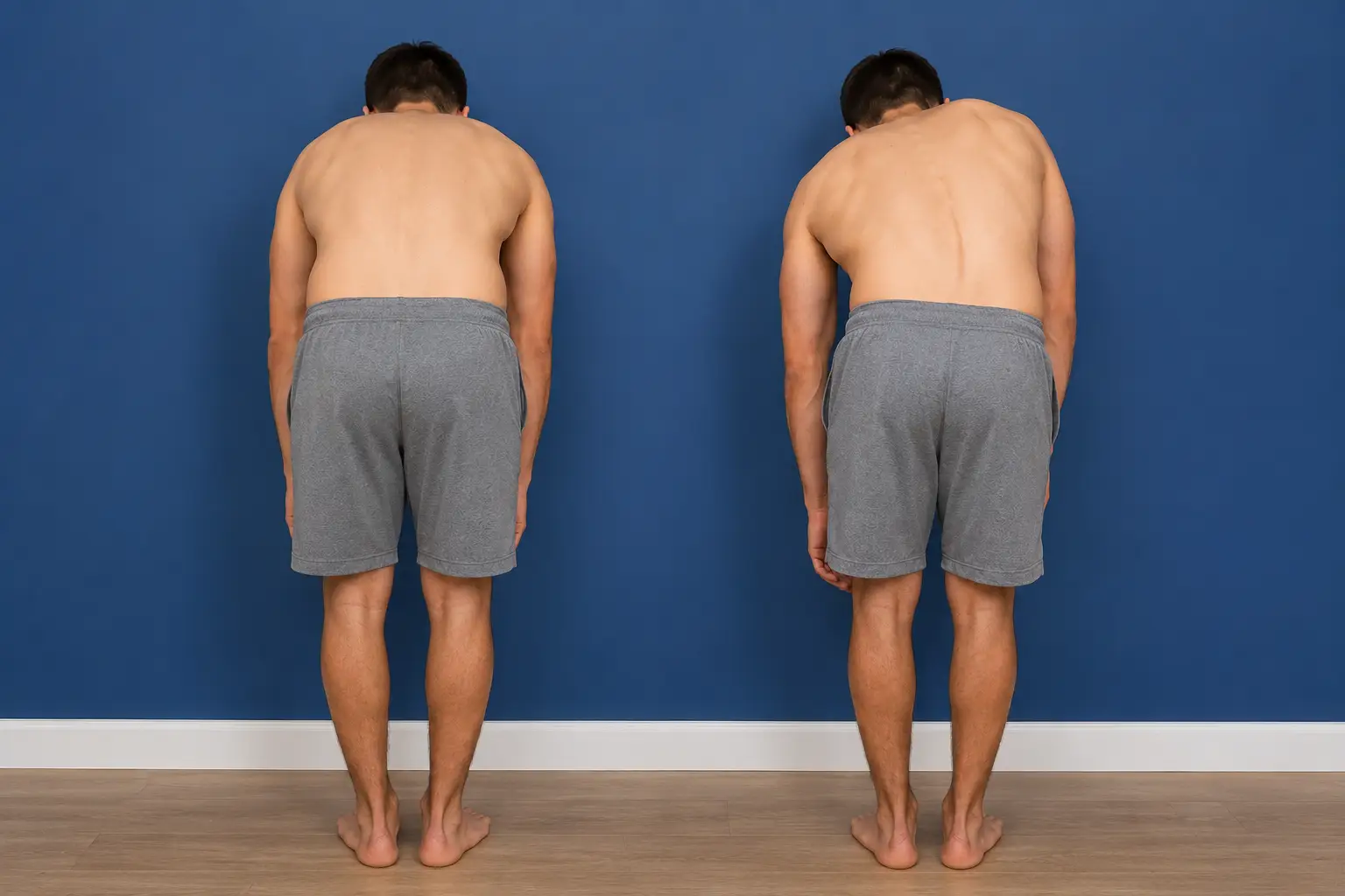
  • Adam’s forward bend test:
    • The child bends forward at the waist while keeping their knees straight.
      • Checks for any asymmetry or curvature in the spine.
    • Positive: elevation of the ribs on one side of the spine
Adam's forward bend test with trunk asymmetry.
Adam's forward bend test
Achievable

Lumbar spine

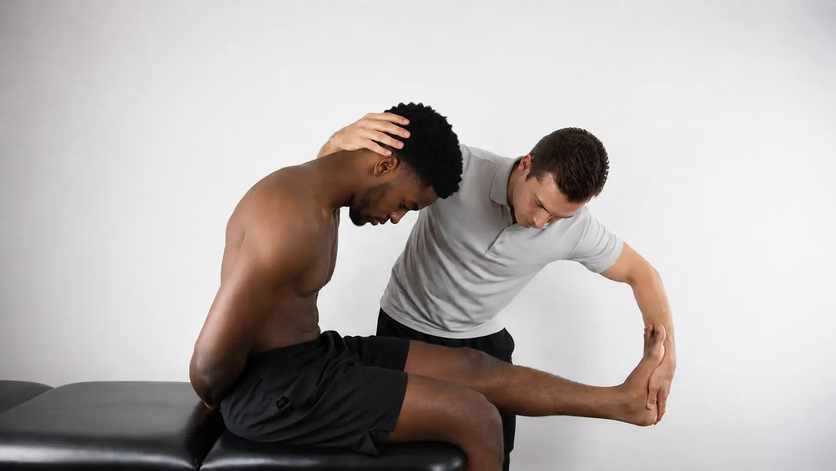
  • Slump test
    • Patient sits with knees flexed and neutral neck and head; progress through the following steps- passively flex head and neck, passively extend knee, passively dorsiflex ankle of limb- repeat with opposite leg
      • The therapist will stop at a step if reproduction of symptoms occurs
        • Tests for neurological dysfunction of the lower extremity
      • Positive: reproduction of pain, paresthesias, numbness/tingling, or other neurological symptoms. This detects disc herniations or screens for sciatica by placing the nervous system under tension.
Slump test with cervical flexion and knee extension.
Slump test
Achievable
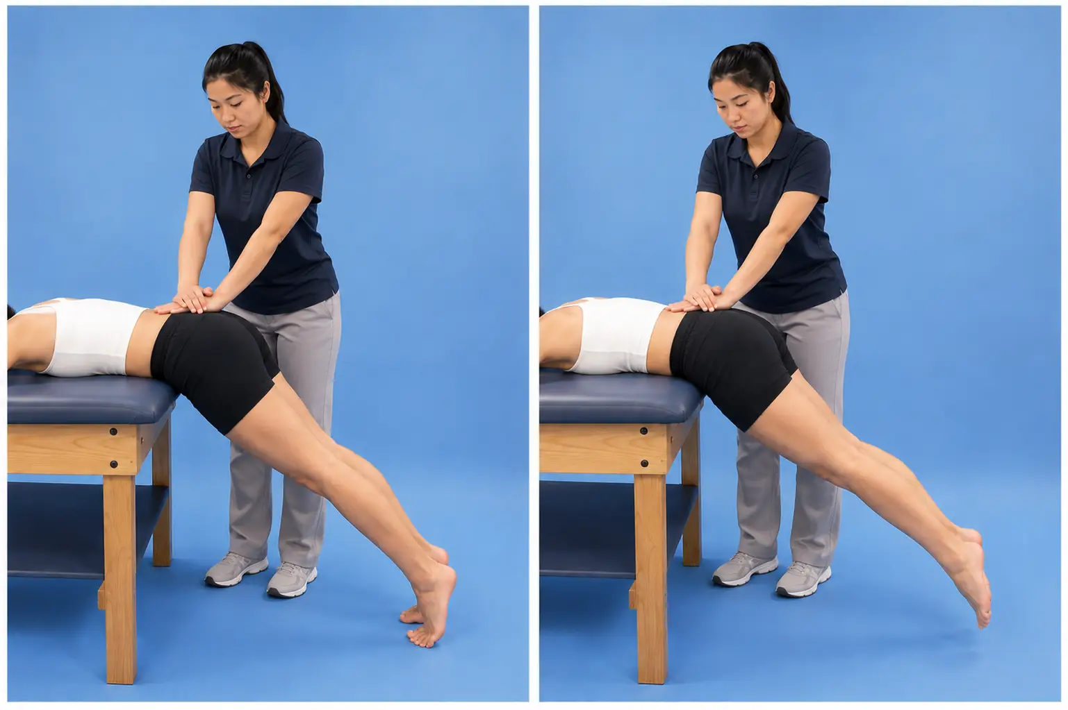
  • Prone instability test
    • Patient prone with torso supported on the mat, legs off the edge, with both feet supported on the ground. The therapist applies posterior/anterior springing throughout the lumbar spine to identify painful segments. The therapist then instructs the patient to lift the legs off the floor and perform the same posterior/anterior springing
      • Tests the instability of the lumbar spine
      • Positive: pain with springing while the feet rest on the ground that decreases or resolves once the patient actively lifts the legs. This shows the patient can stabilize the segment through muscle activation, supporting a diagnosis of clinical lumbar instability that would respond to stabilization exercise.
Prone instability test with trunk extension and lower-extremity activation.
Prone instability test
Achievable
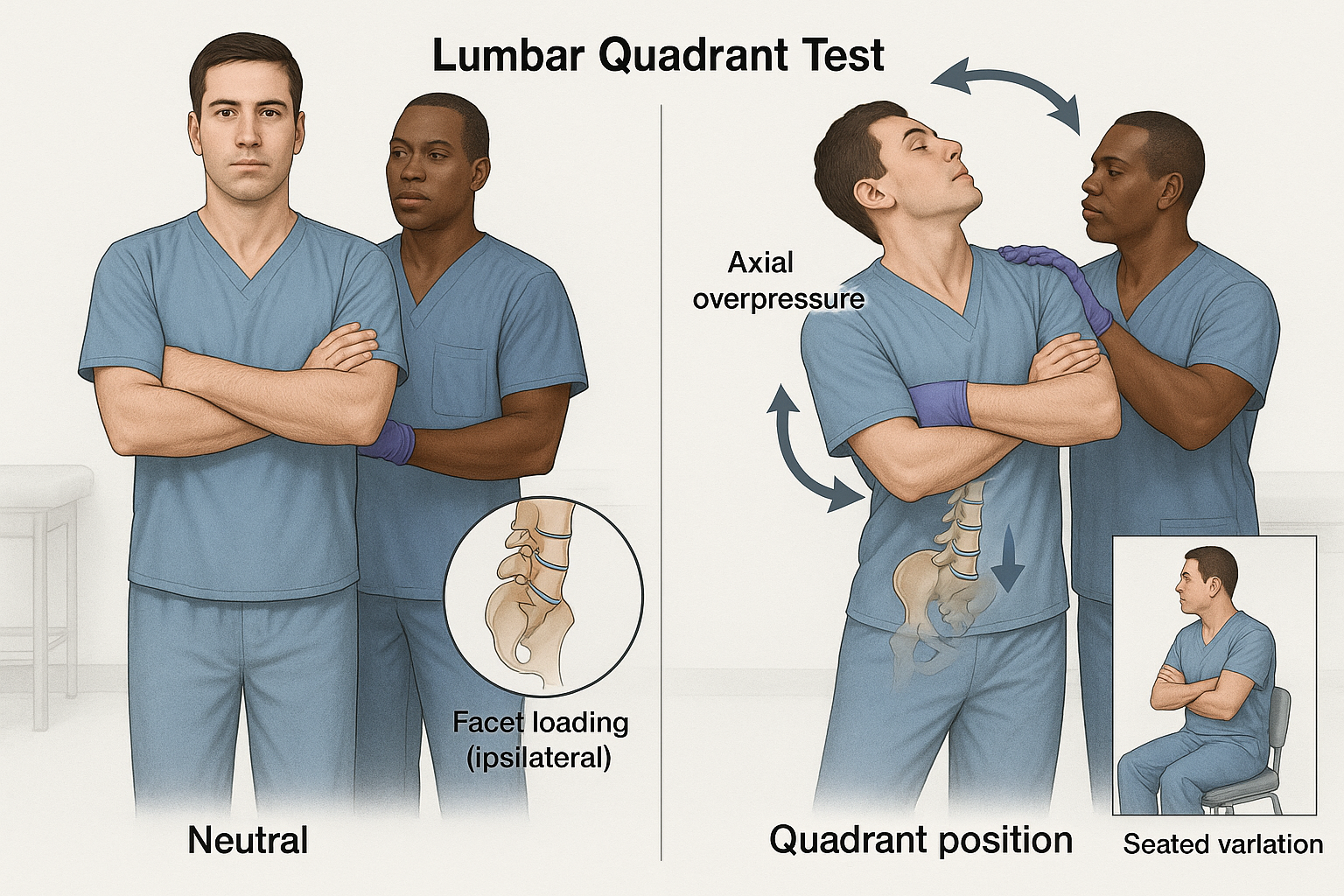
  • Lumbar spine quadrant test (Kemp’s test)
    • Patient standing
      • The patient stands and is guided by the practitioner to maximally extend their spine, then rotate and laterally flex toward the side of the pain. The practitioner applies a gentle downward pressure
      • Tests the compression of neural structures at the facet and intervertebral foramen
      • Positive: pain or paresthesias in a dermatomal pattern or localized pain if facet dysfunction
Lumbar quadrant test
Achievable

Neurological dysfunction

  • Lower limb tension tests assist with identifying peripheral nerve injury by placing the lower limb in positions that will stress the nerve
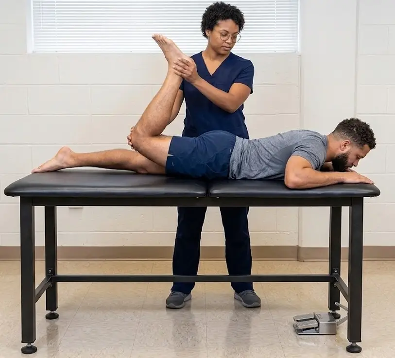
  • Prone knee flexion with hip extension tests femoral nerve
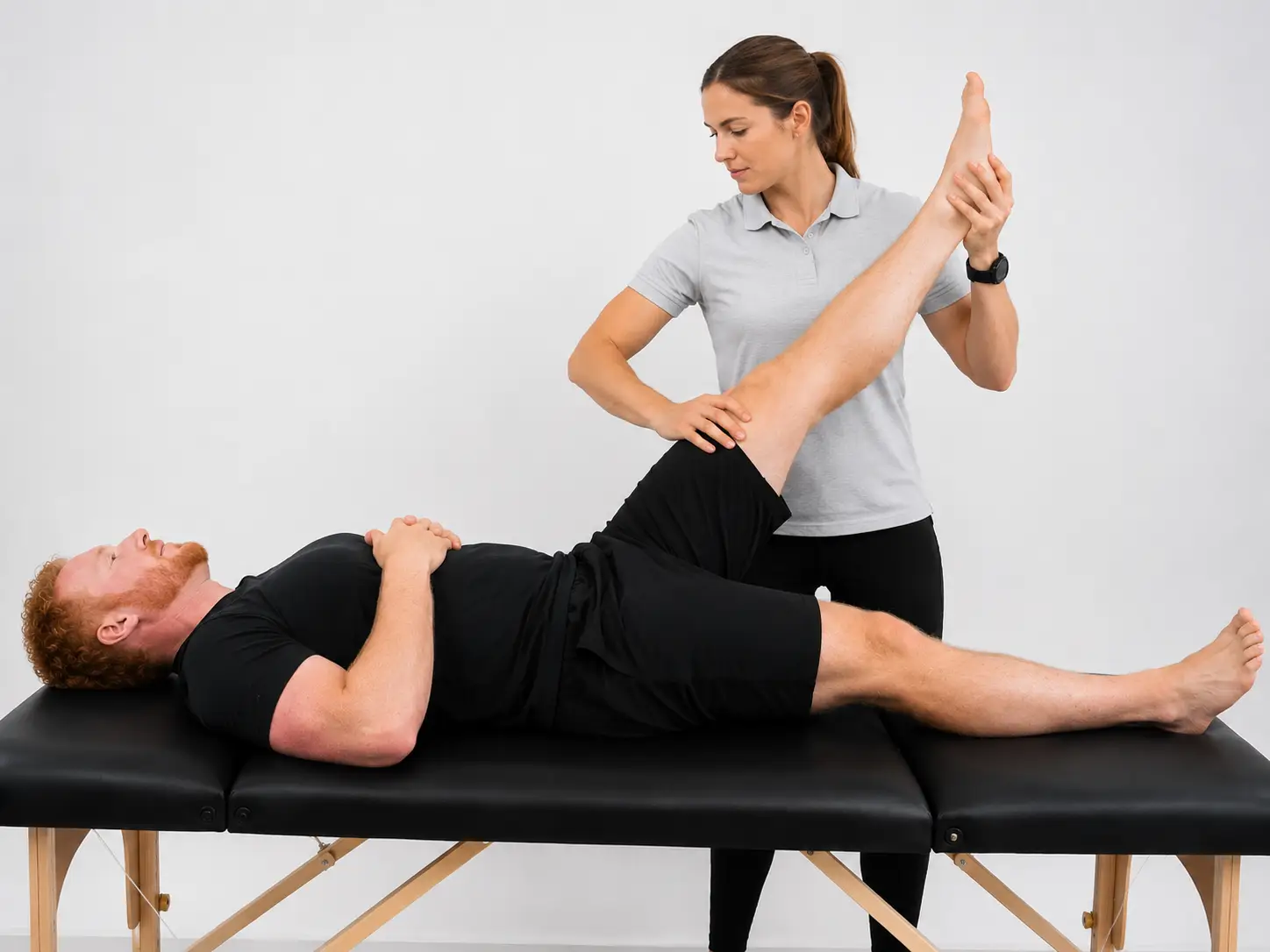
  • Straight leg raise (SLR)- test sciatic and tibial nerve
    • Hip: flexion (neutral rotation)
    • Knee: extension
    • Ankle: dorsiflexion
Straight leg raise test with passive hip flexion and knee extension.
Straight leg raise
Achievable
Femoral nerve stretch test with prone knee flexion and hip extension.
Femoral nerve stretch test
Achievable

Sacroiliac

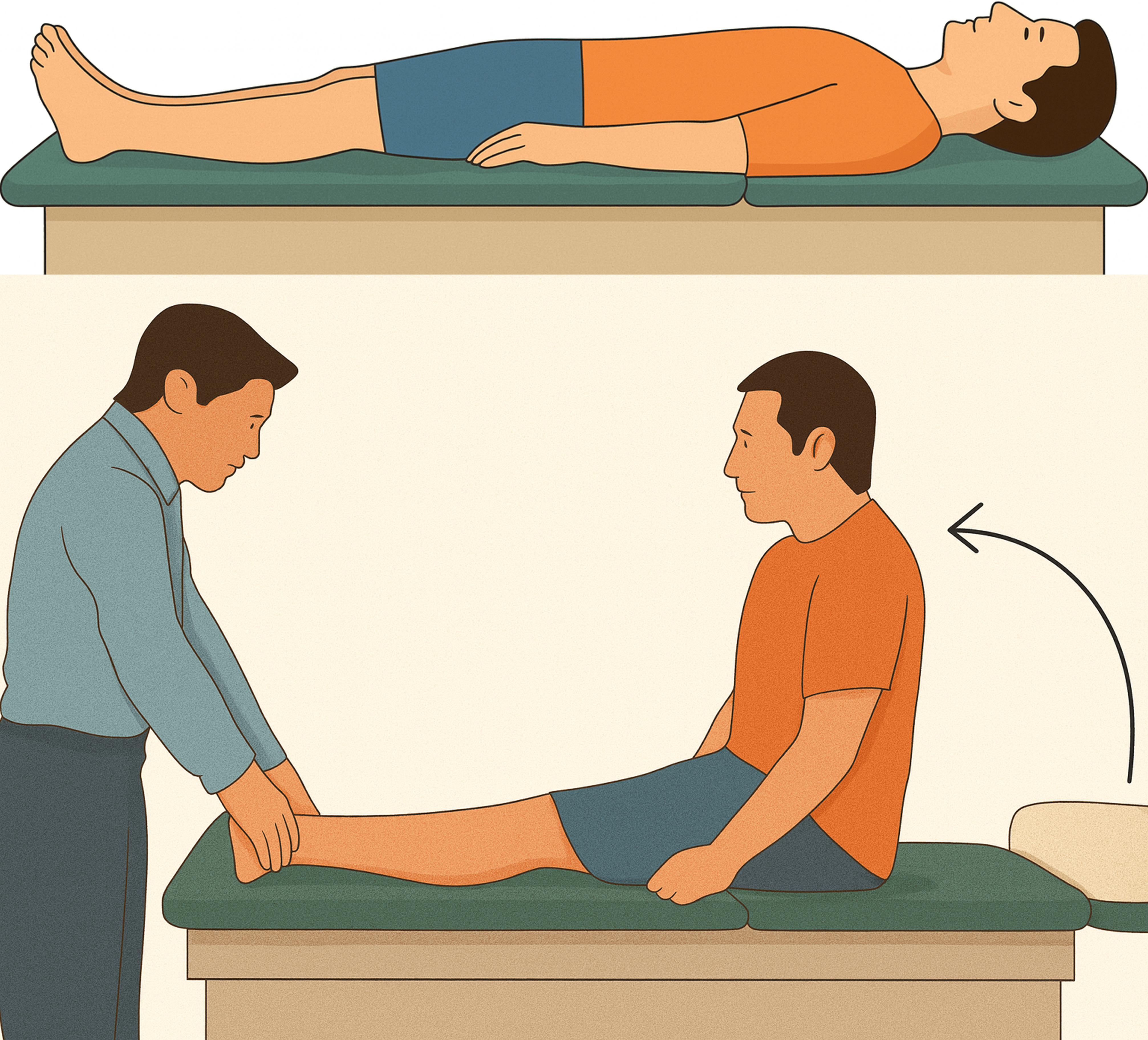
  • Long sitting
    • Patient supine with therapist palpating the medial malleolus- therapist observes alignment. The therapist then asks the patient to come to a long sitting position to observe if alignment at the medial malleolus is still present.
      • Tests for leg length discrepancy due to the sacroiliac joint
    • Positive: one leg observed longer when coming to a long sitting position compared to the supine position
Supine long sitting
Supine long sitting
Achievable

Temporomandibular joint (TMJ)

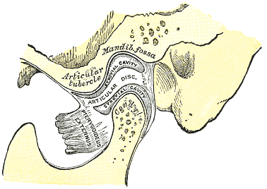
A bilateral synovial joint with articulation between the mandible and the cranium. The articular disc lies between areas of articulation in which there is no vascularization or neural supply. The disc (collateral) ligaments attach the disc to the condyle, so the disc moves anteriorly and posteriorly with the condyle while being kept from displacing medially or laterally. Movements of the TMJ are a combination of axis rotation and sliding movements. Types of movement are seen below:

  • Elevation (closing):
    • Contraction of the masseter, temporalis, and medial pterygoid muscles raises the mandible.
  • Depression (opening):
    • Relaxation of the elevator muscles and contraction of the lateral pterygoid, digastric, geniohyoid, and mylohyoid muscles lowers the mandible.
  • Protrusion (forward movement):
    • Contraction of the lateral pterygoid muscles moves the mandible forward.
  • Retrusion (backward movement):
    • Contraction of the posterior fibers of the temporalis muscles moves the mandible backward.
  • Lateral deviation (side-to-side movement):
    • Contraction of the lateral pterygoid muscle on one side moves the mandible toward the opposite side.

Special tests of TMJ

  • TMJ compression
    • Patient sitting or supine; therapist stabilizes the patient’s head with one hand while the other hand pushes the mandible superiorly, causing a compressive load to the TMJ
      • Assess pain in the retrodiscal tissues
    • Positive: pain in the TMJ
Mandibular joint
Mandibular joint
By - Henry Vandyke Carter (1831-1897), Henry Gray (1918) Anatomy of the Human Body
/
Wikimedia Commons
/
Public domain

Thoracic spine scoliosis screening

  • Adam’s forward bend test: child bends forward, knees straight
  • Checks for spinal asymmetry/curvature
  • Positive: elevation of ribs on one side of spine

Lumbar spine

  • Slump test: sequential head/neck flexion, knee extension, ankle dorsiflexion (bilateral)
    • Tests neurological dysfunction, disc herniation, sciatica via nerve tension
    • Positive: reproduction of pain, paresthesias, numbness/tingling
  • Prone instability test: PA springing with legs on floor vs. lifted
    • Tests lumbar segmental instability
    • Positive: pain resolves/decreases when legs actively lifted (muscle stabilization present)
  • Lumbar quadrant test (Kemp’s test): standing extension + rotation/lateral flexion toward pain side with downward pressure
    • Tests facet/intervertebral foramen compression
    • Positive: dermatomal pain/paresthesias or localized facet pain

Neurological dysfunction

  • Lower limb tension tests stress peripheral nerves via positioning
  • Prone knee flexion with hip extension: tests femoral nerve
  • Straight leg raise (SLR): tests sciatic/tibial nerve
    • Position: hip flexion (neutral rotation), knee extension, ankle dorsiflexion

Sacroiliac

  • Long sitting test: compare medial malleoli alignment supine vs. long sitting
  • Tests leg length discrepancy due to SI joint
  • Positive: one leg appears longer in long sitting vs. supine

Temporomandibular joint (TMJ) anatomy

  • Bilateral synovial joint between mandible and cranium
  • Articular disc: avascular, no neural supply; disc ligaments attach disc to condyle (limits medial/lateral displacement)
  • Movement types:
    • Elevation (closing): masseter, temporalis, medial pterygoid
    • Depression (opening): lateral pterygoid, digastric, geniohyoid, mylohyoid
    • Protrusion: lateral pterygoid (bilateral)
    • Retrusion: posterior temporalis fibers
    • Lateral deviation: unilateral lateral pterygoid (moves mandible to opposite side)

Special tests of TMJ

  • TMJ compression test: stabilize head, push mandible superiorly for compressive load
  • Assesses retrodiscal tissue pain
  • Positive: pain in TMJ

Sign up for free to take 5 quiz questions on this topic

Previous
Next  | 5.12 Comparing clinical presentation and interventions for the spine, pelvis, and temporomandibular joint
All rights reserved ©2016 - 2026 Achievable, Inc.

Thoracolumbar, sacroiliac, and TMJ tests

These special tests cover the thoracic and lumbar spine, the sacroiliac joint, and the temporomandibular joint.

Thoracic spine scoliosis screening

  • Adam’s forward bend test:
    • The child bends forward at the waist while keeping their knees straight.
      • Checks for any asymmetry or curvature in the spine.
    • Positive: elevation of the ribs on one side of the spine

Lumbar spine

  • Slump test
    • Patient sits with knees flexed and neutral neck and head; progress through the following steps- passively flex head and neck, passively extend knee, passively dorsiflex ankle of limb- repeat with opposite leg
      • The therapist will stop at a step if reproduction of symptoms occurs
        • Tests for neurological dysfunction of the lower extremity
      • Positive: reproduction of pain, paresthesias, numbness/tingling, or other neurological symptoms. This detects disc herniations or screens for sciatica by placing the nervous system under tension.
  • Prone instability test
    • Patient prone with torso supported on the mat, legs off the edge, with both feet supported on the ground. The therapist applies posterior/anterior springing throughout the lumbar spine to identify painful segments. The therapist then instructs the patient to lift the legs off the floor and perform the same posterior/anterior springing
      • Tests the instability of the lumbar spine
      • Positive: pain with springing while the feet rest on the ground that decreases or resolves once the patient actively lifts the legs. This shows the patient can stabilize the segment through muscle activation, supporting a diagnosis of clinical lumbar instability that would respond to stabilization exercise.
  • Lumbar spine quadrant test (Kemp’s test)
    • Patient standing
      • The patient stands and is guided by the practitioner to maximally extend their spine, then rotate and laterally flex toward the side of the pain. The practitioner applies a gentle downward pressure
      • Tests the compression of neural structures at the facet and intervertebral foramen
      • Positive: pain or paresthesias in a dermatomal pattern or localized pain if facet dysfunction

Neurological dysfunction

  • Lower limb tension tests assist with identifying peripheral nerve injury by placing the lower limb in positions that will stress the nerve
  • Prone knee flexion with hip extension tests femoral nerve
  • Straight leg raise (SLR)- test sciatic and tibial nerve
    • Hip: flexion (neutral rotation)
    • Knee: extension
    • Ankle: dorsiflexion

Sacroiliac

  • Long sitting
    • Patient supine with therapist palpating the medial malleolus- therapist observes alignment. The therapist then asks the patient to come to a long sitting position to observe if alignment at the medial malleolus is still present.
      • Tests for leg length discrepancy due to the sacroiliac joint
    • Positive: one leg observed longer when coming to a long sitting position compared to the supine position

Temporomandibular joint (TMJ)

A bilateral synovial joint with articulation between the mandible and the cranium. The articular disc lies between areas of articulation in which there is no vascularization or neural supply. The disc (collateral) ligaments attach the disc to the condyle, so the disc moves anteriorly and posteriorly with the condyle while being kept from displacing medially or laterally. Movements of the TMJ are a combination of axis rotation and sliding movements. Types of movement are seen below:

  • Elevation (closing):
    • Contraction of the masseter, temporalis, and medial pterygoid muscles raises the mandible.
  • Depression (opening):
    • Relaxation of the elevator muscles and contraction of the lateral pterygoid, digastric, geniohyoid, and mylohyoid muscles lowers the mandible.
  • Protrusion (forward movement):
    • Contraction of the lateral pterygoid muscles moves the mandible forward.
  • Retrusion (backward movement):
    • Contraction of the posterior fibers of the temporalis muscles moves the mandible backward.
  • Lateral deviation (side-to-side movement):
    • Contraction of the lateral pterygoid muscle on one side moves the mandible toward the opposite side.

Special tests of TMJ

  • TMJ compression
    • Patient sitting or supine; therapist stabilizes the patient’s head with one hand while the other hand pushes the mandible superiorly, causing a compressive load to the TMJ
      • Assess pain in the retrodiscal tissues
    • Positive: pain in the TMJ
Key points

Thoracic spine scoliosis screening

  • Adam’s forward bend test: child bends forward, knees straight
  • Checks for spinal asymmetry/curvature
  • Positive: elevation of ribs on one side of spine

Lumbar spine

  • Slump test: sequential head/neck flexion, knee extension, ankle dorsiflexion (bilateral)
    • Tests neurological dysfunction, disc herniation, sciatica via nerve tension
    • Positive: reproduction of pain, paresthesias, numbness/tingling
  • Prone instability test: PA springing with legs on floor vs. lifted
    • Tests lumbar segmental instability
    • Positive: pain resolves/decreases when legs actively lifted (muscle stabilization present)
  • Lumbar quadrant test (Kemp’s test): standing extension + rotation/lateral flexion toward pain side with downward pressure
    • Tests facet/intervertebral foramen compression
    • Positive: dermatomal pain/paresthesias or localized facet pain

Neurological dysfunction

  • Lower limb tension tests stress peripheral nerves via positioning
  • Prone knee flexion with hip extension: tests femoral nerve
  • Straight leg raise (SLR): tests sciatic/tibial nerve
    • Position: hip flexion (neutral rotation), knee extension, ankle dorsiflexion

Sacroiliac

  • Long sitting test: compare medial malleoli alignment supine vs. long sitting
  • Tests leg length discrepancy due to SI joint
  • Positive: one leg appears longer in long sitting vs. supine

Temporomandibular joint (TMJ) anatomy

  • Bilateral synovial joint between mandible and cranium
  • Articular disc: avascular, no neural supply; disc ligaments attach disc to condyle (limits medial/lateral displacement)
  • Movement types:
    • Elevation (closing): masseter, temporalis, medial pterygoid
    • Depression (opening): lateral pterygoid, digastric, geniohyoid, mylohyoid
    • Protrusion: lateral pterygoid (bilateral)
    • Retrusion: posterior temporalis fibers
    • Lateral deviation: unilateral lateral pterygoid (moves mandible to opposite side)

Special tests of TMJ

  • TMJ compression test: stabilize head, push mandible superiorly for compressive load
  • Assesses retrodiscal tissue pain
  • Positive: pain in TMJ

More from Special tests of the spine, pelvis, and temporomandibular joint

  • Cervical spine tests