


This browser is too old to run Achievable.

Update it, or open this page in a current version of Chrome, Safari, Firefox or Edge. On an iPhone or iPad, update iOS: every browser there runs on the version of Safari that comes with it.

Achievable logoAchievable logo
NPTE-PTA
Sign in
Sign up
Purchase
Textbook
Practice exams
Support
How it works
Resources
Exam catalog
Mountain with a flag at the peak
Textbook
Introduction
1. Cardiovascular system
2. Pulmonary system
3. Neuromuscular system
4. Pediatrics
5. Musculoskeletal system
5.1 Anatomy of musculoskeletal system
5.2 Anatomical terminology and exercise training principles
5.3 Joint mechanics and phases of healing
5.4 Upper extremity anatomy
5.5 Special tests of upper extremity
5.6 Comparing clinical presentation and interventions for upper extremity
5.7 Lower extremity anatomy
5.8 Special tests of lower extremity
5.9 Comparing clinical presentation and interventions for lower extremity
5.10 Spine and pelvis anatomy
5.11 Special tests of the spine, pelvis, and temporomandibular joint
5.12 Comparing clinical presentation and interventions for the spine, pelvis, and temporomandibular joint
5.13 Other musculoskeletal conditions
5.14 Gait
5.15 Prosthetics
5.16 Orthotics
5.17 Medications, imaging, and fractures
5.18 Surgical protocols
6. Other systems
7. Non-systems
Wrapping up
Achievable logoAchievable logo
5.15 Prosthetics
Achievable NPTE-PTA
5. Musculoskeletal system
Our NPTE-PTA course is now in "early access" - get 50% off for a limited time.

Prosthetics

10 min read
Font
Discuss
Share
Feedback

Levels of amputations

Upper extremity

  • Finger or partial hand: Removal of one or more fingers or part of the hand
  • Wrist disarticulation: Removal of the hand at the wrist joint
  • Transradial (below elbow): Removal of the forearm below the elbow joint
  • Elbow disarticulation: Removal of the arm at the elbow joint
  • Transhumeral (above elbow): Removal of the arm above the elbow joint
  • Shoulder disarticulation: Removal of the arm at the shoulder joint

Lower extremity

  • Toe or partial foot: Removal of one or more toes or part of the foot
  • Ankle disarticulation (Syme’s amputation): Removal of the foot at the ankle joint, keeping the heel pad so the residual limb can bear weight at its end
  • Transtibial (below knee): Removal of the leg below the knee joint
  • Knee disarticulation: Removal of the leg at the knee joint
  • Transfemoral (above knee): Removal of the leg above the knee joint
  • Hip disarticulation: Removal of the leg at the hip joint

The NPTE focuses mostly on lower extremity prostheses, but upper extremity prostheses also come up. Body-powered devices use a harness-and-cable system and give sensory feedback through the harness; myoelectric devices use EMG signals from residual muscles for better cosmesis but more weight and cost; hybrid designs combine both. Terminal devices are also classified as voluntary opening (opens with effort, closes passively) or voluntary closing (the reverse).

Components of prosthesis

Primary components

  • Sockets
    • Custom-molded prosthetic that will have total contact with residual limb; goal is equal distribution of tissue load throughout the socket
  • Liners
    • Silicone barrier between residual limb and socket to decrease shear forces
  • Socks
    • Added or removed (by ply) to keep the socket fit snug as residual-limb volume changes during the day and as the limb shrinks after surgery
  • Terminal device
    • Interface between prosthetic limb and external environment
      • Upper limb: the terminal device is a hand or a hook
      • Lower limb: the terminal device is the foot

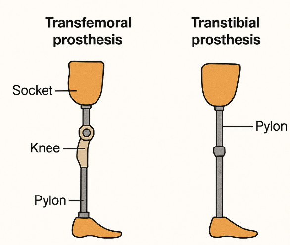
Components of a prosthesis specifically for transtibial amputation

Ankle-foot assembly

  • SACH foot (solid ankle cushioned heel):
    • A simple, durable, and lightweight foot with a solid ankle joint
  • Single-axis foot:
    • Features a hinged ankle joint that allows for up-and-down movement, enhancing knee stability.
  • Multi-axis foot:
    • Similar to a single-axis foot, but also allows for side-to-side movement, making it better suited for uneven terrain
  • Microprocessor-controlled (MPC) feet:
    • These feet use sensors and a computer to adjust the ankle’s position and resistance based on the user’s movements and the environment
  • Energy storing and return (ESAR) feet:
    • These feet use springy components that store mechanical energy during stance and release it during push-off

Shank

  • Endoskeleton prosthesis:
    • Structure: Features a tubular structure connecting the primary components, like the socket, suspension system, foot, and knee unit
    • Appearance: Often covered with cosmetic foam
    • Advantages: Endoskeletal prostheses are adjustable and lighter than exoskeletal prostheses.
    • Disadvantages: Less durable than exoskeletal prostheses
  • Exoskeleton prosthesis:
    • Structure: Has a hard outer shell that provides structural strength and cosmetic shape
    • Durability: More durable than endoskeletal prostheses
    • Advantages: May be preferred by people who do physical labor or are in harsh environments
    • Disadvantages: Less customizable than endoskeletal prostheses.

Socket

  • Patellar tendon bearing (PTB) sockets:
    • Focus: PTB sockets, often used for below-knee amputations, focus weight-bearing stress on pressure-tolerant structures like the patellar tendon and medial tibia flare
      • Advantages:
        • Can be a good choice for patients with shorter or bony residual limbs
        • Easier to don/doff for amputees with poor hand dexterity, poor eyesight, or hemiparesis
        • Can be modified to accommodate changes in the residual limb after amputation
      • Disadvantages:
        • May not be ideal for patients with residual limb scar tissue or chronic skin breakdown
  • Total surface bearing (TSB) sockets:
    • Focus: TSB sockets, also used for below-knee amputations, distribute weight evenly across the entire limb surface, reducing pressure points
      • Advantages:
        • Often used with gel liners, which cushion the limb and protect the skin, making them great for amputees with scar tissue or sensitive skin
        • Offer better suspension than PTB designs, often using locking pins or suction systems
      • Disadvantages:
        • Can be more difficult to don/doff than PTB sockets
Prosthesis components
Achievable

Components of prosthesis specifically for transfemoral amputation

Knee unit

  • Single-axis knees:
    • Mechanism: A single pivot point, resembling a simple hinge
    • Stability: Little built-in stance stability; the user keeps the knee from buckling through alignment and voluntary hip extensor control. The manual-locking type (below) adds stance stability by locking the knee in extension
    • Suitable for: Individuals with lower activity levels, those who can voluntarily stabilize the knee
    • Types:
      • Manual locking: The knee can be locked in extension for stability, and unlocked manually for sitting
      • Constant friction: Provides constant resistance to knee flexion/extension throughout swing, controlling swing speed at a single walking cadence - it does not add stance-phase stability
  • Polycentric (multiaxis) knees:
    • Mechanism: Multiple pivot points, allowing for a more natural and efficient gait, particularly during the swing phase
    • Stability: High inherent stance stability from the shifting center of rotation; also shortens the shank during swing, improving toe clearance
    • Suitable for: Active individuals, those with knee disarticulation or long residual limbs, or those with weak hip extensors who need added stance support
  • Fluid-controlled (hydraulic/pneumatic) swing control:
    • Mechanism: Uses fluid or air resistance to control the speed of knee swing, automatically adjusting to the user’s walking cadence (unlike constant friction, which only controls one speed)
    • Suitable for: Can be paired with single-axis or polycentric knee units for a smoother, more natural, variable-speed gait
  • Microprocessor knees:
    • Mechanism: Use of sensors and electronics to adjust the resistance to knee flexion and extension based on the user’s gait and activity level
    • Benefits: Improve stability, reduce energy expenditure, and allow for a more natural and comfortable gait
    • Suitable for: Highly active individuals, those who require a high level of stability and control, or those who want to maximize their mobility
  • Knee axis alignment: in a transfemoral prosthesis, a knee axis placed behind the weight line (the trochanter-knee-ankle, or TKA, line) is stable in stance because body weight pushes the knee into extension, but a knee placed too far posterior is so stable that it is hard to initiate knee flexion for swing

Socket

  • Ischial containment (IC) sockets
    • These designs feature a narrow medial-lateral dimension with the ischium (part of the pelvic bone) encapsulated within the socket, rather than sitting on the brim
  • Sub-ischial sockets
    • These sockets have a lower proximal trim line than IC sockets, and are often used with vacuum-assisted suspension
  • Quadrilateral (quad) sockets
    • These were historically the most common type, but are less common now, although still used for some patients

Keys for transfemoral socket trim lines

  • In a transfemoral socket, the posterior and medial walls are lower than the anterior and lateral walls
  • This allows room for the groin/adductors (medial) and the gluteals and for sitting (posterior)

Residual limb management and prosthetic training

  • Shrinking the limb: Wrap with a figure-eight elastic bandage or shrinker sock, distal-to-proximal (never circular wrapping, which can act like a tourniquet).
  • Preventing contractures: Transfemoral: avoid prolonged hip flexion/abduction - encourage prone lying, no pillow under the hip. Transtibial: avoid prolonged knee flexion - no pillow under the knee, encourage prone lying.
  • Sock-ply management: Add plies as the limb shrinks or loses volume; remove plies if the socket feels tight or skin shows pressure marks.
  • Skin checks: Inspect the residual limb before and after each wearing session for redness, blistering, or breakdown.
  • Suspension options: pin/lock, suction, vacuum-assisted, sleeve, supracondylar cuff, belt/strap.

K-levels: A functional classification (K0-K4) guiding component selection, from K0 (unable to safely ambulate) to K4 (high-impact/athletic activity); higher levels support advanced components like energy-storing feet and microprocessor knees.

Prosthetic gait deviations

Prosthetic gait deviations refer to any variations from a normal walking pattern that occur in individuals using prosthetic limbs, often due to compensatory strategies or issues with the prosthesis itself.

PTA role and stop signs: The PTA observes and documents gait deviations and reports suspected prosthetic causes (length, alignment, suspension, fit) to the supervising PT, who coordinates with the prosthetist - adjusting alignment is outside the PTA’s role. Stop wear and notify the PT if redness persists beyond ~20 minutes after removal, or if blistering, open skin, or drainage appears. An open wound on a weight-bearing area contraindicates socket wear; fluctuating volume is just a precaution, managed with sock ply. During gait training, also stop and notify the PT for chest pain, marked shortness of breath, dizziness, or an abnormal heart rate or blood pressure response; many people with lower-limb amputations have vascular or heart disease, and walking with a prosthesis raises oxygen use and heart rate.

Vaulting (stance phase, intact limb)

  • Definition: The patient rises up on the toes (plantarflexes) of the intact limb during its mid-stance to functionally lengthen it, so the prosthetic limb can clear the ground during swing.
  • Causes:
    • Limb too long
    • Poorly suspended prosthesis
    • Excessive plantarflexion

Example: Vaulting during gait

A patient with a transfemoral prosthesis rises onto the toes of the intact limb during prosthetic swing. What should the PTA do?

Answer: Document the deviation and consider likely prosthetic causes - the prosthesis may be too long, poorly suspended, or set in excessive plantarflexion. Report these to the supervising PT, who coordinates any adjustment with the prosthetist; the PTA does not adjust the prosthesis directly.

Circumduction (swing phase, mainly transfemoral)

  • Definition: The prosthetic limb travels in a lateral arch during swing.
  • Causes:
    • Excessive abduction of the prosthesis
    • Prosthesis too long
    • Inadequate suspension
    • Excessive plantarflexion of the prosthetic foot
    • Excessive knee friction or a locked knee unit

Abducted gait pattern (swing and stance)

  • Definition: The prosthetic limb is carried in an abducted position throughout the swing and stance phase
  • Causes:
    • Outset prosthetic foot
    • Prosthesis too long
    • Medially placed intact foot

Knee instability (early stance)

  • Definition: Excessive knee flexion on the prosthetic side from initial contact through early stance
  • Causes:
    • Excessive foot dorsiflexion
    • Excessive socket flexion
    • Posterior translation of foot/pylon
    • Excessively hard heel cushion on prosthetic heel
    • Weakness of the knee extensors
    • Pain in the residual limb or socket

Reduced toe clearance (swing)

  • Definition: Prosthetic toe drags or catches during the swing phase
  • Causes:
    • Prosthetic limb too long
    • Inadequate suspension
    • Residual limb not getting into prosthetic socket all the way
    • Excessive plantarflexion of the prosthetic foot

Pelvic drop (initial contact)

  • Definition: The pelvis on the prosthetic side drops on initial contact as if “stepping into a hole.”
  • Causes:
    • Prosthetic limb too short
    • Residual limb has shrunk relative to the socket

Lateral trunk lean over prosthesis (Trendelenburg) (stance)

  • Definition: Trunk bends laterally over the prosthesis (compensated Trendelenburg) during stance
  • Causes:
    • Inadequate adduction of the socket
    • Prosthesis too short
    • Outset foot
    • Medial wall too high, causing pain
    • Gapping at the lateral wall of the socket

Medial/lateral whip (swing)

  • Definition: Abrupt medial or lateral movement of the prosthetic heel during swing
  • Causes:
    • Excessive external or internal rotation of the socket
    • Socket too tight
    • Inadequate suspension
    • Excessive valgus of prosthetic knee

Amputation levels

  • Upper extremity: finger/partial hand → wrist disarticulation → transradial (below elbow) → elbow disarticulation → transhumeral (above elbow) → shoulder disarticulation
  • Lower extremity: toe/partial foot → Syme’s (ankle disarticulation, preserves heel pad) → transtibial (below knee) → knee disarticulation → transfemoral (above knee) → hip disarticulation
  • UE terminal devices: body-powered (harness/cable, sensory feedback) vs myoelectric (EMG-driven, better cosmesis, heavier/costlier) vs hybrid; also classified as voluntary opening or voluntary closing

Core prosthesis components

  • Socket: custom-molded, total contact, even load distribution
  • Liner: silicone layer reducing shear between limb and socket
  • Socks: adjusted by ply to maintain fit as limb volume changes
  • Terminal device: hand/hook (UE) or foot (LE) — interface with environment

Transtibial-specific components

  • Foot types: SACH (solid, simple) → single-axis (sagittal motion, knee stability) → multi-axis (multi-directional, uneven terrain) → microprocessor-controlled (sensor-adjusted)
  • Shank: endoskeletal (tubular, adjustable, lighter, less durable) vs exoskeletal (rigid shell, durable, less adjustable)
  • Socket types: PTB (focuses load on patellar tendon/tibial flare; easier donning, good for short/bony limbs) vs TSB (even surface load, used with gel liners, better suspension but harder to don)

Transfemoral-specific components

  • Knee units: single-axis (simple hinge, little inherent stance stability, relies on alignment and voluntary hip extensor control) → polycentric (multi-pivot, natural swing, good for active users/weak hip extensors) → fluid-controlled (variable-speed swing via hydraulic/pneumatic resistance) → microprocessor (sensor-adjusted, best stability/efficiency)
  • Socket types: ischial containment (ischium encapsulated, narrow M-L), sub-ischial (lower trim line, vacuum suspension), quadrilateral (older design)
  • Trim lines: posterior/medial walls lower than anterior/lateral to allow groin/gluteal clearance and sitting

Residual limb management & training

  • Shrink limb with figure-eight wrap or shrinker sock, distal-to-proximal (never circular/tourniquet-style)
  • Prevent contractures: transfemoral—avoid hip flexion/abduction, prone lie, no hip pillow; transtibial—avoid knee flexion, no knee pillow, prone lie
  • Manage sock plies based on limb volume changes; perform skin checks before/after wear
  • Suspension options: pin/lock, suction, vacuum-assisted, sleeve, supracondylar cuff, belt/strap
  • K-levels (K0–K4): functional classification guiding component choice, from non-ambulatory (K0) to high-impact/athletic (K4)

Gait deviations & PTA role

  • Deviations stem from alignment, suspension, fit, or length issues, categorized by gait phase (e.g., stance-phase: vaulting, lateral trunk lean, knee instability; swing-phase: circumduction, reduced toe clearance, medial/lateral whip; swing and stance: abducted gait; initial contact: pelvic drop)
  • Common causes recur across deviations: prosthesis too long/short, poor suspension, misaligned socket, or abnormal foot plantarflexion/dorsiflexion
  • PTA’s role: observe, document, and report suspected causes to supervising PT (who coordinates with prosthetist) — PTA does not adjust alignment
  • Stop wear/notify PT if redness persists >20 min, or blistering/open skin/drainage occurs; open wounds on weight-bearing areas contraindicate socket use

Sign up for free to take 26 quiz questions on this topic

Previous
Next  | 5.16 Orthotics
All rights reserved ©2016 - 2026 Achievable, Inc.

Prosthetics

Levels of amputations

Upper extremity

  • Finger or partial hand: Removal of one or more fingers or part of the hand
  • Wrist disarticulation: Removal of the hand at the wrist joint
  • Transradial (below elbow): Removal of the forearm below the elbow joint
  • Elbow disarticulation: Removal of the arm at the elbow joint
  • Transhumeral (above elbow): Removal of the arm above the elbow joint
  • Shoulder disarticulation: Removal of the arm at the shoulder joint

Lower extremity

  • Toe or partial foot: Removal of one or more toes or part of the foot
  • Ankle disarticulation (Syme’s amputation): Removal of the foot at the ankle joint, keeping the heel pad so the residual limb can bear weight at its end
  • Transtibial (below knee): Removal of the leg below the knee joint
  • Knee disarticulation: Removal of the leg at the knee joint
  • Transfemoral (above knee): Removal of the leg above the knee joint
  • Hip disarticulation: Removal of the leg at the hip joint

The NPTE focuses mostly on lower extremity prostheses, but upper extremity prostheses also come up. Body-powered devices use a harness-and-cable system and give sensory feedback through the harness; myoelectric devices use EMG signals from residual muscles for better cosmesis but more weight and cost; hybrid designs combine both. Terminal devices are also classified as voluntary opening (opens with effort, closes passively) or voluntary closing (the reverse).

Components of prosthesis

Primary components

  • Sockets
    • Custom-molded prosthetic that will have total contact with residual limb; goal is equal distribution of tissue load throughout the socket
  • Liners
    • Silicone barrier between residual limb and socket to decrease shear forces
  • Socks
    • Added or removed (by ply) to keep the socket fit snug as residual-limb volume changes during the day and as the limb shrinks after surgery
  • Terminal device
    • Interface between prosthetic limb and external environment
      • Upper limb: the terminal device is a hand or a hook
      • Lower limb: the terminal device is the foot

Components of a prosthesis specifically for transtibial amputation

Ankle-foot assembly

  • SACH foot (solid ankle cushioned heel):
    • A simple, durable, and lightweight foot with a solid ankle joint
  • Single-axis foot:
    • Features a hinged ankle joint that allows for up-and-down movement, enhancing knee stability.
  • Multi-axis foot:
    • Similar to a single-axis foot, but also allows for side-to-side movement, making it better suited for uneven terrain
  • Microprocessor-controlled (MPC) feet:
    • These feet use sensors and a computer to adjust the ankle’s position and resistance based on the user’s movements and the environment
  • Energy storing and return (ESAR) feet:
    • These feet use springy components that store mechanical energy during stance and release it during push-off

Shank

  • Endoskeleton prosthesis:
    • Structure: Features a tubular structure connecting the primary components, like the socket, suspension system, foot, and knee unit
    • Appearance: Often covered with cosmetic foam
    • Advantages: Endoskeletal prostheses are adjustable and lighter than exoskeletal prostheses.
    • Disadvantages: Less durable than exoskeletal prostheses
  • Exoskeleton prosthesis:
    • Structure: Has a hard outer shell that provides structural strength and cosmetic shape
    • Durability: More durable than endoskeletal prostheses
    • Advantages: May be preferred by people who do physical labor or are in harsh environments
    • Disadvantages: Less customizable than endoskeletal prostheses.

Socket

  • Patellar tendon bearing (PTB) sockets:
    • Focus: PTB sockets, often used for below-knee amputations, focus weight-bearing stress on pressure-tolerant structures like the patellar tendon and medial tibia flare
      • Advantages:
        • Can be a good choice for patients with shorter or bony residual limbs
        • Easier to don/doff for amputees with poor hand dexterity, poor eyesight, or hemiparesis
        • Can be modified to accommodate changes in the residual limb after amputation
      • Disadvantages:
        • May not be ideal for patients with residual limb scar tissue or chronic skin breakdown
  • Total surface bearing (TSB) sockets:
    • Focus: TSB sockets, also used for below-knee amputations, distribute weight evenly across the entire limb surface, reducing pressure points
      • Advantages:
        • Often used with gel liners, which cushion the limb and protect the skin, making them great for amputees with scar tissue or sensitive skin
        • Offer better suspension than PTB designs, often using locking pins or suction systems
      • Disadvantages:
        • Can be more difficult to don/doff than PTB sockets

Components of prosthesis specifically for transfemoral amputation

Knee unit

  • Single-axis knees:
    • Mechanism: A single pivot point, resembling a simple hinge
    • Stability: Little built-in stance stability; the user keeps the knee from buckling through alignment and voluntary hip extensor control. The manual-locking type (below) adds stance stability by locking the knee in extension
    • Suitable for: Individuals with lower activity levels, those who can voluntarily stabilize the knee
    • Types:
      • Manual locking: The knee can be locked in extension for stability, and unlocked manually for sitting
      • Constant friction: Provides constant resistance to knee flexion/extension throughout swing, controlling swing speed at a single walking cadence - it does not add stance-phase stability
  • Polycentric (multiaxis) knees:
    • Mechanism: Multiple pivot points, allowing for a more natural and efficient gait, particularly during the swing phase
    • Stability: High inherent stance stability from the shifting center of rotation; also shortens the shank during swing, improving toe clearance
    • Suitable for: Active individuals, those with knee disarticulation or long residual limbs, or those with weak hip extensors who need added stance support
  • Fluid-controlled (hydraulic/pneumatic) swing control:
    • Mechanism: Uses fluid or air resistance to control the speed of knee swing, automatically adjusting to the user’s walking cadence (unlike constant friction, which only controls one speed)
    • Suitable for: Can be paired with single-axis or polycentric knee units for a smoother, more natural, variable-speed gait
  • Microprocessor knees:
    • Mechanism: Use of sensors and electronics to adjust the resistance to knee flexion and extension based on the user’s gait and activity level
    • Benefits: Improve stability, reduce energy expenditure, and allow for a more natural and comfortable gait
    • Suitable for: Highly active individuals, those who require a high level of stability and control, or those who want to maximize their mobility
  • Knee axis alignment: in a transfemoral prosthesis, a knee axis placed behind the weight line (the trochanter-knee-ankle, or TKA, line) is stable in stance because body weight pushes the knee into extension, but a knee placed too far posterior is so stable that it is hard to initiate knee flexion for swing

Socket

  • Ischial containment (IC) sockets
    • These designs feature a narrow medial-lateral dimension with the ischium (part of the pelvic bone) encapsulated within the socket, rather than sitting on the brim
  • Sub-ischial sockets
    • These sockets have a lower proximal trim line than IC sockets, and are often used with vacuum-assisted suspension
  • Quadrilateral (quad) sockets
    • These were historically the most common type, but are less common now, although still used for some patients

Keys for transfemoral socket trim lines

  • In a transfemoral socket, the posterior and medial walls are lower than the anterior and lateral walls
  • This allows room for the groin/adductors (medial) and the gluteals and for sitting (posterior)

Residual limb management and prosthetic training

  • Shrinking the limb: Wrap with a figure-eight elastic bandage or shrinker sock, distal-to-proximal (never circular wrapping, which can act like a tourniquet).
  • Preventing contractures: Transfemoral: avoid prolonged hip flexion/abduction - encourage prone lying, no pillow under the hip. Transtibial: avoid prolonged knee flexion - no pillow under the knee, encourage prone lying.
  • Sock-ply management: Add plies as the limb shrinks or loses volume; remove plies if the socket feels tight or skin shows pressure marks.
  • Skin checks: Inspect the residual limb before and after each wearing session for redness, blistering, or breakdown.
  • Suspension options: pin/lock, suction, vacuum-assisted, sleeve, supracondylar cuff, belt/strap.

K-levels: A functional classification (K0-K4) guiding component selection, from K0 (unable to safely ambulate) to K4 (high-impact/athletic activity); higher levels support advanced components like energy-storing feet and microprocessor knees.

Prosthetic gait deviations

Prosthetic gait deviations refer to any variations from a normal walking pattern that occur in individuals using prosthetic limbs, often due to compensatory strategies or issues with the prosthesis itself.

PTA role and stop signs: The PTA observes and documents gait deviations and reports suspected prosthetic causes (length, alignment, suspension, fit) to the supervising PT, who coordinates with the prosthetist - adjusting alignment is outside the PTA’s role. Stop wear and notify the PT if redness persists beyond ~20 minutes after removal, or if blistering, open skin, or drainage appears. An open wound on a weight-bearing area contraindicates socket wear; fluctuating volume is just a precaution, managed with sock ply. During gait training, also stop and notify the PT for chest pain, marked shortness of breath, dizziness, or an abnormal heart rate or blood pressure response; many people with lower-limb amputations have vascular or heart disease, and walking with a prosthesis raises oxygen use and heart rate.

Vaulting (stance phase, intact limb)

  • Definition: The patient rises up on the toes (plantarflexes) of the intact limb during its mid-stance to functionally lengthen it, so the prosthetic limb can clear the ground during swing.
  • Causes:
    • Limb too long
    • Poorly suspended prosthesis
    • Excessive plantarflexion

Example: Vaulting during gait

A patient with a transfemoral prosthesis rises onto the toes of the intact limb during prosthetic swing. What should the PTA do?

Answer: Document the deviation and consider likely prosthetic causes - the prosthesis may be too long, poorly suspended, or set in excessive plantarflexion. Report these to the supervising PT, who coordinates any adjustment with the prosthetist; the PTA does not adjust the prosthesis directly.

Circumduction (swing phase, mainly transfemoral)

  • Definition: The prosthetic limb travels in a lateral arch during swing.
  • Causes:
    • Excessive abduction of the prosthesis
    • Prosthesis too long
    • Inadequate suspension
    • Excessive plantarflexion of the prosthetic foot
    • Excessive knee friction or a locked knee unit

Abducted gait pattern (swing and stance)

  • Definition: The prosthetic limb is carried in an abducted position throughout the swing and stance phase
  • Causes:
    • Outset prosthetic foot
    • Prosthesis too long
    • Medially placed intact foot

Knee instability (early stance)

  • Definition: Excessive knee flexion on the prosthetic side from initial contact through early stance
  • Causes:
    • Excessive foot dorsiflexion
    • Excessive socket flexion
    • Posterior translation of foot/pylon
    • Excessively hard heel cushion on prosthetic heel
    • Weakness of the knee extensors
    • Pain in the residual limb or socket

Reduced toe clearance (swing)

  • Definition: Prosthetic toe drags or catches during the swing phase
  • Causes:
    • Prosthetic limb too long
    • Inadequate suspension
    • Residual limb not getting into prosthetic socket all the way
    • Excessive plantarflexion of the prosthetic foot

Pelvic drop (initial contact)

  • Definition: The pelvis on the prosthetic side drops on initial contact as if “stepping into a hole.”
  • Causes:
    • Prosthetic limb too short
    • Residual limb has shrunk relative to the socket

Lateral trunk lean over prosthesis (Trendelenburg) (stance)

  • Definition: Trunk bends laterally over the prosthesis (compensated Trendelenburg) during stance
  • Causes:
    • Inadequate adduction of the socket
    • Prosthesis too short
    • Outset foot
    • Medial wall too high, causing pain
    • Gapping at the lateral wall of the socket

Medial/lateral whip (swing)

  • Definition: Abrupt medial or lateral movement of the prosthetic heel during swing
  • Causes:
    • Excessive external or internal rotation of the socket
    • Socket too tight
    • Inadequate suspension
    • Excessive valgus of prosthetic knee
Key points

Amputation levels

  • Upper extremity: finger/partial hand → wrist disarticulation → transradial (below elbow) → elbow disarticulation → transhumeral (above elbow) → shoulder disarticulation
  • Lower extremity: toe/partial foot → Syme’s (ankle disarticulation, preserves heel pad) → transtibial (below knee) → knee disarticulation → transfemoral (above knee) → hip disarticulation
  • UE terminal devices: body-powered (harness/cable, sensory feedback) vs myoelectric (EMG-driven, better cosmesis, heavier/costlier) vs hybrid; also classified as voluntary opening or voluntary closing

Core prosthesis components

  • Socket: custom-molded, total contact, even load distribution
  • Liner: silicone layer reducing shear between limb and socket
  • Socks: adjusted by ply to maintain fit as limb volume changes
  • Terminal device: hand/hook (UE) or foot (LE) — interface with environment

Transtibial-specific components

  • Foot types: SACH (solid, simple) → single-axis (sagittal motion, knee stability) → multi-axis (multi-directional, uneven terrain) → microprocessor-controlled (sensor-adjusted)
  • Shank: endoskeletal (tubular, adjustable, lighter, less durable) vs exoskeletal (rigid shell, durable, less adjustable)
  • Socket types: PTB (focuses load on patellar tendon/tibial flare; easier donning, good for short/bony limbs) vs TSB (even surface load, used with gel liners, better suspension but harder to don)

Transfemoral-specific components

  • Knee units: single-axis (simple hinge, little inherent stance stability, relies on alignment and voluntary hip extensor control) → polycentric (multi-pivot, natural swing, good for active users/weak hip extensors) → fluid-controlled (variable-speed swing via hydraulic/pneumatic resistance) → microprocessor (sensor-adjusted, best stability/efficiency)
  • Socket types: ischial containment (ischium encapsulated, narrow M-L), sub-ischial (lower trim line, vacuum suspension), quadrilateral (older design)
  • Trim lines: posterior/medial walls lower than anterior/lateral to allow groin/gluteal clearance and sitting

Residual limb management & training

  • Shrink limb with figure-eight wrap or shrinker sock, distal-to-proximal (never circular/tourniquet-style)
  • Prevent contractures: transfemoral—avoid hip flexion/abduction, prone lie, no hip pillow; transtibial—avoid knee flexion, no knee pillow, prone lie
  • Manage sock plies based on limb volume changes; perform skin checks before/after wear
  • Suspension options: pin/lock, suction, vacuum-assisted, sleeve, supracondylar cuff, belt/strap
  • K-levels (K0–K4): functional classification guiding component choice, from non-ambulatory (K0) to high-impact/athletic (K4)

Gait deviations & PTA role

  • Deviations stem from alignment, suspension, fit, or length issues, categorized by gait phase (e.g., stance-phase: vaulting, lateral trunk lean, knee instability; swing-phase: circumduction, reduced toe clearance, medial/lateral whip; swing and stance: abducted gait; initial contact: pelvic drop)
  • Common causes recur across deviations: prosthesis too long/short, poor suspension, misaligned socket, or abnormal foot plantarflexion/dorsiflexion
  • PTA’s role: observe, document, and report suspected causes to supervising PT (who coordinates with prosthetist) — PTA does not adjust alignment
  • Stop wear/notify PT if redness persists >20 min, or blistering/open skin/drainage occurs; open wounds on weight-bearing areas contraindicate socket use

More from Musculoskeletal system

  • Anatomy of musculoskeletal system
  • Anatomical terminology and exercise training principles
  • Joint mechanics and phases of healing
  • Upper extremity anatomy
  • Lower extremity anatomy