


Achievable logoAchievable logo
NPTE-PTA
Sign in
Sign up
Purchase
Textbook
Practice exams
Support
How it works
Resources
Exam catalog
Mountain with a flag at the peak
Textbook
Introduction
1. Cardiopulmonary system
2. Pulmonary system
3. Neuromuscular system
4. Pediatrics
5. Musculoskeletal system
5.1 Anatomy of musculoskeletal system
5.2 Anatomical terminology and exercise training principles
5.3 Joint mechanics and phases of healing
5.4 Upper extremity anatomy
5.5 Special tests of upper extremity
5.6 Comparing clinical presentation and interventions for upper extremity
5.7 Lower extremity anatomy
5.8 Special tests of lower extremity
5.9 Comparing clinical presentation and interventions of lower extremity
5.9.1 Hip and knee pathologies
5.9.2 Lower leg, ankle, and foot pathologies
5.10 Spine and pelvis anatomy
5.11 Special tests of the spine, pelvis, and temporomandibular joint
5.12 Comparing clinical presentation and interventions for the spine, pelvis, and temporomandibular joint
5.13 Other MSK conditions
5.14 Gait
5.15 Prosthetics
5.16 Orthotics
5.17 Medications, imaging, and fractures
5.18 Surgical protocols
6. Other system
7. Non systems
Wrapping up
Achievable logoAchievable logo
5.9.2 Lower leg, ankle, and foot pathologies
Achievable NPTE-PTA
5. Musculoskeletal system
5.9. Comparing clinical presentation and interventions of lower extremity
Our NPTE-PTA course is now in "early access" - get 50% off for a limited time.

Lower leg, ankle, and foot pathologies

6 min read
Font
Discuss
Share
Feedback

As with the hip and knee, acetaminophen and NSAIDs are the standard first-line analgesic/anti-inflammatory management here; additional or different medical management is called out where it applies.

Conditions of the lower leg

  • Acute compartment syndrome

    • Increased compartment pressure of lower leg (specifically- anterior, lateral, and posterior areas)
      • Commonly caused by direct trauma or fracture
  • Symptoms (six P’s)

    • Pain in the lower leg
    • Palpable tenderness in the lower leg
    • Paresthesia to the lower leg - specifically to the deep peroneal nerve
    • Paresis of the lower leg, specifically of the deep peroneal nerve
    • Pallor to the lower leg
    • Pulselessness in the dorsal pedal artery
  • Diagnosis

    • Clinical presentation
  • Medical management

    • Medical emergency- immediate fasciotomy is indicated to relieve pressure and decrease the likelihood of prolonged neurovascular compromise
  • Physical therapy management

    • Post-operatively, physical therapy to ensure return to normal function

PTA role and pitfall: Acute compartment syndrome is a medical emergency, not a condition to treat with exercise. If any of the six P’s emerge or worsen during a session, stop the activity, take vital/neurovascular measurements, and notify the supervising PT or physician immediately rather than continuing the plan of care. This is different from chronic exertional compartment syndrome (below), which develops gradually with activity and resolves with rest.

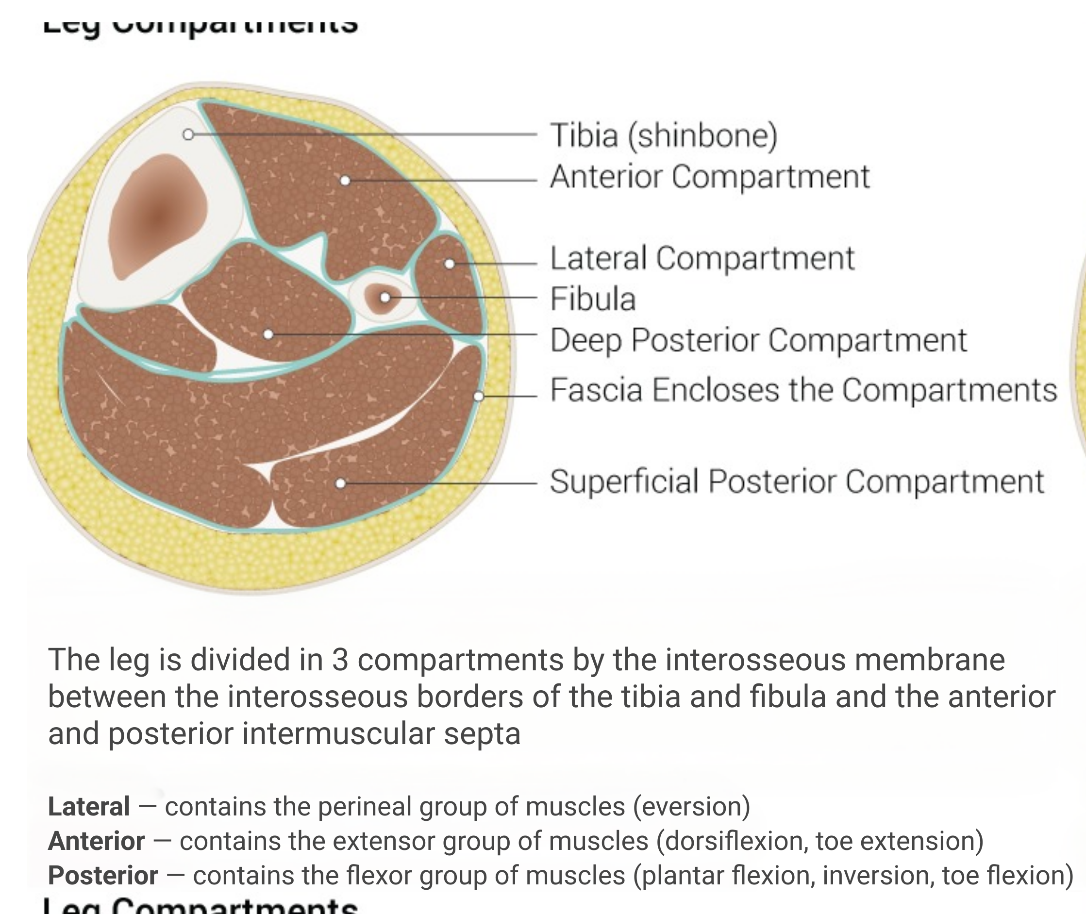
Cross-sectional view of lower leg compartments and bones.
Cross-sectional view of lower leg
Achievable
  • Chronic exertional compartment syndrome

    • Increased compartment pressure of the lower leg (specifically, the anterior, lateral, and posterior areas), causing a restriction in blood flow
      • Develops gradually over a period of time due to repetitive movements and exertional activities
      • Typically occurring in only one compartment of the lower leg
  • Symptoms

    • Pain
    • Numbness and tingling
    • Weakness
    • Swelling
    • Tightness
    • Foot drop
  • Diagnosis

    • Clinical presentation
  • Physical therapy management

    • Functional training, proprioceptive training
    • Decrease exacerbating activity
    • May require the use of orthoses
  • Stress fractures

    • Microfractures to the tibia, fibula, or bones of the foot and ankle due to overuse (repetitive micro-traumas to the tibia or fibula)
      • Commonly due to poor alignment of the lower extremity when performing activity, deconditioning, or improper training
  • Symptoms

    • Localized sharp pain that gradually worsens with activity
    • Mild swelling may occur around the fracture site
    • The affected area may feel weak or unstable
    • Difficult to put weight on the injured area
    • Pain may be more severe at night or when resting
  • Diagnosis

    • Clinical presentation reproduced with percussion
    • Bone scan
    • MRI
  • Physical therapy management

    • Initially decreased exacerbating activity
    • Correction of malalignment and biomechanical issues
    • Strengthening and coordinating activities

Ankle and foot conditions

  • Ligament sprains

    • Classification of sprains
      • Grade 1
        • A mild sprain where the ligament is stretched or slightly torn
        • Symptoms include minor swelling, tenderness, and bruising
        • Recovery time is usually 1-3 weeks
      • Grade 2
        • A moderate sprain where the ligament is partially torn
        • Symptoms include swelling, bruising, pain, and difficulty walking
        • Recovery time is usually 3-6 weeks
      • Grade 3
        • A severe sprain where the ligament is completely torn
        • Symptoms include severe pain, swelling, bruising, and instability
        • Recovery time can be several months
  • Symptoms

    • See specific grade for symptoms associated
  • Diagnosis

    • Clinical presentation, ligament laxity
      • Special tests:
        • Anterior drawer (Anterior talofibular ligament)
        • Talar tilt (inversion stresses the calcaneofibular ligament; eversion stresses the deltoid ligament)
  • Medical management

    • Grade 2- immobilization device may be recommended
    • Grade 3 sprains are usually managed conservatively with immobilization/bracing and functional rehabilitation; surgery is reserved for chronic instability or failed conservative care
  • Physical therapy management

    • Grade 1- conservative management of rest, ice, compression, and elevation acutely; then functional mobility training
    • Grades 2 and 3:
      • Functional mobility training
      • Biomechanical fault realignment
      • Joint protection and mobility
      • Post-surgical protocols if surgery is indicated
  • Achilles tendinopathy

    • Chronic degeneration (tendinosis) of the Achilles tendon due to overuse, age, rheumatoid arthritis, or gout
  • Symptoms

    • Pain, typically located in the back of the heel or just above it
    • Tenderness and stiffness in the Achilles tendon
    • Swelling and redness around the tendon
    • Pain that worsens with activity and improves with rest
  • Diagnosis

    • Clinical presentation
      • Thompson’s test to rule out Achilles tendon rupture
  • Medical management

    • Corticosteroids
    • Management of rheumatoid arthritis or gout
  • Physical therapy management

    • Determine the phase (acute vs subacute vs chronic) and make an appropriate selection of interventions based on the phase of healing
  • Tarsal tunnel syndrome

    • Entrapment of the posterior tibial nerve within the tarsal tunnel
      • Overuse injury due to tendonitis of the posterior tibialis
  • Symptoms

    • Numbness and tingling in the sole, arch, or toes.
    • Burning or shooting pain may radiate from the ankle to the foot or calf
    • Swelling or redness
    • Pain that improves with rest
  • Diagnosis

    • Clinical presentation
      • Special test- Tinel’s sign
    • Electrodiagnostic testing
  • Medical management

    • Surgical intervention if conservative management is not effective
  • Physical therapy management

    • Joint mobility
    • Functional exercise training
    • Orthoses
    • Neurotension stretching
  • Foot deformities

    • Pes cavus- high arch foot
      • Due to genetics or biomechanical faults
    • Pes planus- absent arch foot, overpronation
      • Due to genetics, age, obesity, or biomechanical faults
    • Equinus- plantar flexed foot
      • Can be due to genetics, bone deformity, or neurological conditions
    • Talipes equinovarus- club foot (plantar flexion and inversion)
      • Due to congenital deformity
  • Symptoms

    • Observation of the foot as noted above
  • Diagnosis

    • Clinical presentation and observation
  • Medical management

    • Management of underlying causes as appropriate
  • Physical therapy management

    • Improved biomechanical alignment
    • Orthoses as appropriate
    • Joint mobility
    • Patient education
  • Charcot-Marie-Tooth disease

    • Progressive disorder causing peroneal muscular atrophy and involvement of the peroneal nerves - causing motor and sensory deficits
      • Genetic disorder
      • Begins in the lower legs but can progress to the forearm and hands
    • Symptoms
      • Foot and leg weakness
      • Foot abnormalities: high arches, hammertoes, and flat feet
      • Foot drop with a steppage gait due to weakness of the ankle dorsiflexors
      • Numbness, tingling, and burning sensations in the lower leg
      • Muscle atrophy in the feet and legs
      • Clumsiness and difficulty with fine motor skills
  • Diagnosis

    • Clinical presentation
    • Electrodiagnostic exam
    • Genetic testing
  • Medical management

    • Surgical correction of joint abnormalities as deemed appropriate
  • Physical therapy management

    • Progressive disorder with no cure, so physical therapy interventions are centered around patient education, skin assessments for possible development of wounds, contracture prevention/management, and functional mobility assessment/training
  • Foot deformities

    • Rearfoot varus
      • Inversion of calcaneus when the subtalar joint is in neutral
    • Rearfoot valgus
      • Eversion of calcaneus when the subtalar joint is in neutral
    • Forefoot varus
      • Inversion of the forefoot when the subtalar is in neutral
    • Forefoot valgus
      • Eversion of forefoot when the subtalar is in neutral
  • Diagnosis of foot deformities

    • Clinical presentation and observation
  • Symptoms

    • Abnormalities in gait due to uneven levers
  • Medical management

    • None indicated
  • Physical therapy management

    • Improving foot alignment
    • Orthoses as appropriate
    • Strengthening as appropriate

Acute compartment syndrome

  • Increased pressure in lower leg compartments (anterior, lateral, posterior); caused by trauma/fracture
  • Six P’s: Pain, Palpable tenderness, Paresthesia (deep peroneal nerve), Paresis, Pallor, Pulselessness (dorsal pedal artery)
  • Medical emergency — requires immediate fasciotomy
  • PTA pitfall: never treat with exercise; if six P’s appear/worsen, stop and notify PT/physician immediately

Chronic exertional compartment syndrome

  • Gradual pressure increase from repetitive activity, usually one compartment
  • Symptoms: pain, numbness/tingling, weakness, swelling, tightness, foot drop
  • PT management: functional/proprioceptive training, activity modification, orthoses
  • Resolves with rest (unlike acute form)

Stress fractures

  • Microfractures of tibia/fibula/foot bones from overuse, malalignment, deconditioning
  • Symptoms: localized sharp pain worsening with activity, mild swelling, instability, worse at night
  • Diagnosis: percussion reproduces pain, bone scan, MRI
  • PT management: decrease aggravating activity, correct malalignment, strengthening/coordination

Ligament sprains

  • Graded 1 (mild, stretch) to 3 (severe, complete tear); recovery ranges 1-3 weeks to several months
  • Diagnosis: anterior drawer (ATFL), talar tilt (inversion=calcaneofibular, eversion=deltoid)
  • Medical management: Grade 2 may need immobilization; Grade 3 usually conservative, surgery only for chronic instability
  • PT management: RICE acutely (Grade 1), functional mobility training, biomechanical correction, post-surgical protocols as needed

Achilles tendinopathy

  • Chronic tendon degeneration (tendinosis) from overuse, age, RA, or gout
  • Symptoms: heel pain/tenderness/stiffness, swelling, worsens with activity
  • Diagnosis: Thompson’s test (rules out rupture)
  • Medical management: corticosteroids, treat underlying RA/gout
  • PT management: intervention selection based on healing phase (acute/subacute/chronic)

Tarsal tunnel syndrome

  • Posterior tibial nerve entrapment, often from posterior tibialis tendonitis
  • Symptoms: numbness/tingling in sole/arch/toes, burning pain radiating ankle to foot/calf
  • Diagnosis: Tinel’s sign, electrodiagnostic testing
  • Medical management: surgery if conservative care fails
  • PT management: joint mobility, functional training, orthoses, neurotension stretching

Foot deformities (general)

  • Types: pes cavus (high arch), pes planus (flat/overpronation), equinus (plantar flexed), talipes equinovarus (clubfoot)
  • Diagnosis: clinical observation
  • PT management: biomechanical alignment, orthoses, joint mobility, patient education

Charcot-Marie-Tooth disease

  • Genetic, progressive peroneal muscular atrophy causing motor/sensory deficits
  • Symptoms: foot/leg weakness, high arches/hammertoes/flat feet, foot drop with steppage gait, numbness/tingling, muscle atrophy
  • Diagnosis: electrodiagnostic exam, genetic testing
  • No cure — PT focuses on education, skin checks, contracture prevention, functional mobility

Foot deformities (rearfoot/forefoot varus-valgus)

  • Rearfoot varus/valgus: calcaneal inversion/eversion with subtalar neutral
  • Forefoot varus/valgus: forefoot inversion/eversion with subtalar neutral
  • Symptoms: gait abnormalities from uneven levers
  • No medical management indicated
  • PT management: alignment correction, orthoses, strengthening

Sign up for free to take 7 quiz questions on this topic

Previous
Next  | 5.10 Spine and pelvis anatomy
All rights reserved ©2016 - 2026 Achievable, Inc.

Lower leg, ankle, and foot pathologies

As with the hip and knee, acetaminophen and NSAIDs are the standard first-line analgesic/anti-inflammatory management here; additional or different medical management is called out where it applies.

Conditions of the lower leg

  • Acute compartment syndrome

    • Increased compartment pressure of lower leg (specifically- anterior, lateral, and posterior areas)
      • Commonly caused by direct trauma or fracture
  • Symptoms (six P’s)

    • Pain in the lower leg
    • Palpable tenderness in the lower leg
    • Paresthesia to the lower leg - specifically to the deep peroneal nerve
    • Paresis of the lower leg, specifically of the deep peroneal nerve
    • Pallor to the lower leg
    • Pulselessness in the dorsal pedal artery
  • Diagnosis

    • Clinical presentation
  • Medical management

    • Medical emergency- immediate fasciotomy is indicated to relieve pressure and decrease the likelihood of prolonged neurovascular compromise
  • Physical therapy management

    • Post-operatively, physical therapy to ensure return to normal function

PTA role and pitfall: Acute compartment syndrome is a medical emergency, not a condition to treat with exercise. If any of the six P’s emerge or worsen during a session, stop the activity, take vital/neurovascular measurements, and notify the supervising PT or physician immediately rather than continuing the plan of care. This is different from chronic exertional compartment syndrome (below), which develops gradually with activity and resolves with rest.

  • Chronic exertional compartment syndrome

    • Increased compartment pressure of the lower leg (specifically, the anterior, lateral, and posterior areas), causing a restriction in blood flow
      • Develops gradually over a period of time due to repetitive movements and exertional activities
      • Typically occurring in only one compartment of the lower leg
  • Symptoms

    • Pain
    • Numbness and tingling
    • Weakness
    • Swelling
    • Tightness
    • Foot drop
  • Diagnosis

    • Clinical presentation
  • Physical therapy management

    • Functional training, proprioceptive training
    • Decrease exacerbating activity
    • May require the use of orthoses
  • Stress fractures

    • Microfractures to the tibia, fibula, or bones of the foot and ankle due to overuse (repetitive micro-traumas to the tibia or fibula)
      • Commonly due to poor alignment of the lower extremity when performing activity, deconditioning, or improper training
  • Symptoms

    • Localized sharp pain that gradually worsens with activity
    • Mild swelling may occur around the fracture site
    • The affected area may feel weak or unstable
    • Difficult to put weight on the injured area
    • Pain may be more severe at night or when resting
  • Diagnosis

    • Clinical presentation reproduced with percussion
    • Bone scan
    • MRI
  • Physical therapy management

    • Initially decreased exacerbating activity
    • Correction of malalignment and biomechanical issues
    • Strengthening and coordinating activities

Ankle and foot conditions

  • Ligament sprains

    • Classification of sprains
      • Grade 1
        • A mild sprain where the ligament is stretched or slightly torn
        • Symptoms include minor swelling, tenderness, and bruising
        • Recovery time is usually 1-3 weeks
      • Grade 2
        • A moderate sprain where the ligament is partially torn
        • Symptoms include swelling, bruising, pain, and difficulty walking
        • Recovery time is usually 3-6 weeks
      • Grade 3
        • A severe sprain where the ligament is completely torn
        • Symptoms include severe pain, swelling, bruising, and instability
        • Recovery time can be several months
  • Symptoms

    • See specific grade for symptoms associated
  • Diagnosis

    • Clinical presentation, ligament laxity
      • Special tests:
        • Anterior drawer (Anterior talofibular ligament)
        • Talar tilt (inversion stresses the calcaneofibular ligament; eversion stresses the deltoid ligament)
  • Medical management

    • Grade 2- immobilization device may be recommended
    • Grade 3 sprains are usually managed conservatively with immobilization/bracing and functional rehabilitation; surgery is reserved for chronic instability or failed conservative care
  • Physical therapy management

    • Grade 1- conservative management of rest, ice, compression, and elevation acutely; then functional mobility training
    • Grades 2 and 3:
      • Functional mobility training
      • Biomechanical fault realignment
      • Joint protection and mobility
      • Post-surgical protocols if surgery is indicated
  • Achilles tendinopathy

    • Chronic degeneration (tendinosis) of the Achilles tendon due to overuse, age, rheumatoid arthritis, or gout
  • Symptoms

    • Pain, typically located in the back of the heel or just above it
    • Tenderness and stiffness in the Achilles tendon
    • Swelling and redness around the tendon
    • Pain that worsens with activity and improves with rest
  • Diagnosis

    • Clinical presentation
      • Thompson’s test to rule out Achilles tendon rupture
  • Medical management

    • Corticosteroids
    • Management of rheumatoid arthritis or gout
  • Physical therapy management

    • Determine the phase (acute vs subacute vs chronic) and make an appropriate selection of interventions based on the phase of healing
  • Tarsal tunnel syndrome

    • Entrapment of the posterior tibial nerve within the tarsal tunnel
      • Overuse injury due to tendonitis of the posterior tibialis
  • Symptoms

    • Numbness and tingling in the sole, arch, or toes.
    • Burning or shooting pain may radiate from the ankle to the foot or calf
    • Swelling or redness
    • Pain that improves with rest
  • Diagnosis

    • Clinical presentation
      • Special test- Tinel’s sign
    • Electrodiagnostic testing
  • Medical management

    • Surgical intervention if conservative management is not effective
  • Physical therapy management

    • Joint mobility
    • Functional exercise training
    • Orthoses
    • Neurotension stretching
  • Foot deformities

    • Pes cavus- high arch foot
      • Due to genetics or biomechanical faults
    • Pes planus- absent arch foot, overpronation
      • Due to genetics, age, obesity, or biomechanical faults
    • Equinus- plantar flexed foot
      • Can be due to genetics, bone deformity, or neurological conditions
    • Talipes equinovarus- club foot (plantar flexion and inversion)
      • Due to congenital deformity
  • Symptoms

    • Observation of the foot as noted above
  • Diagnosis

    • Clinical presentation and observation
  • Medical management

    • Management of underlying causes as appropriate
  • Physical therapy management

    • Improved biomechanical alignment
    • Orthoses as appropriate
    • Joint mobility
    • Patient education
  • Charcot-Marie-Tooth disease

    • Progressive disorder causing peroneal muscular atrophy and involvement of the peroneal nerves - causing motor and sensory deficits
      • Genetic disorder
      • Begins in the lower legs but can progress to the forearm and hands
    • Symptoms
      • Foot and leg weakness
      • Foot abnormalities: high arches, hammertoes, and flat feet
      • Foot drop with a steppage gait due to weakness of the ankle dorsiflexors
      • Numbness, tingling, and burning sensations in the lower leg
      • Muscle atrophy in the feet and legs
      • Clumsiness and difficulty with fine motor skills
  • Diagnosis

    • Clinical presentation
    • Electrodiagnostic exam
    • Genetic testing
  • Medical management

    • Surgical correction of joint abnormalities as deemed appropriate
  • Physical therapy management

    • Progressive disorder with no cure, so physical therapy interventions are centered around patient education, skin assessments for possible development of wounds, contracture prevention/management, and functional mobility assessment/training
  • Foot deformities

    • Rearfoot varus
      • Inversion of calcaneus when the subtalar joint is in neutral
    • Rearfoot valgus
      • Eversion of calcaneus when the subtalar joint is in neutral
    • Forefoot varus
      • Inversion of the forefoot when the subtalar is in neutral
    • Forefoot valgus
      • Eversion of forefoot when the subtalar is in neutral
  • Diagnosis of foot deformities

    • Clinical presentation and observation
  • Symptoms

    • Abnormalities in gait due to uneven levers
  • Medical management

    • None indicated
  • Physical therapy management

    • Improving foot alignment
    • Orthoses as appropriate
    • Strengthening as appropriate
Key points

Acute compartment syndrome

  • Increased pressure in lower leg compartments (anterior, lateral, posterior); caused by trauma/fracture
  • Six P’s: Pain, Palpable tenderness, Paresthesia (deep peroneal nerve), Paresis, Pallor, Pulselessness (dorsal pedal artery)
  • Medical emergency — requires immediate fasciotomy
  • PTA pitfall: never treat with exercise; if six P’s appear/worsen, stop and notify PT/physician immediately

Chronic exertional compartment syndrome

  • Gradual pressure increase from repetitive activity, usually one compartment
  • Symptoms: pain, numbness/tingling, weakness, swelling, tightness, foot drop
  • PT management: functional/proprioceptive training, activity modification, orthoses
  • Resolves with rest (unlike acute form)

Stress fractures

  • Microfractures of tibia/fibula/foot bones from overuse, malalignment, deconditioning
  • Symptoms: localized sharp pain worsening with activity, mild swelling, instability, worse at night
  • Diagnosis: percussion reproduces pain, bone scan, MRI
  • PT management: decrease aggravating activity, correct malalignment, strengthening/coordination

Ligament sprains

  • Graded 1 (mild, stretch) to 3 (severe, complete tear); recovery ranges 1-3 weeks to several months
  • Diagnosis: anterior drawer (ATFL), talar tilt (inversion=calcaneofibular, eversion=deltoid)
  • Medical management: Grade 2 may need immobilization; Grade 3 usually conservative, surgery only for chronic instability
  • PT management: RICE acutely (Grade 1), functional mobility training, biomechanical correction, post-surgical protocols as needed

Achilles tendinopathy

  • Chronic tendon degeneration (tendinosis) from overuse, age, RA, or gout
  • Symptoms: heel pain/tenderness/stiffness, swelling, worsens with activity
  • Diagnosis: Thompson’s test (rules out rupture)
  • Medical management: corticosteroids, treat underlying RA/gout
  • PT management: intervention selection based on healing phase (acute/subacute/chronic)

Tarsal tunnel syndrome

  • Posterior tibial nerve entrapment, often from posterior tibialis tendonitis
  • Symptoms: numbness/tingling in sole/arch/toes, burning pain radiating ankle to foot/calf
  • Diagnosis: Tinel’s sign, electrodiagnostic testing
  • Medical management: surgery if conservative care fails
  • PT management: joint mobility, functional training, orthoses, neurotension stretching

Foot deformities (general)

  • Types: pes cavus (high arch), pes planus (flat/overpronation), equinus (plantar flexed), talipes equinovarus (clubfoot)
  • Diagnosis: clinical observation
  • PT management: biomechanical alignment, orthoses, joint mobility, patient education

Charcot-Marie-Tooth disease

  • Genetic, progressive peroneal muscular atrophy causing motor/sensory deficits
  • Symptoms: foot/leg weakness, high arches/hammertoes/flat feet, foot drop with steppage gait, numbness/tingling, muscle atrophy
  • Diagnosis: electrodiagnostic exam, genetic testing
  • No cure — PT focuses on education, skin checks, contracture prevention, functional mobility

Foot deformities (rearfoot/forefoot varus-valgus)

  • Rearfoot varus/valgus: calcaneal inversion/eversion with subtalar neutral
  • Forefoot varus/valgus: forefoot inversion/eversion with subtalar neutral
  • Symptoms: gait abnormalities from uneven levers
  • No medical management indicated
  • PT management: alignment correction, orthoses, strengthening

More from Comparing clinical presentation and interventions of lower extremity

  • Hip and knee pathologies