Achievable logoAchievable logo
USMLE/1
Sign in
Sign up
Purchase
Textbook
Support
How it works
Resources
Exam catalog
Mountain with a flag at the peak
Textbook
Introduction
1. Anatomy
2. Microbiology
2.1 General bacteriology
2.2 Introduction to systemic bacteriology
2.3 Gram positive cocci
2.4 Gram negative cocci
2.5 Gram positive bacilli
2.6 Gram negative bacilli
2.7 Other important bacteria
2.8 Virology
2.9 Parasitology
2.9.1 Entamoeba histolytica
2.9.2 Acanthamoeba
2.9.3 Naegleria fowleri
2.9.4 Giardia lamblia
2.9.5 Cryptosporidium parvum
2.9.6 Trichomonas
2.9.7 Plasmodium spp.
2.9.8 Babesia
2.9.9 Toxoplasma gondii
2.9.10 Taenia
2.9.11 Hymenolepis nana
2.9.12 Enterobius vermicularis
2.9.13 Trichuris
2.9.14 Trichinella
2.9.15 Ascaris
2.9.16 Hookworms
2.9.17 Strongyloides
2.9.18 Additional information
2.10 Mycology
3. Physiology
4. Pathology
5. Pharmacology
6. Immunology
7. Biochemistry
8. Cell and molecular biology
9. Biostatistics and epidemiology
10. Genetics
11. Behavioral science
Wrapping up
Achievable logoAchievable logo
2.9.6 Trichomonas
Achievable USMLE/1
2. Microbiology
2.9. Parasitology

Trichomonas

1 min read
Font
Discuss
Share
Feedback

Trichomonas

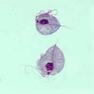
Trichomonas exists only in the trophozoite form and shows characteristic wobbling motility. It’s transmitted as an STD and can also spread through contaminated towels and underwear.

Clinically, it presents as vaginitis with:

  • Local burning and itching
  • Frothy, foul-smelling, yellowish vaginal discharge

It’s usually accompanied by:

  • Dysuria
  • Increased urinary frequency
  • Dyspareunia

Diagnosis is made by direct microscopy showing motile trophozoites. Acridine orange or Papanicolaou staining can also be used. Indirect haemagglutination can detect antibodies. ELISA is used for antigen detection. DNA probes and PCR are used for nucleic acid detection.

Trichomonas
Trichomonas

Two trophozoites of T. vaginalis obtained from in vitro culture, stained with Giemsa.

All rights reserved ©2016 - 2026 Achievable, Inc.