Achievable logoAchievable logo
USMLE/1
Sign in
Sign up
Purchase
Textbook
Support
How it works
Resources
Exam catalog
Mountain with a flag at the peak
Textbook
Introduction
1. Anatomy
2. Microbiology
2.1 General bacteriology
2.2 Introduction to systemic bacteriology
2.3 Gram positive cocci
2.4 Gram negative cocci
2.5 Gram positive bacilli
2.6 Gram negative bacilli
2.7 Other important bacteria
2.8 Virology
2.9 Parasitology
2.9.1 Entamoeba histolytica
2.9.2 Acanthamoeba
2.9.3 Naegleria fowleri
2.9.4 Giardia lamblia
2.9.5 Cryptosporidium parvum
2.9.6 Trichomonas
2.9.7 Plasmodium spp.
2.9.8 Babesia
2.9.9 Toxoplasma gondii
2.9.10 Taenia
2.9.11 Hymenolepis nana
2.9.12 Enterobius vermicularis
2.9.13 Trichuris
2.9.14 Trichinella
2.9.15 Ascaris
2.9.16 Hookworms
2.9.17 Strongyloides
2.9.18 Additional information
2.10 Mycology
3. Physiology
4. Pathology
5. Pharmacology
6. Immunology
7. Biochemistry
8. Cell and molecular biology
9. Biostatistics and epidemiology
10. Genetics
11. Behavioral science
Wrapping up
Achievable logoAchievable logo
2.9.2 Acanthamoeba
Achievable USMLE/1
2. Microbiology
2.9. Parasitology

Acanthamoeba

2 min read
Font
Discuss
Share
Feedback

Acanthamoeba

Acanthamoeba is a free-living amoeba that can infect the eye, skin, and central nervous system (CNS). CNS infection can be rapidly fatal. The trophozoite is the infective form.

Acanthamoeba keratitis can occur after contact lens use and may lead to blindness. The organism typically enters through minor cuts in the skin or is inhaled into the lungs.

Acanthamoeba has been isolated from soil, air, sewage, seawater, chlorinated swimming pools, domestic tap water, bottled water, dental treatment units, hospitals, air-conditioning units, and contact lens cases.

Granulomatous amoebic encephalitis is a severe form that presents acutely with fever, headache, seizures, delirium, and cranial nerve palsies, and it can rapidly progress to death.

Keratitis presents with eye pain, redness, photosensitivity, a foreign body sensation, and eye watering.

Skin lesions present as granulomas.

Diagnosis is made by demonstrating trophozoites in CSF and on brain biopsy. Culture is difficult and is performed as a co-culture with E. coli. For keratitis, corneal scrapings and confocal microscopy can be used for diagnosis.

Acanthamoeba
Acanthamoeba

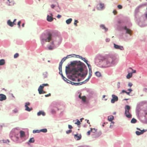
Cyst of Acanthamoeba sp. from brain tissue, stained with hematoxylin and eosin (H&E).

All rights reserved ©2016 - 2026 Achievable, Inc.