Achievable logoAchievable logo
USMLE/1
Sign in
Sign up
Purchase
Textbook
Support
How it works
Resources
Exam catalog
Mountain with a flag at the peak
Textbook
Introduction
1. Anatomy
2. Microbiology
2.1 General bacteriology
2.2 Introduction to systemic bacteriology
2.3 Gram positive cocci
2.4 Gram negative cocci
2.5 Gram positive bacilli
2.6 Gram negative bacilli
2.7 Other important bacteria
2.8 Virology
2.9 Parasitology
2.9.1 Entamoeba histolytica
2.9.2 Acanthamoeba
2.9.3 Naegleria fowleri
2.9.4 Giardia lamblia
2.9.5 Cryptosporidium parvum
2.9.6 Trichomonas
2.9.7 Plasmodium spp.
2.9.8 Babesia
2.9.9 Toxoplasma gondii
2.9.10 Taenia
2.9.11 Hymenolepis nana
2.9.12 Enterobius vermicularis
2.9.13 Trichuris
2.9.14 Trichinella
2.9.15 Ascaris
2.9.16 Hookworms
2.9.17 Strongyloides
2.9.18 Additional information
2.10 Mycology
3. Physiology
4. Pathology
5. Pharmacology
6. Immunology
7. Biochemistry
8. Cell and molecular biology
9. Biostatistics and epidemiology
10. Genetics
11. Behavioral science
Wrapping up
Achievable logoAchievable logo
2.9.5 Cryptosporidium parvum
Achievable USMLE/1
2. Microbiology
2.9. Parasitology

Cryptosporidium parvum

1 min read
Font
Discuss
Share
Feedback

Cryptosporidium parvum

Cryptosporidium parvum causes watery diarrhea. Disease is often severe in immunocompromised individuals (for example, people with AIDS or patients receiving cancer therapy).

Transmission

Infection is acquired by ingesting oocysts.

Clinical features

It typically presents with:

  • Watery diarrhea
  • Abdominal cramps
  • Dehydration
  • Fever
  • Vomiting

Diagnosis

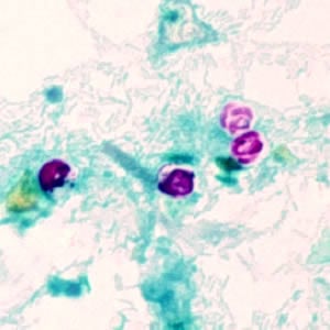
  • Oocysts can be visualized in stool using modified acid-fast staining.
  • Nuclei of sporozoites can be stained with DAPI.
  • Immunofluorescence and ELISA can be used to detect antigens and antibodies.
  • PCR is mainly used for research and epidemiological purposes.
Cryptosporidium
Cryptosporidium

Cryptosporidium parvum oocysts stained with modified acid-fast. Against a blue-green background, the oocysts stand out in a bright red stain. Sporozoites are visible inside the two oocysts to the right in this image.

All rights reserved ©2016 - 2026 Achievable, Inc.