Meninges of the brain
Meninges of the brain
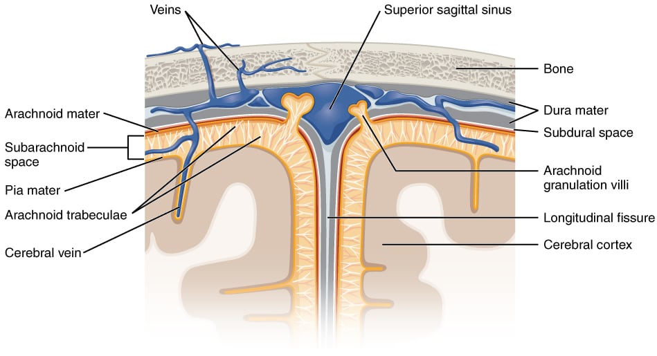
The meninges are three connective tissue layers that surround the brain:
- Pia mater (innermost)
- Arachnoid mater (middle)
- Dura mater (outermost)
The pia and arachnoid together are called the leptomeninges. The dura is called the pachymeninx.
The pia mater is tightly attached to the brain and follows the gyri, grooves, and sulci. Beneath the arachnoid mater is the subarachnoid space, which contains cerebrospinal fluid (CSF).
Meningeal nociceptors are located in the dura mater. These pain-sensitive fibers detect pain in headaches and in meningeal irritation.
CSF and ventricles of the brain
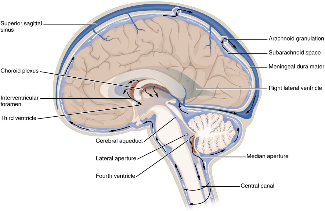
Cerebrospinal fluid (CSF) is produced mainly by the choroid plexuses and partly by ependymal cells. It circulates through the ventricles and the subarachnoid space.
About 150 mL of CSF is present in the ventricles at any time. It is continuously generated and reabsorbed. Reabsorption occurs at the arachnoid granulations.
The cells of the choroid plexus are joined by tight junctions, forming the blood-CSF barrier. CSF is essentially an ultrafiltrate of plasma:
- Higher concentration of sodium, chloride, and magnesium than plasma
- Lower levels of potassium and calcium than plasma
CSF functions include:
- Acting as a cushion, protecting the soft brain from injury against the hard skull
- Helping remove waste
- Helping maintain homeostasis in the brain
The brain has 4 ventricles:
- Two lateral ventricles (one in each cerebral hemisphere)
- One third ventricle
- One fourth ventricle
The lateral ventricles communicate with the third ventricle through the interventricular foramina. The third ventricle is located in the diencephalon.
The cerebral aqueduct of Sylvius is located in the midbrain and connects the third ventricle to the fourth ventricle.
The fourth ventricle is diamond-shaped. It is located posterior to the pons and upper medulla and anterior to the cerebellum. It connects to the subarachnoid space via:
- Two lateral foramina of Luschka
- One median foramen of Magendie
CSF flow follows this path:
- Lateral ventricles
- Interventricular foramina
- Third ventricle
- Cerebral aqueduct of Sylvius
- Fourth ventricle
- Median foramen of Magendie and lateral foramina of Luschka
- Subarachnoid space (distributed over the surface of the brain and spinal cord)
Basal Nuclei or Ganglia
The basal nuclei (basal ganglia) include:
- Caudate nucleus
- Putamen
- Globus pallidus
- Subthalamic nucleus
- Substantia nigra
The putamen and caudate nucleus together are called the striatum (or neostriatum). The putamen and globus pallidus together form the lentiform (or lenticular) nuclei.
The main function of the basal nuclei is motor control, with additional effects on cognition and eye movements.
The caudate nucleus is a C-shaped structure closely associated with the lateral wall of the lateral ventricle. The anterior limb of the internal capsule separates it from the putamen.
The globus pallidus is divided into:
- A medial internal segment
- A lateral external segment
The subthalamic nucleus is located under the thalamus.
The substantia nigra is a midbrain structure with two distinct parts:
- Pars compacta
- Pars reticulata
The pars compacta is the source of a clinically important dopaminergic pathway to the striatum. Loss of neurons in this area causes Parkinson’s disease. A functionally analogous area is the ventral tegmental area, located nearby, which makes a dopaminergic projection to the nucleus accumbens. The pars reticulata contains GABA neurons.
The basal nuclei receive excitatory afferents from the cerebral cortex and thalamus:
- Frontal lobe projects to the head of the caudate nucleus and putamen
- Temporal lobe projects to the tail of the caudate
- Parietal and occipital lobes project to the body of the caudate nucleus
Limbic system
The limbic system includes:
- Amygdala
- Hippocampus
- Fornix
- Mammillary bodies
- Cingulate gyrus
- Parahippocampal gyrus
It is involved in memory and emotions.
The hippocampus is also called Ammon’s horn because it is shaped like two horns of a ram. It is located in the medial temporal lobe and is important for:
- Declarative memory (memory for facts and events)
- Spatial memory
- Formation of new memories
Long-term potentiation is vital for memory formation in the hippocampus. The hippocampus is one of the earliest structures involved in Alzheimer’s disease. Hippocampal atrophy is seen in schizophrenia, PTSD, depression, and chronic stress. Pyramidal cells of the hippocampus have been implicated in temporal lobe epilepsy.
The amygdala is an almond-shaped group of neurons located deep in the medial temporal lobe. It is involved in motivation, mediating the emotional aspects of memory, and processing emotions such as fear.
The mammillary bodies are involved in spatial and episodic memory consolidation and storage (as part of the Papez circuit), recollective memory, emotion and reward behaviors, and goal-directed behaviors. Damage to the mammillary bodies from alcoholism and thiamine deficiency causes Wernicke- Korsakoff syndrome, presenting with anterograde amnesia and confabulation.
The Papez circuit
The Papez circuit is the most important relay in the limbic system. It begins and ends at the hippocampus.
From the hippocampus, signals are relayed:
- Via the fornix to the mammillary bodies
- From the mammillary bodies to the anterior nucleus of the thalamus via the mammillothalamic tract
- From the thalamus to the cingulate gyrus
- From the cingulate gyrus back to the hippocampus