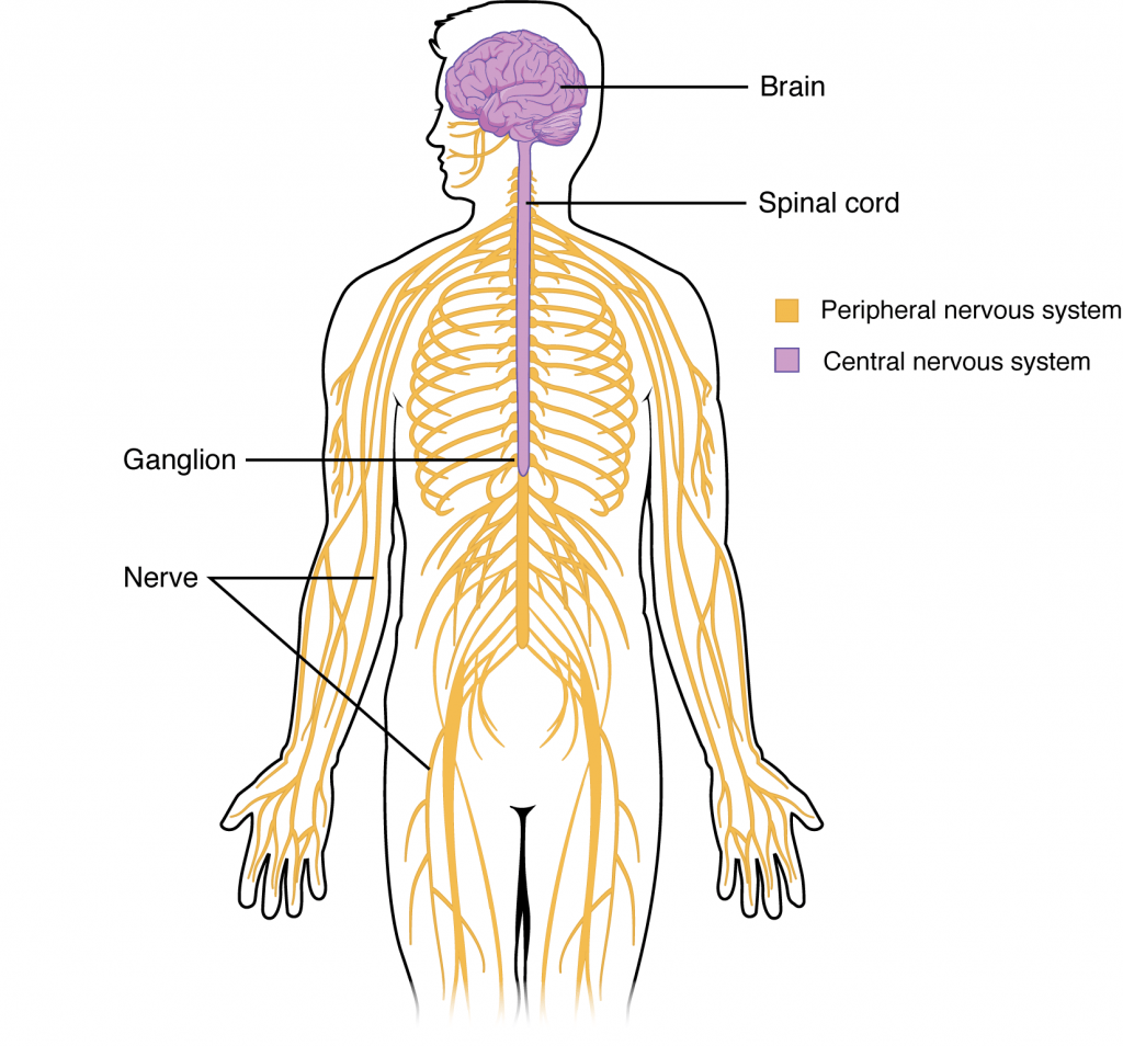
Central nervous system
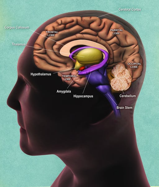
The central nervous system (CNS) is composed of the brain and the spinal cord. The brain consists of the cerebrum, brainstem, and cerebellum. The brainstem is made up of the midbrain, pons, and medulla.
The CNS contains the brain and spinal cord, while the PNS includes nerves.
Cerebrum
The cerebrum is made of two cerebral hemispheres connected by the corpus callosum. It contains:
- Grey matter, made of neuron cell bodies
- White matter, made of connecting fibers (the axons of neurons)
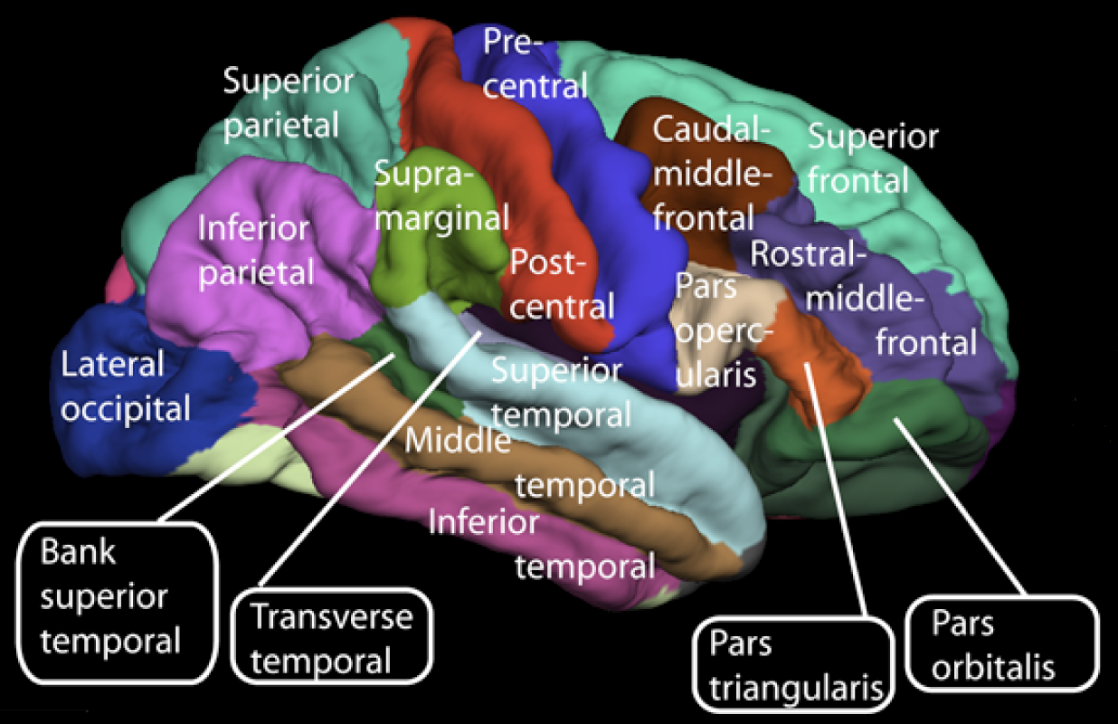
Each hemisphere is divided into four lobes by deep fissures called sulci:
- Frontal lobe
- Parietal lobe
- Temporal lobe
- Occipital lobe
Because the brain grows rapidly within a relatively small cranial cavity, brain tissue bulges between sulci, forming gyri. Gyri can atrophy in Alzheimer’s disease, some cases of psychosis, and frontotemporal (Pick’s) dementia.
Key sulci:
- The central sulcus divides the frontal lobe from the parietal lobe.
- The lateral sulcus (Sylvian fissure) separates the frontal and parietal lobes (superior) from the temporal lobe (inferior).
The cerebrum can be further divided into:
- Diencephalon (thalamus, hypothalamus, epithalamus, and subthalamus)
- Telencephalon (the cortex and basal ganglia)
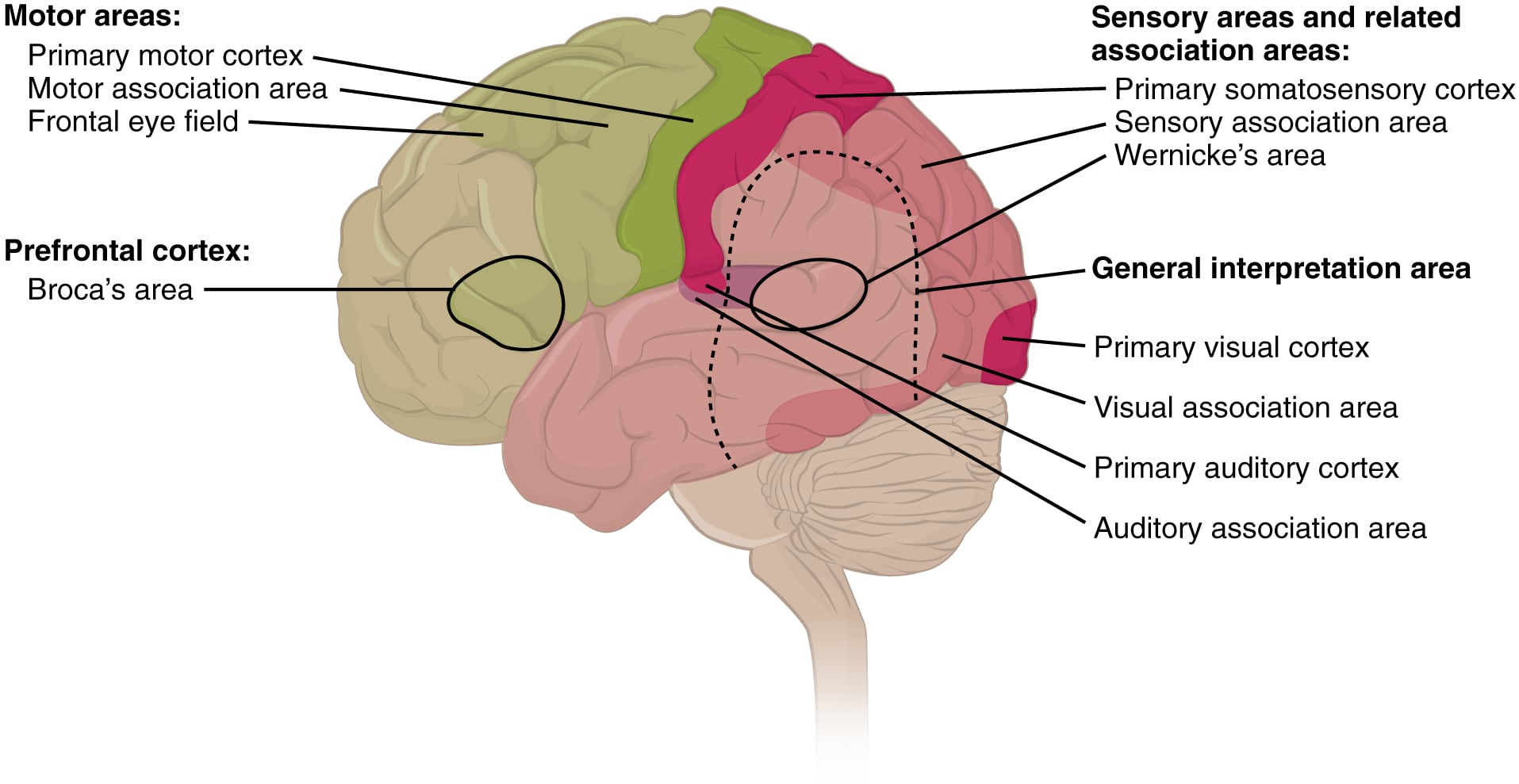
Functional anatomy of the brain
Primary motor cortex (M1)
The primary motor cortex is located in the precentral gyrus of the frontal lobe (Brodmann area 4). It executes simple movements of contralateral muscles.
Premotor cortex
The premotor cortex lies anterior to the primary motor cortex in the frontal lobe (Brodmann area 6). It’s associated with more complex movements and movement planning. It has two regions:
- Lateral region: involved in conditional motor tasks and selecting movements appropriate to the context of the action
- Medial region: involved in initiating movements specified by internal cues (for example, executing motor movements from memory)
Supplemental motor cortex (MII)
The supplemental motor cortex is located on the lateral and medial sides of the frontal lobe. It’s involved in complex movements, such as performing movements in a sequence (e.g., dancing).
The cerebral cortex can be described as containing three types of processing regions: primary, association, and integration areas. The primary cortical areas are where sensory information is initially processed, or where motor commands emerge to go to the brain stem or spinal cord. Association areas are adjacent to primary areas and further process the modality-specific input. Multimodal integration areas are found where the modality-specific regions meet; they can process multiple modalities together or different modalities on the basis of similar functions, such as spatial processing in vision or somatosensation.
Primary somatosensory area (S1)
The primary somatosensory area is located in the postcentral gyrus of the parietal lobe (Brodmann areas 3, 1, and 2). S1 ultimately receives sensory input from the VPL and VPM nuclei of the thalamus and processes pain, temperature, touch, vibration, and position from contralateral body areas.
Secondary somatosensory area (SII)
The secondary somatosensory area is located in the inferior part of the parietal lobe (Brodmann area 40). It receives inputs from S1 and the thalamus. It’s involved in:
- Sensorimotor integration
- Integration of information from the two body halves
- Attention
- Learning and memory
Somatosensory association cortex
The somatosensory association cortex is located in the superior parietal lobule (Brodmann areas 5 and 7). These areas are involved in complex associations. Damage causes tactile agnosia (inability to recognize an object even though the object can be felt when it is touched).
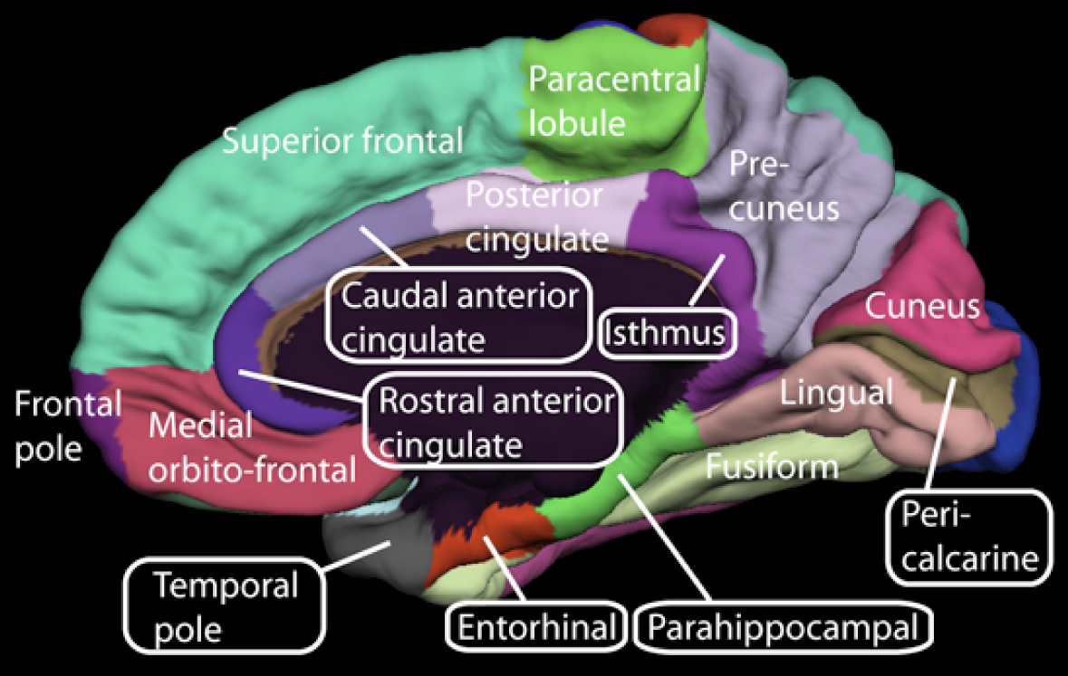
Primary visual cortex (V1 or striate cortex)
The primary visual cortex lies in the occipital lobe (Brodmann area 17), surrounding the calcarine sulcus.
- The upper parts of the world project to the lower part of the visual cortex.
- The macula is represented at the posterior tip of the striate cortex.
The visual association areas surround the primary visual cortex and are involved in recognition of form, color, and movement.
Primary auditory cortex (A1)
The primary auditory cortex is located in the superior temporal gyrus of the temporal lobe (Brodmann area 41). The auditory association area is Brodmann area 42. Their function is interpretation of sound.
Broca’s area
Broca’s area is located in the inferior frontal gyrus of the frontal lobe in the dominant hemisphere (Brodmann areas 44 and 45). It was originally thought to be involved in language production, but it has also been associated (partly) with language comprehension.
It’s involved in:
- Language repetition
- Gesture production
- Grammar
- Language fluidity
- Interpretation of actions
- Understanding the meaning of words
- Understanding how words sound/accent
Hypoactivity in Broca’s area is seen in stammering/stuttering.
Wernicke’s area
Wernicke’s area is located in the left posterior-superior temporal lobe, approximately Brodmann areas 39 and 40. Traditionally, it has mainly been associated with speech comprehension (understanding).
A lesion in this area causes Wernicke’s aphasia (fluent or receptive aphasia). The ability to understand the meaning of spoken words is affected, but talking (language production) is not impaired, so speech may seem nonsensical.
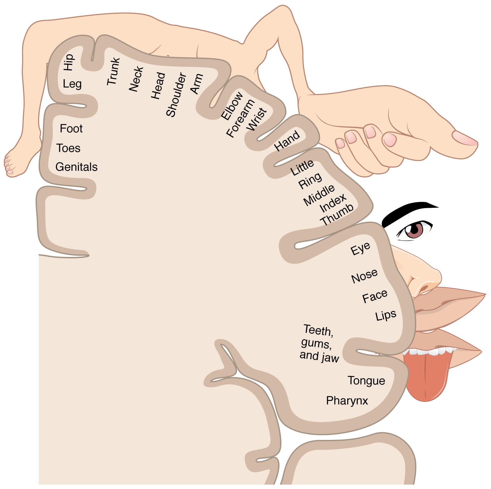
Motor and sensory homunculus
The body is represented upside down in the cerebral cortex:
- Toes are represented superiorly
- Face is represented inferiorly
Neocortex and cortical laminae
The neocortex is the most recently evolved area of the cerebral cortex, and it makes up the majority of the human cerebral cortex. It’s arranged in six layers, from superficial to deep:
Layer I
Molecular layer. It is deficient in cells but has an interconnected network of dendrites and axons called the neuropil.
Layer II
External granular layer. It is composed of inhibitory granule cells.
Layer III
External pyramidal layer. It has smaller pyramidal cells.
Layer IV
Internal granular layer. It is rich in stellate cells, which receive inputs from the thalamus in the primary sensory cortex.
Layer V
Internal pyramidal layer. Larger pyramidal neurons in layers V and VI form the corticospinal and corticobulbar tracts. A “Betz cell” is a large pyramidal cell that has a long dendrite at the apex that extends all the way up to layer I.
Layer VI
Multiform or fusiform layer. It forms association and projection fibers.
Glial cells
Glial cells provide support to neurons. They include microglia, astrocytes, and oligodendrocytes.
Microglia are like monocytes and are involved in phagocytosis. They are scavenger cells of the CNS. They fuse to form multinucleated giant cells in HIV.
Astrocytes are multifunctional and are involved in:
- Formation of the blood brain barrier via foot processes
- Laying down scar tissue after local injury
- Buffering extracellular K+
- Removing excess neurotransmitters
- Forming the external and internal glial limiting membrane
- Undergoing hypertrophy and hyperplasia in response to injury
GFAP (glial fibrillary acidic protein) and glutamine synthetase are markers for astrocytes.
Oligodendrocytes lay down myelin in the CNS. (Remember that Schwann cells lay down myelin in the peripheral nervous system; they are injured in GBS.) Oligodendrocytes have a “fried egg” appearance on histology. They are injured in multiple sclerosis, leukodystrophies, and progressive multifocal leukoencephalopathy.
Ependymal cells line the ventricles of the brain. Choroid epithelial cells line the villi of the choroid plexuses and secrete CSF.