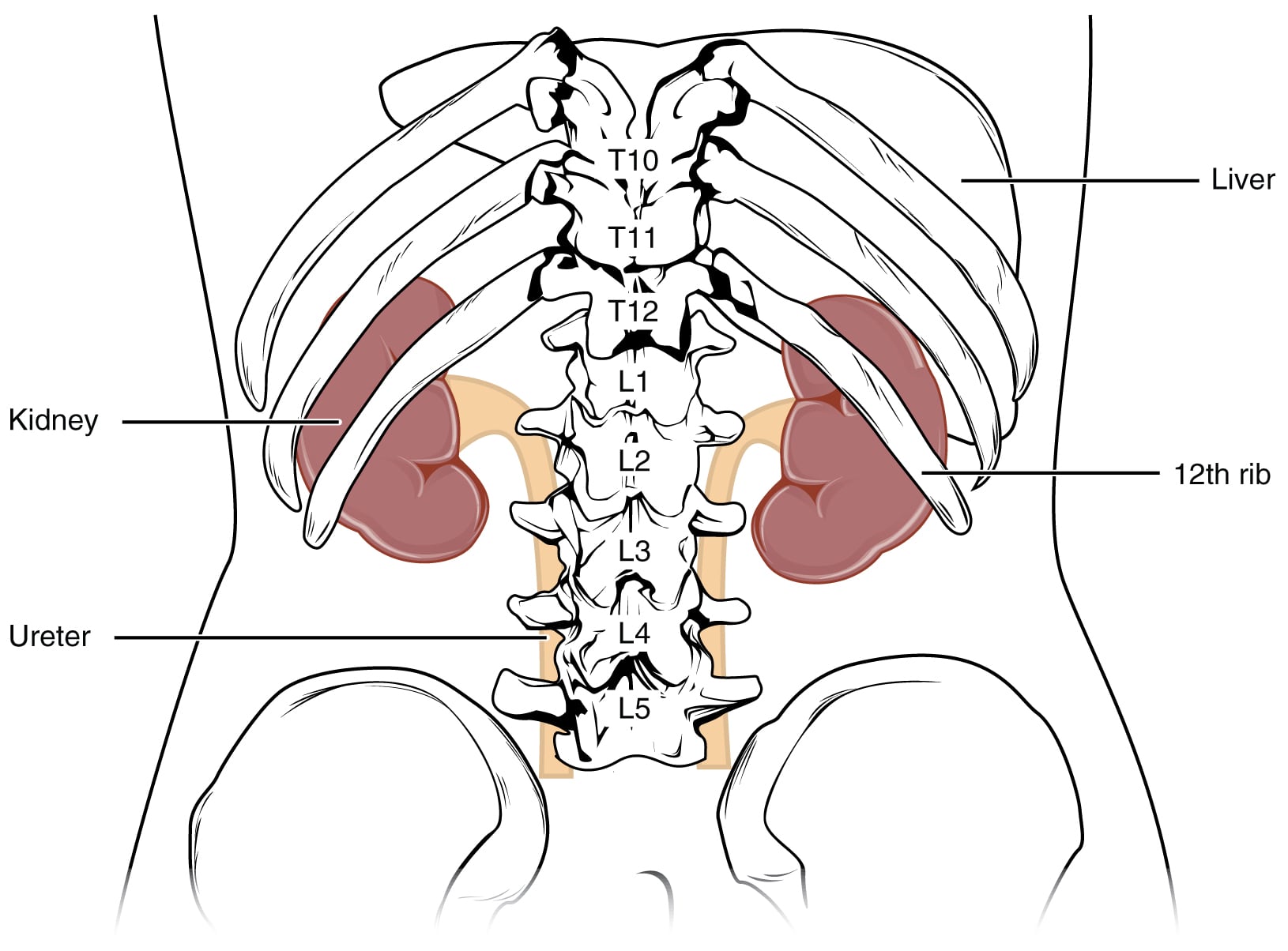
The kidneys are bean-shaped, retroperitoneal organs located on either side of the vertebral column. They extend from vertebrae T12-L3, with the left kidney positioned slightly higher than the right. Ribs 11 and 12 are related to the posterior surface of the kidneys.
Each kidney is covered by a fibrous capsule and surrounded by a renal fat pad. Gerota’s fascia encloses the kidney and the adrenal gland. The kidneys lie anterior to the quadratus lumborum muscle and lateral to the psoas major.

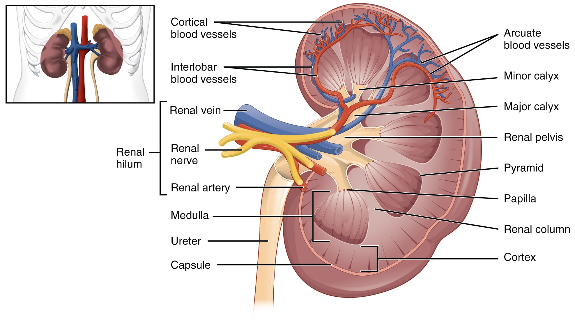
On cross section, the kidney is divided into:
The base of each pyramid faces the cortex, while the tip points medially and forms a renal papilla. The papillae open into the minor calyces.
Renal lobes correspond to the number of pyramids. Each lobe consists of:
Each lobe contains multiple lobules. Each lobule drains into its corresponding collecting duct.

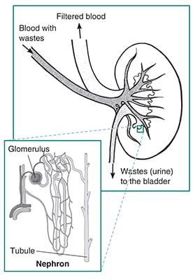
The functional unit of the kidney is the nephron. A nephron includes:
Some collecting ducts are located in the cortex, while others are located in the medulla. In the medulla, collecting ducts merge into larger papillary ducts of Bellini, which open at the renal papillae.
Urine flow follows this path:
Both types of calyces and the renal pelvis are lined by transitional epithelium and contain smooth muscle in their walls.

The proximal and distal convoluted tubules, along with the short loops of Henle of cortical nephrons, are located in the cortex. The collecting tubules (which connect the DCT to the collecting ducts) and the long loops of Henle are located in the medulla. Some collecting ducts are located in the cortex while others are in the medulla.
The glomerulus is a tuft of capillaries with an afferent arteriole entering on one end and an efferent arteriole exiting on the other. The glomerular capillaries are continuous, fenestrated capillaries without diaphragms, which supports filtration of plasma. The fenestrae are about 70-100 nm in diameter.
The glomerular basement membrane contains type IV collagen, proteoglycans, laminins, and fibronectins. The capillaries are covered by podocytes (visceral epithelial cells).
The glomerular filtration barrier is composed of:
Mesangial matrix and mesangial cells (pericytes) lie between the capillaries.
The glomerulus is surrounded by Bowman’s capsule, which has:
From Bowman’s capsule, the tubule continues as the proximal convoluted tubule.
The proximal convoluted tubule (PCT) is lined by cuboidal epithelium, and the cells are connected by tight junctions (zonula occludens).
The loop of Henle has four parts:
The distal convoluted tubule contains the macula densa.
The collecting duct has three important cell types:
Principal cells have ADH receptors. Intercalated cells are predominant in the cortical collecting ducts. Type A cells have H+ATPase at the luminal surface, while type B cells have H+ATPase in the basolateral membrane.
The renal artery and renal vein supply the kidneys. The renal artery arises from the abdominal aorta at the level of the L1-L2 vertebrae, just below the origin of the SMA. The renal arteries run posterior to the renal veins.
At the hilum, each renal artery divides into anterior and posterior divisions. Overall, each renal artery divides into five segmental arteries:
Segmental arteries give rise to lobar arteries, then interlobar arteries, followed by arcuate and interlobular arteries. The arcuate arteries travel along the base of the renal pyramids at the corticomedullary junction. Interlobular arteries give rise to afferent arterioles that lead to the glomerulus. The efferent arteriole drains the glomerulus.

The efferent arterioles of cortical nephrons form a peritubular capillary network around the cortical tubules. The efferent arterioles of juxtamedullary nephrons are arranged in a “hairpin loop” pattern and give rise to straight vasa recta that run deep into the medulla.
The descending vasa recta form a peritubular network around the medullary tubules and then join to form the ascending vasa recta. This system is integral to the countercurrent multiplier system.
Accessory renal arteries are present in about 30% of the population; some may obstruct the ureteropelvic junction. Rarely, aberrant renal arteries enter the renal capsule rather than the hilum. Ectopic kidneys may be supplied by renal arteries arising from the celiac, SMA, or iliac arteries.
Both renal veins (right and left) drain into the IVC. The right renal vein is much shorter than the left. The left renal vein courses anterior to the abdominal aorta and inferior to the SMA. The left adrenal vein, left inferior phrenic vein, left gonadal vein, and (in some cases) lumbar veins drain into the left renal vein. In contrast, the right renal vein typically has no such tributaries, except for an accessory adrenal vein in a few cases.
At the renal pelvis, the renal vein lies anterior to the renal artery.
Stellate veins of the kidney drain into interlobular veins, which drain into arcuate veins. The arcuate veins drain into interlobar veins, which form segmental veins that unite into the renal vein. The ascending vasa recta drain into the interlobular and arcuate veins.
Anatomical variations in the renal veins occur in about 30% of the population. Typically, the left kidney is preferred for live organ donation because the longer left renal vein allows easier technical manipulation.
Sign up for free to take 4 quiz questions on this topic