
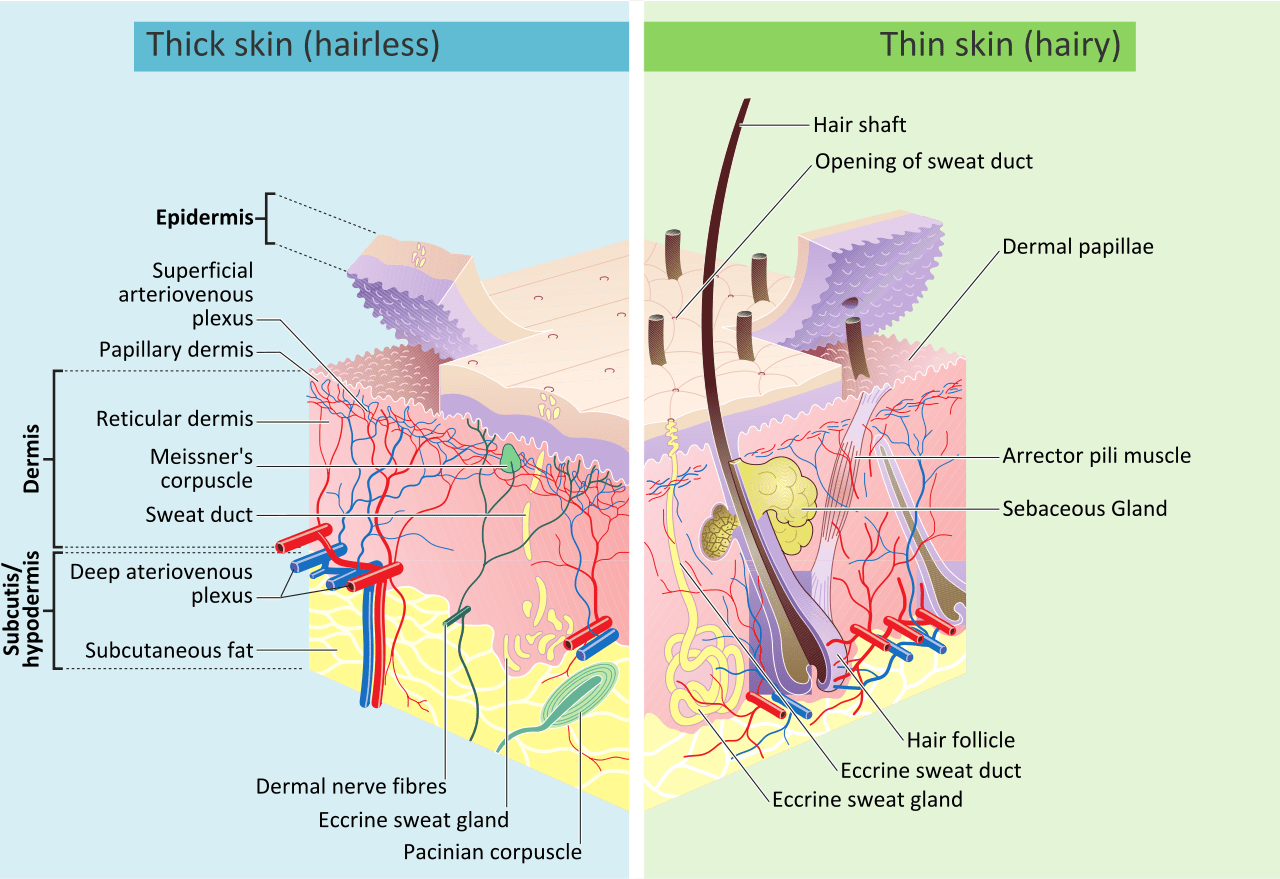
Skin and subcutaneous tissue
The skin is the largest organ in the body. It has three layers: epidermis, dermis, and subcutaneous tissue.
Epidermis
The epidermis is stratified squamous epithelium. It’s made mainly of keratinocytes and dendritic cells. It renews continuously as new cells form from basal cells.
The epidermis is organized into five layers, from deepest to most superficial:
Stratum basale (Stratum germinativum)
This layer consists of cuboidal to columnar, mitotically active stem cells called basal cells. They form a single layer that continuously produces keratinocytes.
- Basal cells adhere to each other by desmosomes and to the basement membrane by hemidesmosomes.
- Basal cells have dark-staining, elongated nuclei.
This layer also contains melanocytes, which are clear cells with long, branching cytoplasmic projections.
- Melanin produced by melanocytes is taken up by adjacent keratinocytes by phagocytosis.
- Melanosome number is the same among the sexes and races.
- Differences in skin color depend on:
- how much melanin is produced
- the size of the melanosomes (melanin-containing vesicles)
- the level of aggregation (aggregates in white-skinned persons; more dispersed in dark-skinned individuals)
This layer also contains Merkel cells, which are mechanoreceptors for light touch and are numerous in the fingertips.
Stratum spinosum
This layer is also called the squamous cell layer or prickle cell layer because the cells show spine-like cytoplasmic projections.
- It’s made of squamous cells that become progressively flatter and larger toward the more superficial layers.
- Cells are connected by desmosomes and gap junctions.
- These cells are metabolically very active and provide tensile strength to the skin.
This layer also contains Langerhans cells (dendritic cells of the skin).
- They function in antigen presentation.
- They can be identified by characteristic tennis racket-shaped intracytoplasmic Birbeck granules.
Stratum granulosum
This layer is named for its diamond-shaped cells containing basophilic keratohyalin granules.
- Keratohyalin granules contain keratin precursors.
- This layer is thick in the palms and soles, and thin in psoriasis.
- It is rich in lysosomal enzymes.
Stratum lucidum
This layer is made of dead keratinocytes and is seen in thick skin (for example, palms and soles).
- It appears as a clear layer.
- It consists of eleidin, a breakdown product of keratohyalin.
Stratum corneum
This is the outermost layer of the skin. It consists of dead keratinocytes called horny cells (corneocytes).
- It is rich in keratin.
- It acts as an environmental barrier by preventing water loss and providing a first line of defense against immunogens.
- Its thickness varies by body region.
In thick skin (such as palms and soles), the stratum corneum and stratum lucidum are thicker.
The basal lamina (basal membrane) separates the epidermis from the dermis and contains type IV collagen.
Dermis
The dermis lies beneath the epidermis and makes up most of the skin’s bulk. It makes the skin supple and elastic and provides tensile strength.
- The dermis atrophies with age, leading to thinning of the skin.
- Estrogen helps maintain dermal suppleness.
The dermis has:
- an upper papillary layer composed of loose connective tissue
- a deeper, thicker reticular layer composed of collagen
Fibroblasts produce and secrete procollagen in the dermis. The dermis contains primarily type I collagen and some type III collagen, along with a small amount of elastic fibers.
It contains sweat glands, hair follicles, blood vessels, sensory neurons, and muscles. Mast cells are present in the dermis.
Hypodermis
This layer lies beneath the dermis and contains subcutaneous fascia and fat.
Skin blood supply
The skin has two vascular plexuses:
- a subpapillary (superficial) plexus between the papillary and reticular layers of the dermis
- a deeper plexus between the dermis and subcutaneous tissue
The superficial plexus is important for thermoregulation:
- When body temperature is high, vasodilation increases heat loss.
- In cold conditions, vasoconstriction reduces heat loss.
Changes in blood flow and sweating help regulate body temperature. This mainly involves sympathetic innervation of cutaneous blood vessels:
- sympathetic stimulation causes vasoconstriction and conserves body heat
- sympathetic inhibition causes vasodilation and increases heat loss
Appendages of the skin
These include sweat and sebaceous glands, hair, and nails.
Sweat glands
Sweat glands include apocrine, eccrine, and apoeccrine glands.
-
Apocrine glands:
- are responsible for body odor
- are present in the axillae and perineum
- open into pilosebaceous follicles
- become active just before puberty
-
Eccrine sweat glands:
- are involved in thermoregulation
- are most abundant on the soles
- have ducts that open onto the surface of the skin
- contain clear secretory cells, darker mucoidal cells, and contractile myoepithelial cells
Postganglionic sympathetic fibers innervating eccrine glands for thermoregulation use acetylcholine as a neurotransmitter, while postganglionic sympathetics causing emotional sweating release norepinephrine.
- Apoeccrine glands:
- resemble apocrine glands
- open onto the surface of the skin
- have a very high rate of secretion
- are associated with hyperhidrosis
Sebaceous glands
Sebaceous glands secrete oily sebum.
- They are found on most areas of the skin except the palms and soles.
- They are abundant on the face and scalp.
- Some open into hair follicles, while a few open directly onto the skin surface.
- They are involved in the development of acne.