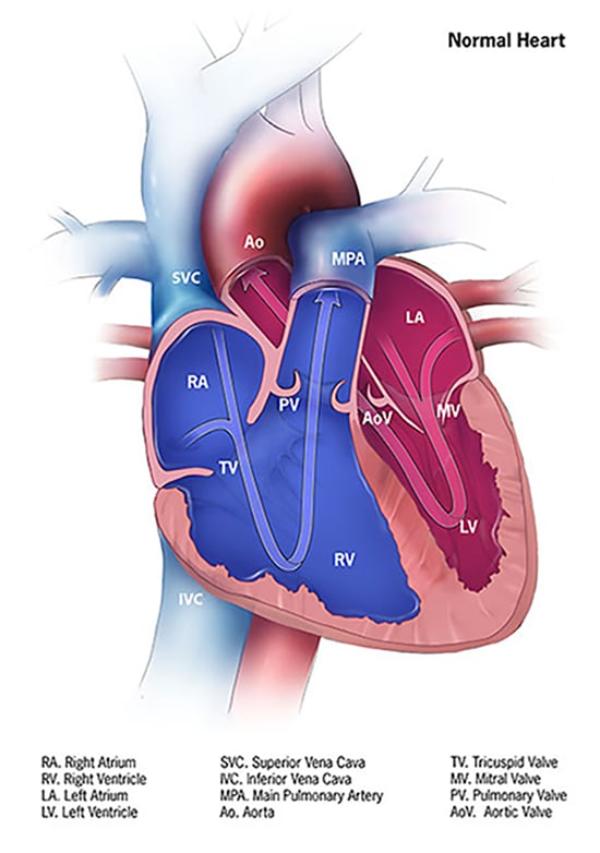
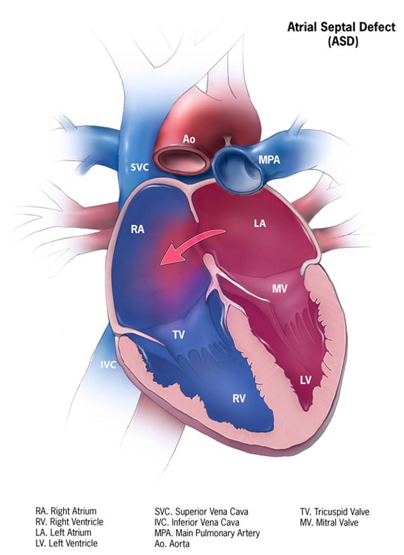
The heart is derived mainly from mesoderm and partly from neural crest. Early in development, a heart tube forms and then undergoes looping. This looping helps place the atria and ventricles into their adult anatomical positions.
Two atrioventricular (AV) cushions, also called endocardial cushions, grow from the endocardium of the heart tube. They fuse in the middle to divide the AV canal into right and left parts. The endocardial cushions contribute to formation of:
The AV canal must be partitioned correctly to allow normal circulation and oxygen delivery. Retinoic acid plays a major role in development of the heart tube and AV cushions. Disruption (e.g., intake of retinoic acid for acne) can lead to AV canal defects, hypoplastic right heart, etc. AV canal (endocardial cushion) defects are frequently seen in Down’s syndrome.

The septum primum extends from the roof of the atrium toward the endocardial cushions. The opening between them is the ostium (foramen) primum. Eventually, the septum primum and endocardial cushions fuse, closing the foramen primum.
At the same time, a new opening forms in the septum primum. This occurs by apoptosis of a few cells within the septum. This new opening is called the foramen secundum.
Next, the septum secundum develops in the right atrium, adjacent to the septum primum. It grows down toward the endocardial cushions but does not fuse with them completely. This leaves an oval opening called the foramen ovale.
[Note that the foramen ovale is located inferiorly while the foramen secundum is located superiorly near the roof of the right atrium].
Immediately after birth, the foramen ovale closes functionally, stopping shunting of blood from the right atrium to the left atrium. This occurs because pressure increases in the left heart and decreases in the right heart (blood won’t flow from low to high pressures). Over the next few months, anatomical fusion occurs, leading to complete closure of the foramen ovale.

The primitive ventricle is divided by the interventricular septum into right and left ventricles. This septum is:
The membranous part has contributions from the endocardial cushions and conotruncal cushions (read below).
Neural crest cells from pharyngeal arches 4 and 6 migrate into the truncus arteriosus and bulbus cordis. This migration forms the conotruncal cushions (ridges). The conotruncal ridges divide the truncus arteriosus into the pulmonary trunk and aorta. They fuse and then spiral so that the pulmonary trunk ends up anterior to the aorta.
The conotruncal ridges also give rise to the semilunar valves:
The atrioventricular valves (tricuspid and mitral) are formed from the AV cushions.
Failure of neural crest cell migration and differentiation can cause persistent truncus arteriosus, transposition of great vessels, and tetralogy of Fallot.
The right vitelline vein forms the hepatic, splenic, superior mesenteric, and inferior mesenteric veins.
The respiratory diverticulum, which eventually forms the lower respiratory tract and lungs, develops as a ventral outgrowth from the endoderm of the foregut. Mesodermal ridges called tracheo-esophageal ridges give rise to the trachea. Epithelium and glands of the larynx and trachea are derived from endoderm. Surfactant production begins between weeks 20-22.
The diaphragm is formed by the septum transversum and the right and left pleuroperitoneal membranes.
Sign up for free to take 3 quiz questions on this topic