


Achievable logoAchievable logo
NPTE-PTA
Sign in
Sign up
Purchase
Textbook
Practice exams
Support
How it works
Resources
Exam catalog
Mountain with a flag at the peak
Textbook
Introduction
1. Cardiopulmonary system
2. Pulmonary system
3. Neuromuscular system
4. Pediatrics
5. Musculoskeletal system
5.1 Anatomy of musculoskeletal system
5.2 Anatomical terminology and exercise training principles
5.3 Joint mechanics and phases of healing
5.4 Upper extremity anatomy
5.5 Special tests of upper extremity
5.6 Comparing clinical presentation and interventions for upper extremity
5.7 Lower extremity anatomy
5.8 Special tests of lower extremity
5.8.1 Hip special tests
5.8.2 Knee special tests
5.8.3 Ankle and foot special tests
5.9 Comparing clinical presentation and interventions of lower extremity
5.10 Spine and pelvis anatomy
5.11 Special tests of the spine, pelvis, and temporomandibular joint
5.12 Comparing clinical presentation and interventions for the spine, pelvis, and temporomandibular joint
5.13 Other MSK conditions
5.14 Gait
5.15 Prosthetics
5.16 Orthotics
5.17 Medications, imaging, and fractures
5.18 Surgical protocols
6. Other system
7. Non systems
Wrapping up
Achievable logoAchievable logo
5.8.2 Knee special tests
Achievable NPTE-PTA
5. Musculoskeletal system
5.8. Special tests of lower extremity
Our NPTE-PTA course is now in "early access" - get 50% off for a limited time.

Knee special tests

9 min read
Font
Discuss
Share
Feedback

Distinguishing overlapping tests:

  • ACL: Lachman (knee flexed 20-30°) and the anterior drawer (hip flexed 45°, knee flexed 90°) both check for excessive anterior tibial translation, but Lachman has higher sensitivity and specificity and is the preferred test.
  • Collateral ligaments: valgus stress checks the MCL; varus stress checks the LCL.
  • Meniscus: McMurray and Apley’s compression both use tibial rotation to reproduce clicking or pain; Thessaly uses weight-bearing rotation at 5° and 20° of knee flexion instead.
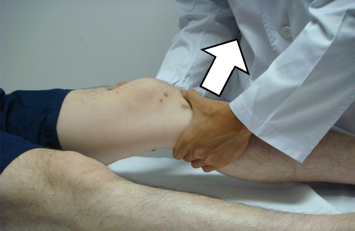
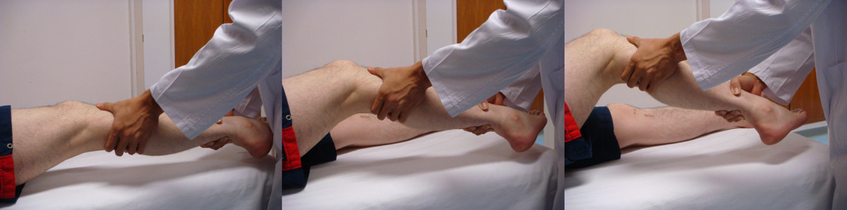
  • Lachman test
    • Patient supine with knee flexed to 20-30 degrees; stabilize the femur and passively translate the tibia anteriorly
      • Tests the integrity of the anterior cruciate ligament
    • Positive: excessive anterior translation on the tibia compared to the uninvolved limb
Lachman test
Lachman test
By - Mak-Ham Lam, CC BY-SA 2.0
/
Wikimedia Commons
/
CC BY-SA 2.0
  • Anterior drawer test
    • Patient supine with hip flexed to 45 degrees and knee flexed to 90 degrees; therapist passively translates the tibia anteriorly
      • Tests the integrity of the anterior cruciate ligament
    • Positive: excessive anterior translation on the tibia compared to the uninvolved limb
Anterior drawer test of the knee with the knee flexed to 90 degrees and the tibia pulled forward to assess ACL integrity.
Anterior drawer test
Achievable
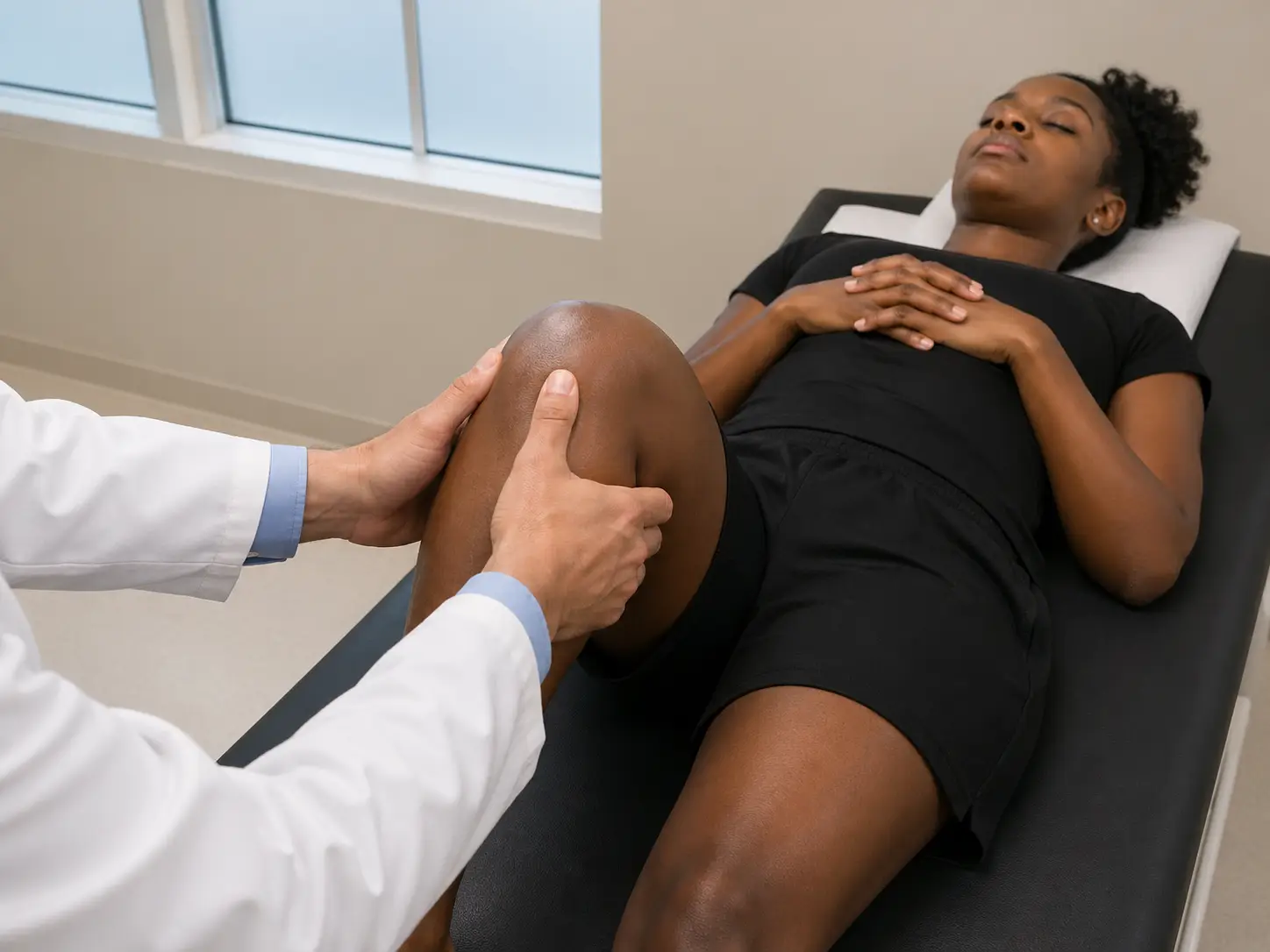
  • Posterior drawer test
    • Patient in supine with hip flexed to 45 degrees and knee flexed to 90 degrees; therapist passively translates tibia posteriorly
      • Tests the integrity of the posterior cruciate ligament
    • Positive: excessive posterior translation on the tibia compared to the uninvolved limb
Posterior drawer test of the knee with the knee flexed to 90 degrees and the tibia pushed posteriorly to assess PCL integrity.
Posterior drawer test
Achievable
  • Valgus stress test
    • Patient supine with knee resting at the edge of the mat; therapist applies valgus stress to the knee with the knee flexed at 0 and 30 degrees
      • Tests the integrity of the medial collateral ligament
    • Positive: laxity and pain compared to the uninvolved side
Valgus stress test being performed on a patient's knee to assess medial ligament stability.
Valgus stress test
Achievable
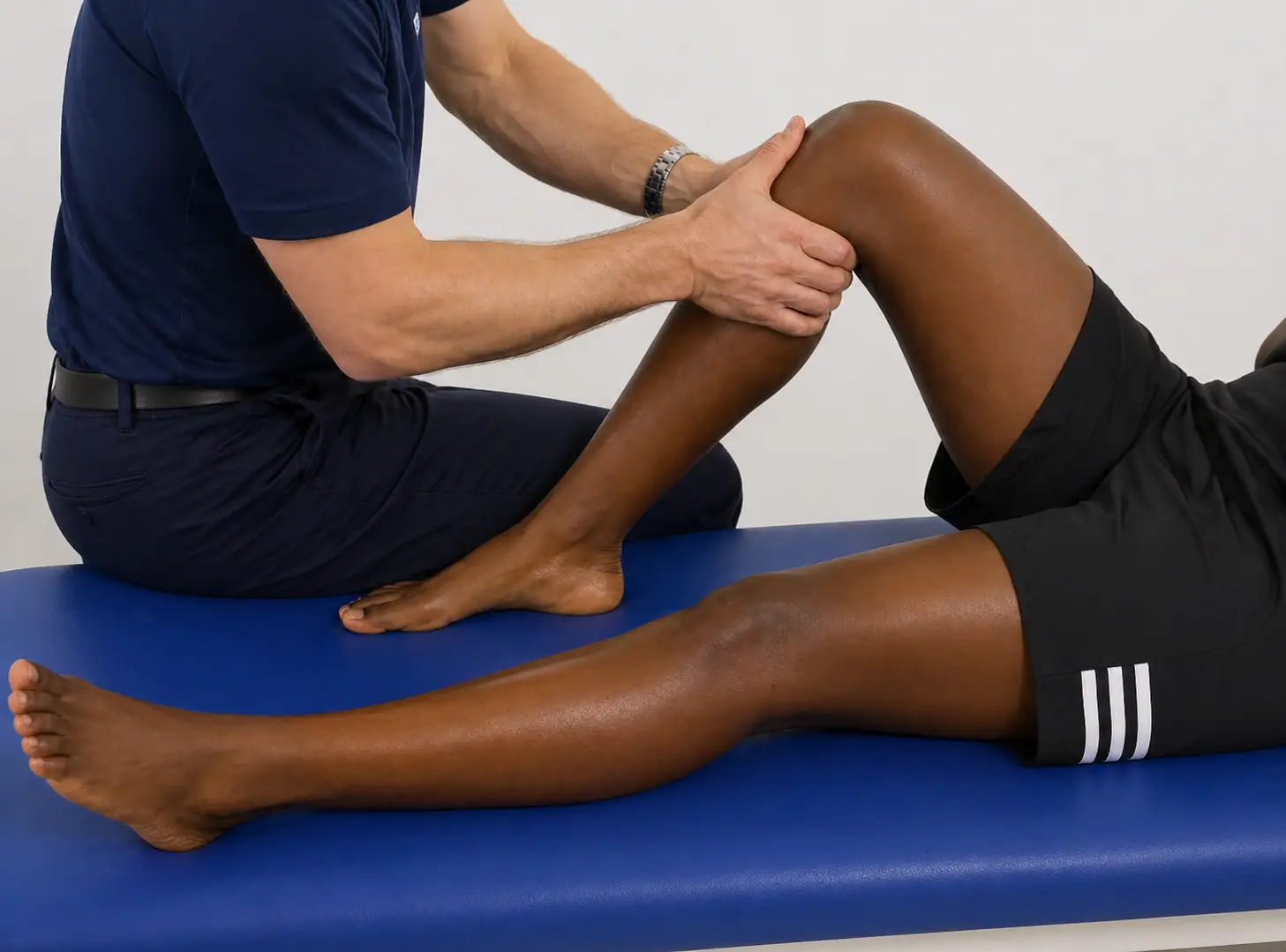
  • Varus stress test
    • Patient supine with knee resting at the edge of the mat; therapist applies varus stress to the knee with the knee flexed at 0 and 30 degrees
      • Tests the integrity of the lateral collateral ligament
    • Positive: laxity and pain compared to the uninvolved side
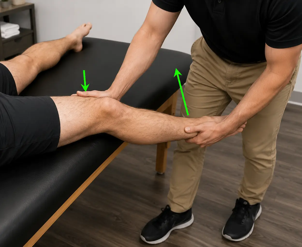
Clinician performing a varus stress test on a patient's knee while stabilizing the leg.
Varus stress test
Achievable
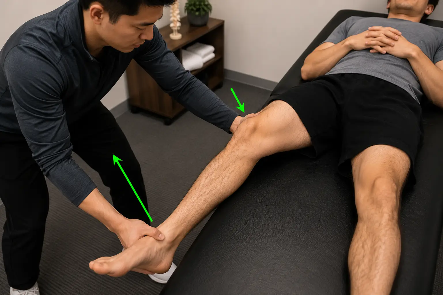
  • Pivot shift test
    • Patient supine with knee extended, hip flexed, and abducted to 30 degrees and slight internal rotation; therapist holding knee with one hand and the foot with the other applies valgus force through a flexed knee
      • Tests the integrity of the anterior cruciate ligament
    • Positive: tibia reduction during the test by the iliotibial band
Pivot shift test
Pivot shift test
Achievable
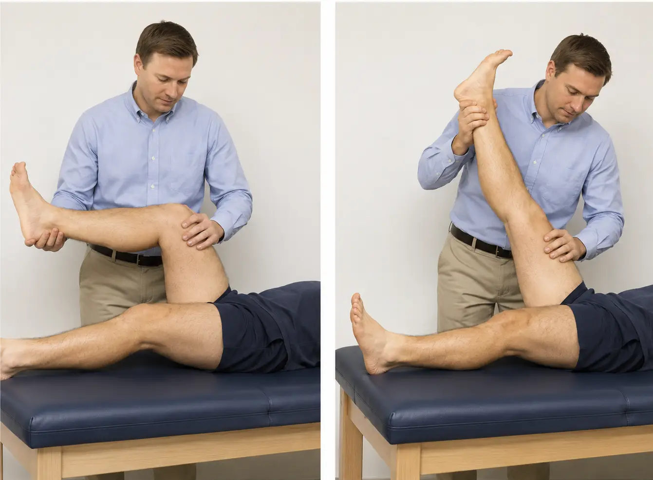
  • McMurray test
    • Patient supine with knee maximally flexed; therapist passively internally rotates and extends the knee, then moves to externally rotating and extending the knee
      • Test the lateral meniscus (internal rotation) and the medial meniscus (external rotation)
    • Positive: reproduction of click, popping, or pain in the knee
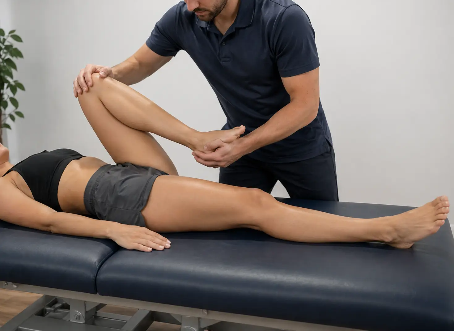
McMurray's test of the knee using combined flexion, rotation, and extension to assess for meniscal tears.
McMurray's test
Achievable
  • Apley’s compression test
    • Patient positioned prone with the knee flexed to 90 degrees. The therapist applies a downward compressive force through the heel while medially and laterally rotating the tibia.
      • Tests the integrity of the menisci of the knee
    • Positive: reproduction of pain
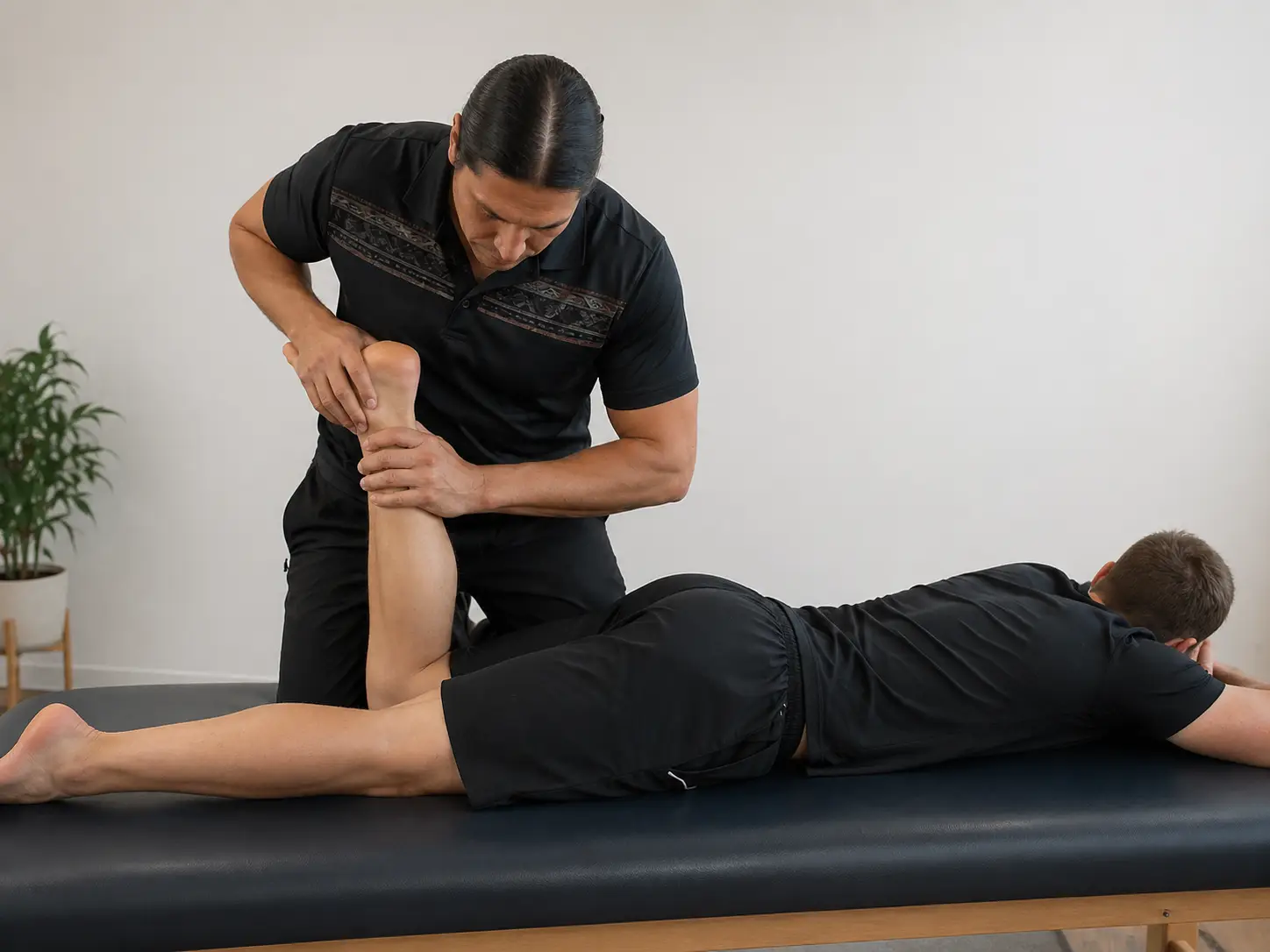
Apley's compression test with the patient prone and the knee flexed to 90 degrees while applying downward pressure with rotation to assess meniscal injury.
Apley's compression test
Achievable
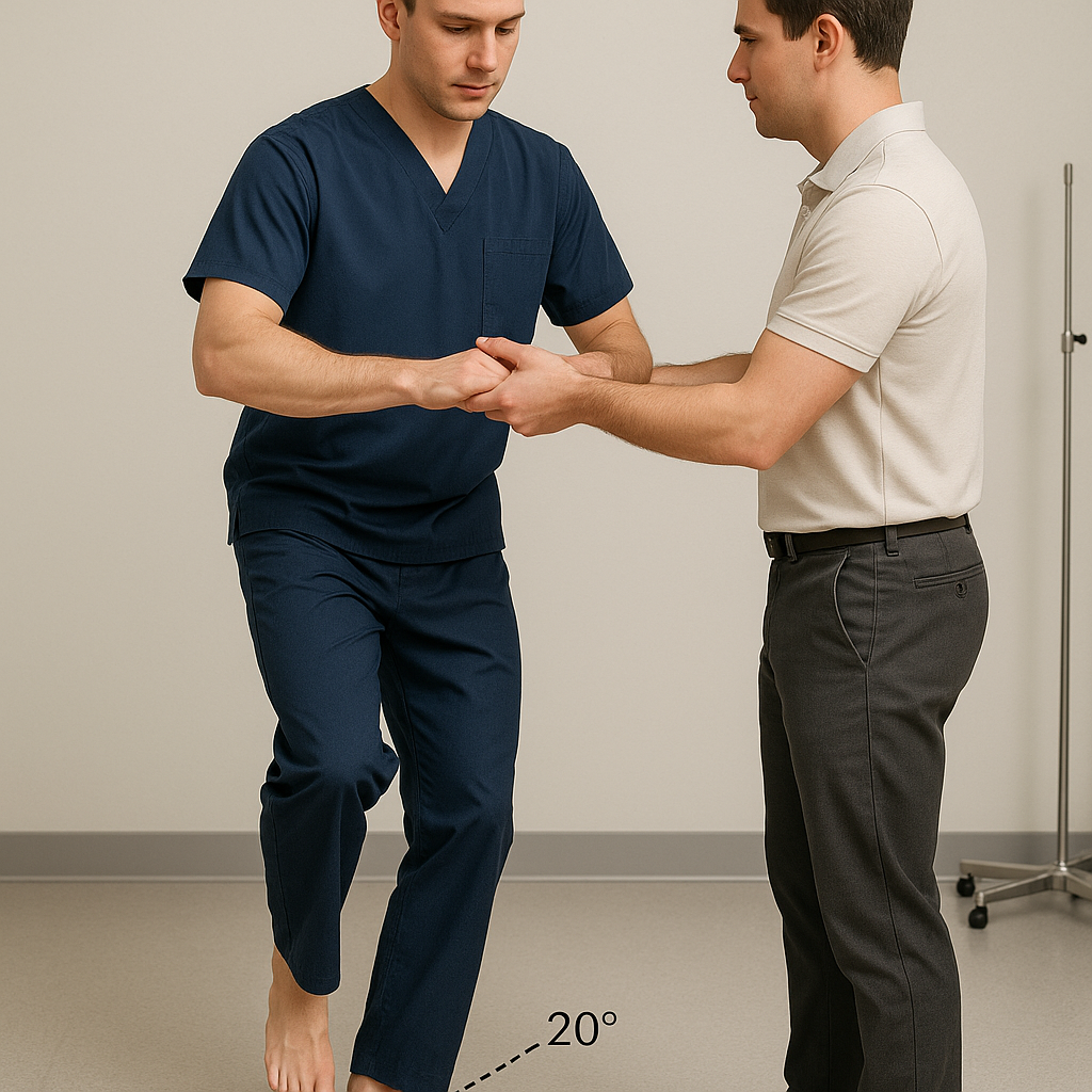
  • Thessaly test
    • Patient standing on involved leg while holding therapist’s hands; patient rotates body and leg internally and externally with knee flexed to 5 degrees and then at 20 degrees
      • Test the lateral meniscus (internal rotation) and the medial meniscus (external rotation)
    • Positive: reproduction of click, popping, or pain in the knee
Thessaly test
Thessaly test
Achievable
  • Patellofemoral instability

    • Patient supine with knee flexed to 30 degrees and quadriceps relaxed; therapist passively translates the patella laterally
      • Test for patellar instability
    • Positive: patient expresses apprehension or contracts the quadriceps muscle to prevent patellar dislocation
  • Noble compression test

    • Patient supine with knee flexed to 90 degrees and hip flexion; therapist applies pressure 1-2cm proximal to the lateral femoral epicondyle; with pressure maintained, the patient’s knee is passively extended
      • Tests the iliotibial band
    • Positive: patient experiences pain over the lateral femoral condyle
Clinician performing the Noble compression test on a patient's knee while applying pressure to the outer leg.
Noble compression test
Achievable
  • Ottawa knee rules
    • Apply the Ottawa knee rules to:
      • Rule out fracture after acute knee injury
      • Refer for imaging with one or more positive answers
      • A negative test result states there is an absence of fracture
    • If the answer is yes to any of these questions, the finding is reported to the supervising PT, who determines whether imaging is needed to rule out a fracture
      • Age 55 years or older
      • Isolated patellar tenderness without bone tenderness
      • Tenderness of the fibula head
      • Inability to actively flex knee to 90 degrees
      • Inability to bear weight immediately after injury

Distinguishing overlapping tests

  • ACL: Lachman (knee flexed 20-30°) vs anterior drawer (hip 45°, knee 90°) — both check anterior tibial translation; Lachman more sensitive/specific
  • Collateral ligaments: valgus stress = MCL; varus stress = LCL
  • Meniscus: McMurray/Apley’s use tibial rotation; Thessaly uses weight-bearing rotation at 5° and 20° flexion

Lachman test

  • Supine, knee flexed 20-30°; femur stabilized, tibia translated anteriorly
  • Tests ACL integrity
  • Positive: excessive anterior tibial translation vs uninvolved side

Anterior drawer test

  • Supine, hip flexed 45°, knee flexed 90°; tibia translated anteriorly
  • Tests ACL integrity
  • Positive: excessive anterior tibial translation vs uninvolved side

Posterior drawer test

  • Supine, hip flexed 45°, knee flexed 90°; tibia translated posteriorly
  • Tests PCL integrity
  • Positive: excessive posterior tibial translation vs uninvolved side

Valgus stress test

  • Supine, knee at mat edge; valgus stress applied at 0° and 30° flexion
  • Tests MCL integrity
  • Positive: laxity and pain vs uninvolved side

Varus stress test

  • Supine, knee at mat edge; varus stress applied at 0° and 30° flexion
  • Tests LCL integrity
  • Positive: laxity and pain vs uninvolved side

Pivot shift test

  • Supine, knee extended, hip flexed/abducted 30° with slight internal rotation; valgus force applied through flexing knee
  • Tests ACL integrity
  • Positive: tibial reduction during test via IT band

McMurray test

  • Supine, knee maximally flexed; passive internal rotation/extension (lateral meniscus) then external rotation/extension (medial meniscus)
  • Positive: click, pop, or pain reproduction

Apley’s compression test

  • Prone, knee flexed 90°; downward compression through heel with medial/lateral tibial rotation
  • Tests menisci integrity
  • Positive: reproduction of pain

Thessaly test

  • Standing on involved leg holding therapist’s hands; body/leg rotated internally/externally at 5° and 20° knee flexion
  • Internal rotation = lateral meniscus; external rotation = medial meniscus
  • Positive: click, pop, or pain reproduction

Patellofemoral instability test

  • Supine, knee flexed 30°, quadriceps relaxed; patella passively translated laterally
  • Tests patellar instability
  • Positive: apprehension or quad contraction to prevent dislocation

Noble compression test

  • Supine, knee flexed 90°, hip flexed; pressure applied 1-2cm proximal to lateral femoral epicondyle while knee passively extended
  • Tests IT band
  • Positive: pain over lateral femoral condyle

Ottawa knee rules

  • Used to rule out fracture after acute knee injury; positive finding(s) prompt imaging referral
  • Negative result = absence of fracture
  • Red flags requiring supervising PT notification:
    • Age ≥55 years
    • Isolated patellar tenderness without bone tenderness
    • Fibular head tenderness
    • Inability to actively flex knee to 90°
    • Inability to bear weight immediately after injury

Sign up for free to take 5 quiz questions on this topic

Previous
Next  | 5.8.3 Ankle and foot special tests
All rights reserved ©2016 - 2026 Achievable, Inc.

Knee special tests

Distinguishing overlapping tests:

  • ACL: Lachman (knee flexed 20-30°) and the anterior drawer (hip flexed 45°, knee flexed 90°) both check for excessive anterior tibial translation, but Lachman has higher sensitivity and specificity and is the preferred test.
  • Collateral ligaments: valgus stress checks the MCL; varus stress checks the LCL.
  • Meniscus: McMurray and Apley’s compression both use tibial rotation to reproduce clicking or pain; Thessaly uses weight-bearing rotation at 5° and 20° of knee flexion instead.
  • Lachman test
    • Patient supine with knee flexed to 20-30 degrees; stabilize the femur and passively translate the tibia anteriorly
      • Tests the integrity of the anterior cruciate ligament
    • Positive: excessive anterior translation on the tibia compared to the uninvolved limb
  • Anterior drawer test
    • Patient supine with hip flexed to 45 degrees and knee flexed to 90 degrees; therapist passively translates the tibia anteriorly
      • Tests the integrity of the anterior cruciate ligament
    • Positive: excessive anterior translation on the tibia compared to the uninvolved limb
  • Posterior drawer test
    • Patient in supine with hip flexed to 45 degrees and knee flexed to 90 degrees; therapist passively translates tibia posteriorly
      • Tests the integrity of the posterior cruciate ligament
    • Positive: excessive posterior translation on the tibia compared to the uninvolved limb
  • Valgus stress test
    • Patient supine with knee resting at the edge of the mat; therapist applies valgus stress to the knee with the knee flexed at 0 and 30 degrees
      • Tests the integrity of the medial collateral ligament
    • Positive: laxity and pain compared to the uninvolved side
  • Varus stress test
    • Patient supine with knee resting at the edge of the mat; therapist applies varus stress to the knee with the knee flexed at 0 and 30 degrees
      • Tests the integrity of the lateral collateral ligament
    • Positive: laxity and pain compared to the uninvolved side
  • Pivot shift test
    • Patient supine with knee extended, hip flexed, and abducted to 30 degrees and slight internal rotation; therapist holding knee with one hand and the foot with the other applies valgus force through a flexed knee
      • Tests the integrity of the anterior cruciate ligament
    • Positive: tibia reduction during the test by the iliotibial band
  • McMurray test
    • Patient supine with knee maximally flexed; therapist passively internally rotates and extends the knee, then moves to externally rotating and extending the knee
      • Test the lateral meniscus (internal rotation) and the medial meniscus (external rotation)
    • Positive: reproduction of click, popping, or pain in the knee
  • Apley’s compression test
    • Patient positioned prone with the knee flexed to 90 degrees. The therapist applies a downward compressive force through the heel while medially and laterally rotating the tibia.
      • Tests the integrity of the menisci of the knee
    • Positive: reproduction of pain
  • Thessaly test
    • Patient standing on involved leg while holding therapist’s hands; patient rotates body and leg internally and externally with knee flexed to 5 degrees and then at 20 degrees
      • Test the lateral meniscus (internal rotation) and the medial meniscus (external rotation)
    • Positive: reproduction of click, popping, or pain in the knee
  • Patellofemoral instability

    • Patient supine with knee flexed to 30 degrees and quadriceps relaxed; therapist passively translates the patella laterally
      • Test for patellar instability
    • Positive: patient expresses apprehension or contracts the quadriceps muscle to prevent patellar dislocation
  • Noble compression test

    • Patient supine with knee flexed to 90 degrees and hip flexion; therapist applies pressure 1-2cm proximal to the lateral femoral epicondyle; with pressure maintained, the patient’s knee is passively extended
      • Tests the iliotibial band
    • Positive: patient experiences pain over the lateral femoral condyle
  • Ottawa knee rules
    • Apply the Ottawa knee rules to:
      • Rule out fracture after acute knee injury
      • Refer for imaging with one or more positive answers
      • A negative test result states there is an absence of fracture
    • If the answer is yes to any of these questions, the finding is reported to the supervising PT, who determines whether imaging is needed to rule out a fracture
      • Age 55 years or older
      • Isolated patellar tenderness without bone tenderness
      • Tenderness of the fibula head
      • Inability to actively flex knee to 90 degrees
      • Inability to bear weight immediately after injury
Key points

Distinguishing overlapping tests

  • ACL: Lachman (knee flexed 20-30°) vs anterior drawer (hip 45°, knee 90°) — both check anterior tibial translation; Lachman more sensitive/specific
  • Collateral ligaments: valgus stress = MCL; varus stress = LCL
  • Meniscus: McMurray/Apley’s use tibial rotation; Thessaly uses weight-bearing rotation at 5° and 20° flexion

Lachman test

  • Supine, knee flexed 20-30°; femur stabilized, tibia translated anteriorly
  • Tests ACL integrity
  • Positive: excessive anterior tibial translation vs uninvolved side

Anterior drawer test

  • Supine, hip flexed 45°, knee flexed 90°; tibia translated anteriorly
  • Tests ACL integrity
  • Positive: excessive anterior tibial translation vs uninvolved side

Posterior drawer test

  • Supine, hip flexed 45°, knee flexed 90°; tibia translated posteriorly
  • Tests PCL integrity
  • Positive: excessive posterior tibial translation vs uninvolved side

Valgus stress test

  • Supine, knee at mat edge; valgus stress applied at 0° and 30° flexion
  • Tests MCL integrity
  • Positive: laxity and pain vs uninvolved side

Varus stress test

  • Supine, knee at mat edge; varus stress applied at 0° and 30° flexion
  • Tests LCL integrity
  • Positive: laxity and pain vs uninvolved side

Pivot shift test

  • Supine, knee extended, hip flexed/abducted 30° with slight internal rotation; valgus force applied through flexing knee
  • Tests ACL integrity
  • Positive: tibial reduction during test via IT band

McMurray test

  • Supine, knee maximally flexed; passive internal rotation/extension (lateral meniscus) then external rotation/extension (medial meniscus)
  • Positive: click, pop, or pain reproduction

Apley’s compression test

  • Prone, knee flexed 90°; downward compression through heel with medial/lateral tibial rotation
  • Tests menisci integrity
  • Positive: reproduction of pain

Thessaly test

  • Standing on involved leg holding therapist’s hands; body/leg rotated internally/externally at 5° and 20° knee flexion
  • Internal rotation = lateral meniscus; external rotation = medial meniscus
  • Positive: click, pop, or pain reproduction

Patellofemoral instability test

  • Supine, knee flexed 30°, quadriceps relaxed; patella passively translated laterally
  • Tests patellar instability
  • Positive: apprehension or quad contraction to prevent dislocation

Noble compression test

  • Supine, knee flexed 90°, hip flexed; pressure applied 1-2cm proximal to lateral femoral epicondyle while knee passively extended
  • Tests IT band
  • Positive: pain over lateral femoral condyle

Ottawa knee rules

  • Used to rule out fracture after acute knee injury; positive finding(s) prompt imaging referral
  • Negative result = absence of fracture
  • Red flags requiring supervising PT notification:
    • Age ≥55 years
    • Isolated patellar tenderness without bone tenderness
    • Fibular head tenderness
    • Inability to actively flex knee to 90°
    • Inability to bear weight immediately after injury

More from Special tests of lower extremity

  • Hip special tests
  • Ankle and foot special tests