Update it, or open this page in a current version of Chrome, Safari, Firefox or Edge. On an iPhone or iPad, update iOS: every browser there runs on the version of Safari that comes with it.
5.8.1 Hip special tests
Achievable NPTE-PTA
5. Musculoskeletal system
5.8. Special tests of lower extremity
Our NPTE-PTA course is now in "early access" - get 50% off for a limited time.
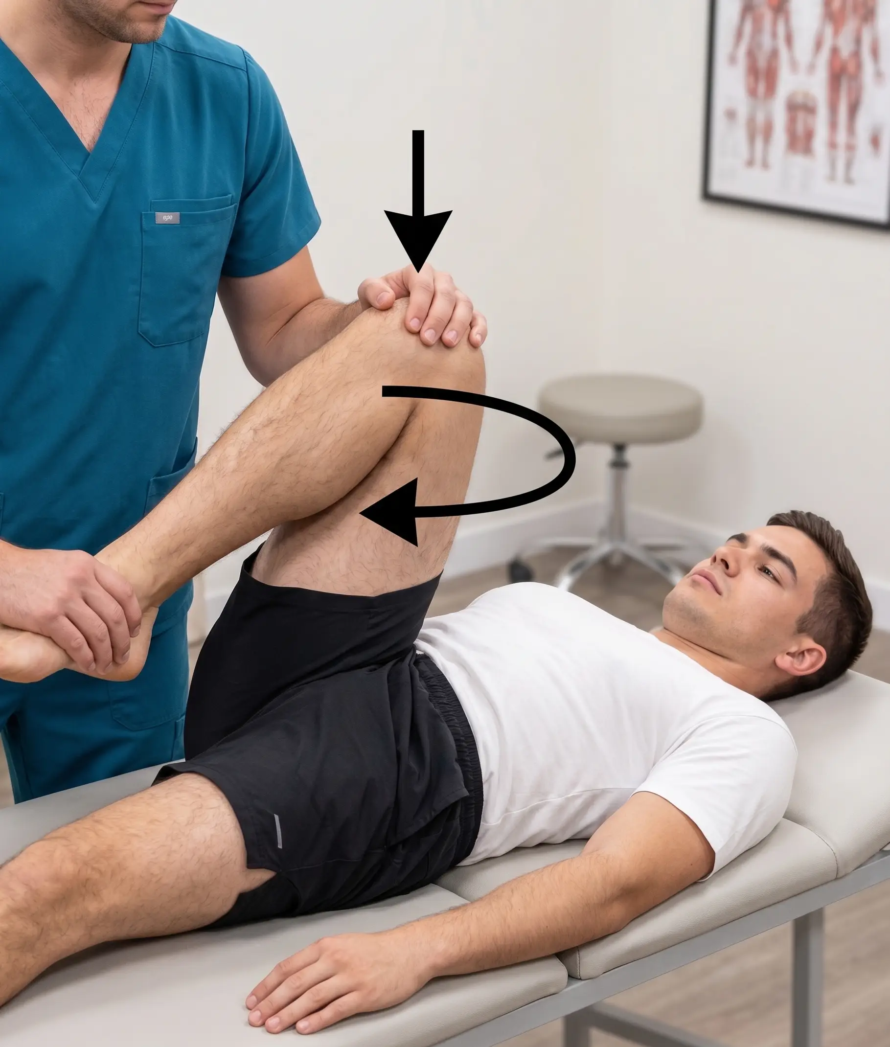
Patient supine; the examiner flexes and adducts the hip, applies axial compression down the femoral shaft, and sweeps the hip through an arc from flexion/adduction to abduction (adding rotation)
Tests for general hip pathology and degenerative joint disease
Positive: reproduction of pain symptoms or apprehension to perform
Hip scour test
Achievable
Patrick (FABER) test
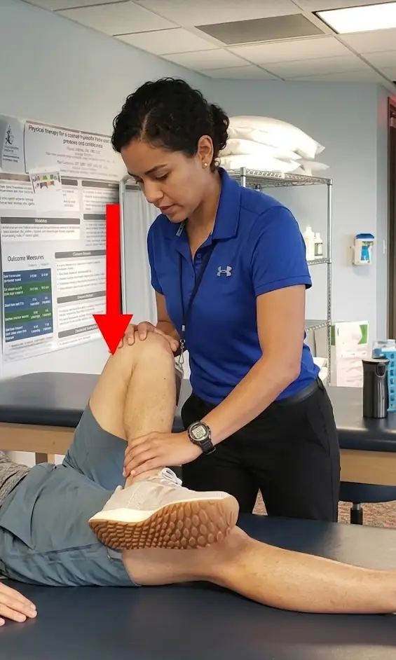
Patient supine, passively flex, abduct, and externally rotate the test leg so the foot is resting above the knee on the opposite leg; then slowly lower the leg toward the table
Identifiers: dysfunction of the hip, specifically mobility dysfunction
Also screens for iliopsoas tightness and SI joint dysfunction: anterior/groin pain points to the hip, while posterior pain near the SI joint points to the SI joint
Positive: involved knee is unable to assume a relaxed position and/or reproduce painful symptoms
Patrick (FABER) test
Achievable
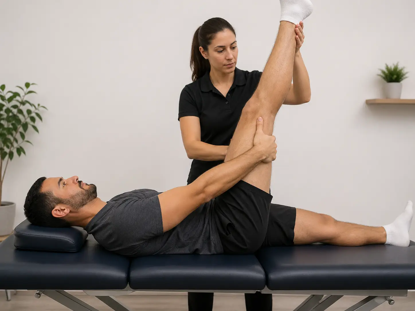
Thomas test
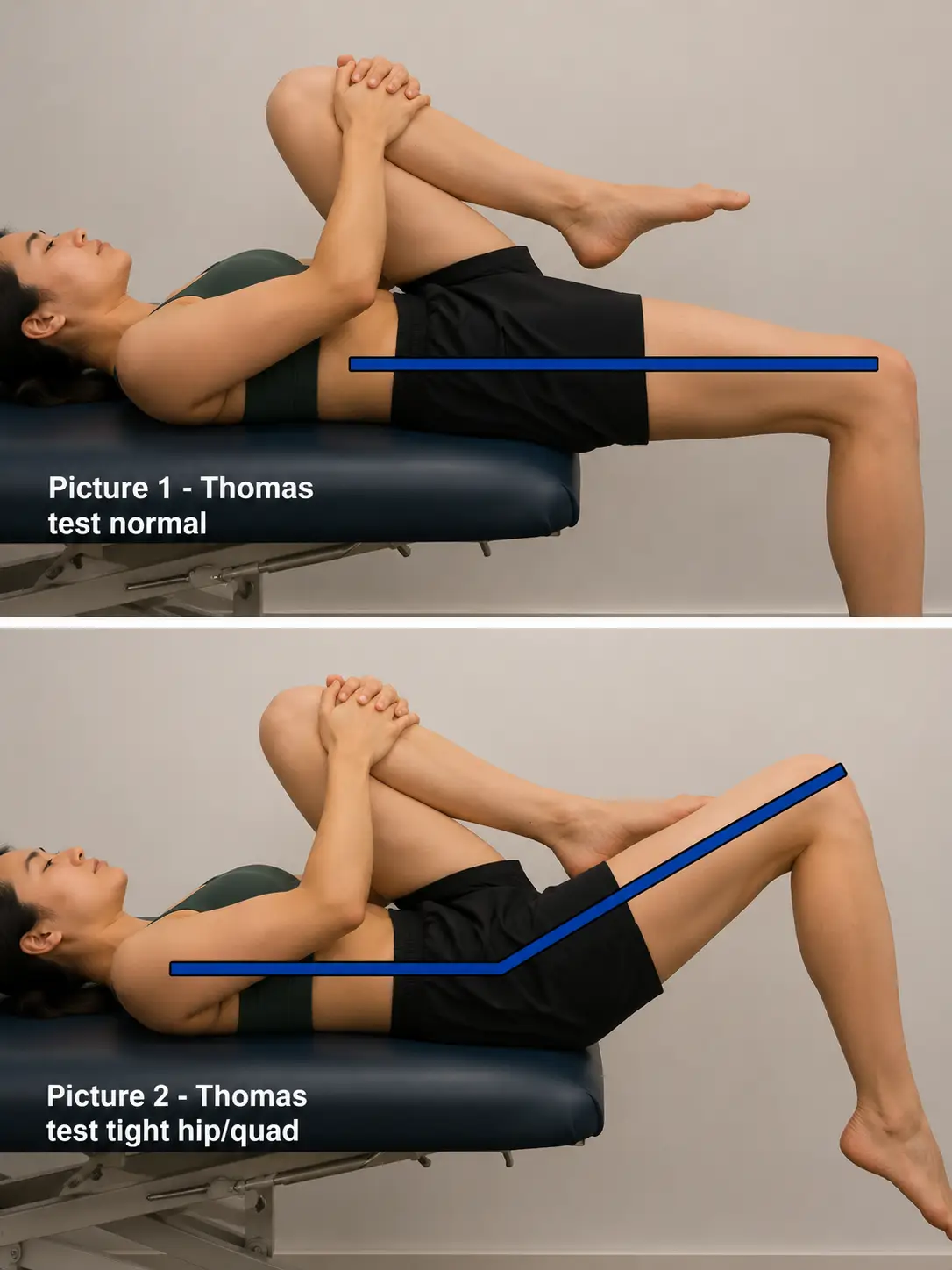
Patient supine; one hip and knee maximally flexed to the chest with hold; opposite limb is kept straight on the table
Tests for the tightness of hip flexors
Positive: straight limb flexes, and the patient is unable to keep this leg straight on the table
If the thigh rises off the table, this implicates the iliopsoas (a one-joint hip flexor); if the thigh stays down but the knee extends instead, this implicates the rectus femoris
Thomas test
Achievable
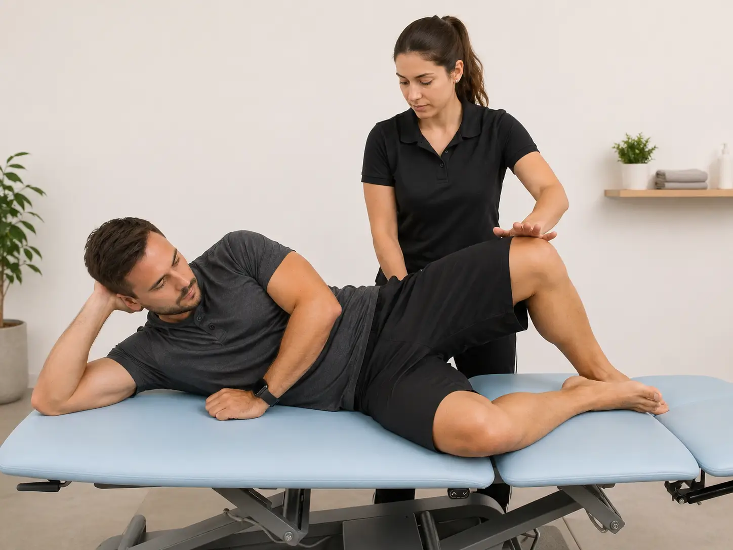
Ober’s test
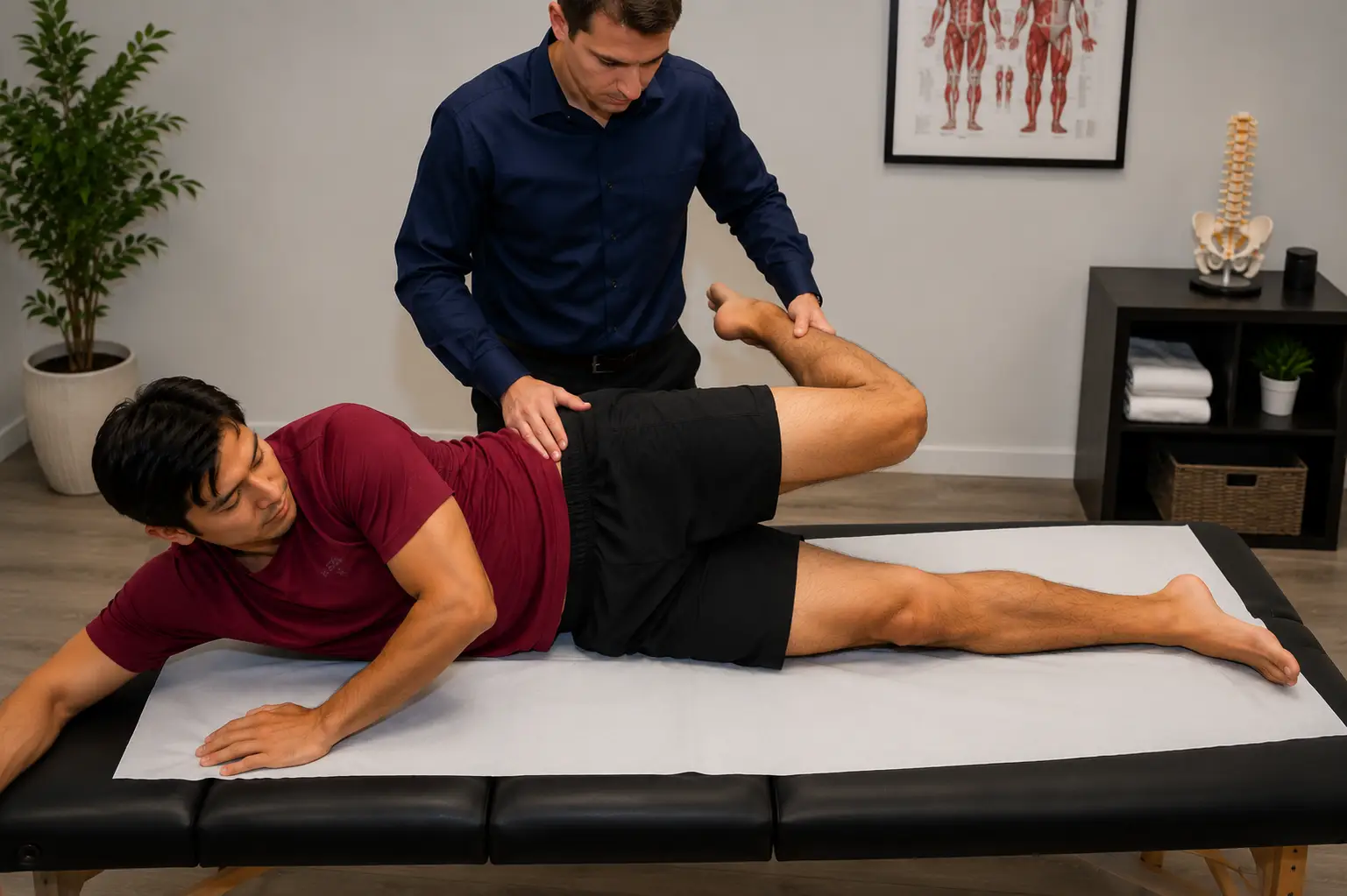
Patient side-lying on the untested side with the bottom leg flexed at the hip and knee for stability; the examiner stabilizes the pelvis, passively abducts and extends the top (tested) leg with its knee flexed to 90 degrees, then slowly lowers it toward the table
Tests for the tightness of the tensor fascia lata or the iliotibial band
Positive: uppermost leg remains above horizontal
Ober's test
Achievable
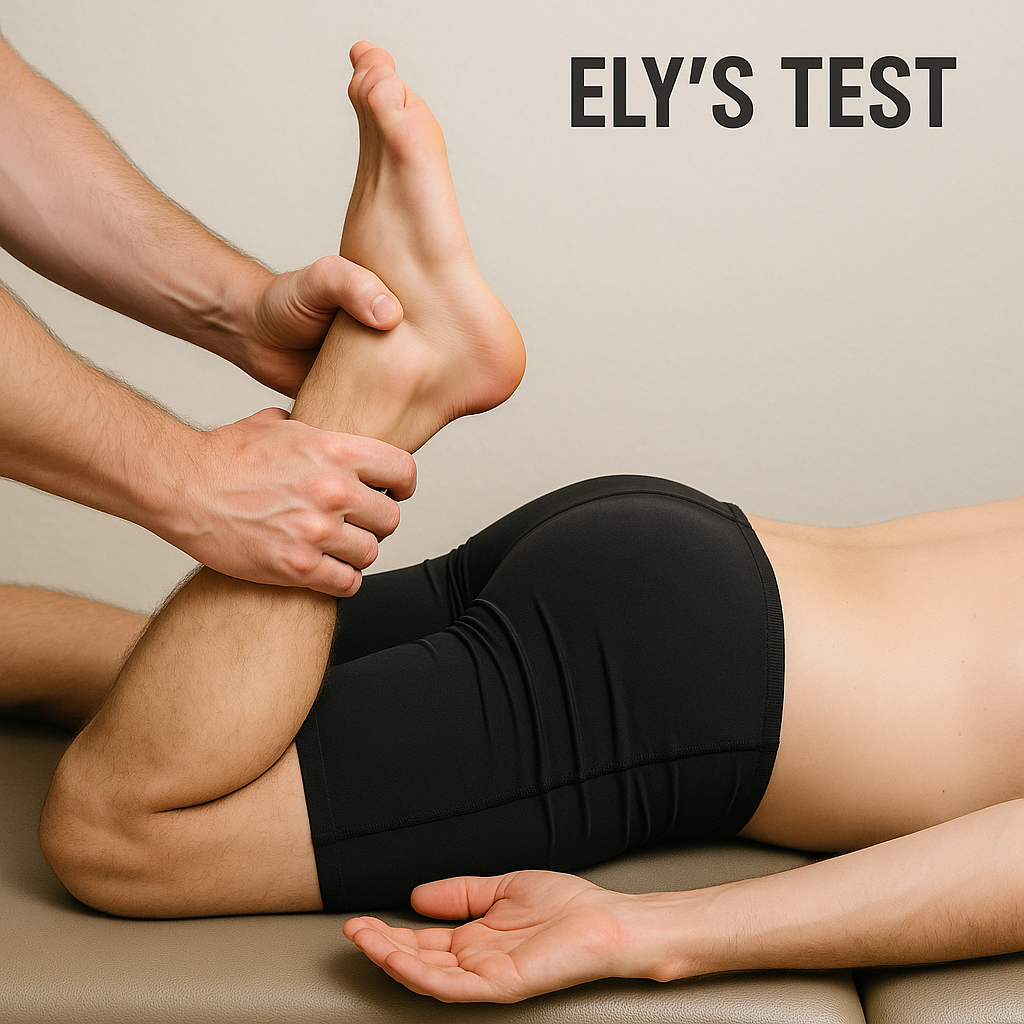
Ely’s test
Patient is prone; flex the knee of the tested limb
Tests for the tightness of the rectus femoris
Positive: hip of the tested limb flexes
Ely's test
Achievable
90-90 hamstring test
Patient position in supine; hip and knee supported in 90 degrees flexion; passively extend the knee until end feel is encountered
Test for the tightness of the hamstrings
Positive: knee lacks 10 degrees or greater of knee extension
90-90 hamstring test
Achievable
Piriformis test
Patient supine with the tested hip and knee flexed so the foot rests lateral to the opposite knee; the tested hip is then passively adducted (moved across midline)
Tests for piriformis tightness and syndrome
Positive: tested knee is unable to pass over the resting knee, or reproduction of pain in the buttock or along the sciatic nerve
Piriformis test
Achievable
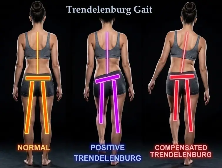
Trendelenburg test
Patient standing and asked to stand on one leg; observe the pelvis on the swing (unsupported) side.
Tests for gluteus medius weakness
Watch the pelvis on the swing (unsupported) side: a drop there indicates weakness of the stance-leg gluteus medius (the hip abductor on the side the patient is standing on)
A leg length discrepancy commonly produces a compensatory pelvic tilt (pelvic obliquity), which is different from the contralateral pelvic drop during single-leg stance that defines a positive Trendelenburg sign
Positive: swing side pelvis drops when in single leg stance, or compensation by leaning the trunk over the stance leg
Trendelenburg test
Achievable
PTA scope of practice
Special tests are data-collection tools within an existing plan of care, not diagnostic tools
Positive findings/red flags must be reported to supervising PT, who decides referral/imaging
Some positive findings (e.g., confirmed hip flexor tightness) can still guide same-day treatment already in the plan; others (e.g., pain on scour test) warrant reporting before continued loading
General hip joint pathology tests
Scour test: supine, hip flexed/adducted with axial compression swept through arc; positive = pain reproduction or apprehension; screens for degenerative joint disease
Patrick (FABER) test: figure-4 position, leg lowered toward table; positive = knee can’t relax or pain reproduced
Anterior/groin pain = hip dysfunction; posterior/SI pain = SI joint dysfunction
Also screens iliopsoas tightness
Muscle tightness/flexibility tests
Thomas test: supine, opposite hip/knee flexed to chest, test leg stays straight; positive = test leg flexes/can’t stay straight
Patient supine; the examiner flexes and adducts the hip, applies axial compression down the femoral shaft, and sweeps the hip through an arc from flexion/adduction to abduction (adding rotation)
Tests for general hip pathology and degenerative joint disease
Positive: reproduction of pain symptoms or apprehension to perform
Patrick (FABER) test
Patient supine, passively flex, abduct, and externally rotate the test leg so the foot is resting above the knee on the opposite leg; then slowly lower the leg toward the table
Identifiers: dysfunction of the hip, specifically mobility dysfunction
Also screens for iliopsoas tightness and SI joint dysfunction: anterior/groin pain points to the hip, while posterior pain near the SI joint points to the SI joint
Positive: involved knee is unable to assume a relaxed position and/or reproduce painful symptoms
Thomas test
Patient supine; one hip and knee maximally flexed to the chest with hold; opposite limb is kept straight on the table
Tests for the tightness of hip flexors
Positive: straight limb flexes, and the patient is unable to keep this leg straight on the table
If the thigh rises off the table, this implicates the iliopsoas (a one-joint hip flexor); if the thigh stays down but the knee extends instead, this implicates the rectus femoris
Ober’s test
Patient side-lying on the untested side with the bottom leg flexed at the hip and knee for stability; the examiner stabilizes the pelvis, passively abducts and extends the top (tested) leg with its knee flexed to 90 degrees, then slowly lowers it toward the table
Tests for the tightness of the tensor fascia lata or the iliotibial band
Positive: uppermost leg remains above horizontal
Ely’s test
Patient is prone; flex the knee of the tested limb
Tests for the tightness of the rectus femoris
Positive: hip of the tested limb flexes
90-90 hamstring test
Patient position in supine; hip and knee supported in 90 degrees flexion; passively extend the knee until end feel is encountered
Test for the tightness of the hamstrings
Positive: knee lacks 10 degrees or greater of knee extension
Piriformis test
Patient supine with the tested hip and knee flexed so the foot rests lateral to the opposite knee; the tested hip is then passively adducted (moved across midline)
Tests for piriformis tightness and syndrome
Positive: tested knee is unable to pass over the resting knee, or reproduction of pain in the buttock or along the sciatic nerve
Trendelenburg test
Patient standing and asked to stand on one leg; observe the pelvis on the swing (unsupported) side.
Tests for gluteus medius weakness
Watch the pelvis on the swing (unsupported) side: a drop there indicates weakness of the stance-leg gluteus medius (the hip abductor on the side the patient is standing on)
A leg length discrepancy commonly produces a compensatory pelvic tilt (pelvic obliquity), which is different from the contralateral pelvic drop during single-leg stance that defines a positive Trendelenburg sign
Positive: swing side pelvis drops when in single leg stance, or compensation by leaning the trunk over the stance leg