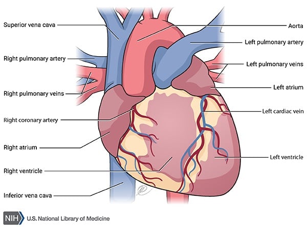
The heart
Surface anatomy
The heart extends from approximately the 2nd costal cartilage on the left and the 3rd costal cartilage on the right down to the 5th intercostal space on the left and the 6th right costal cartilage on the right. The apex lies in the left 5th intercostal space at the midclavicular line.
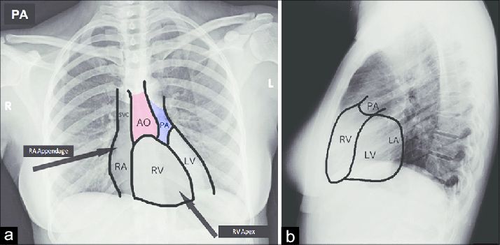
Borders of the heart
- Right border - Right atrium (extends from the 3rd to the 6th costal cartilages just to the right of the sternum)
- Left border - Left ventricle (extends from the 2nd left costal cartilage to the 5th left intercostal space)
- Apex - tip of the left ventricle
- Superior border - Right and left auricles and conus arteriosus of the right ventricle (extends from the 3rd right to the 2nd left costal cartilage)
- Inferior border - Mostly right ventricle, resting on the diaphragm (extends from the 6th right costal cartilage to the apex)
Surfaces of the heart
- Anterior surface - Right ventricle
- Posterior surface - Left atrium
- Diaphragmatic surface - Mainly left ventricle
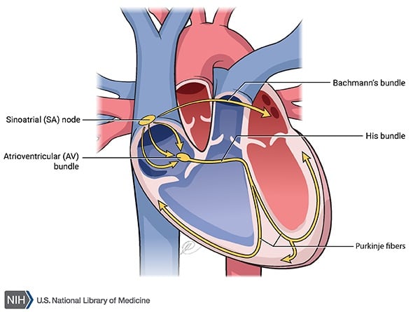
Conducting system
The SA node is located in the upper part of the crista terminalis (which runs from the SVC to the IVC) in the right atrium. The AV node is located in the interatrial septum near the opening of the coronary sinus.
Electrical impulses travel in this sequence:
- SA node → AV node
- AV node → Bundle of His
- Bundle of His → right and left bundle branches (one for each ventricle)
- Bundle branches → Purkinje fibres
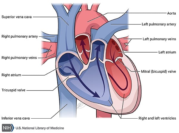
Cardiac valves
Tricuspid valve; Mitral or Bicuspid valve; Semilunar valves are Aortic and Pulmonary. Tricuspid and mitral are also called atrioventricular valves.
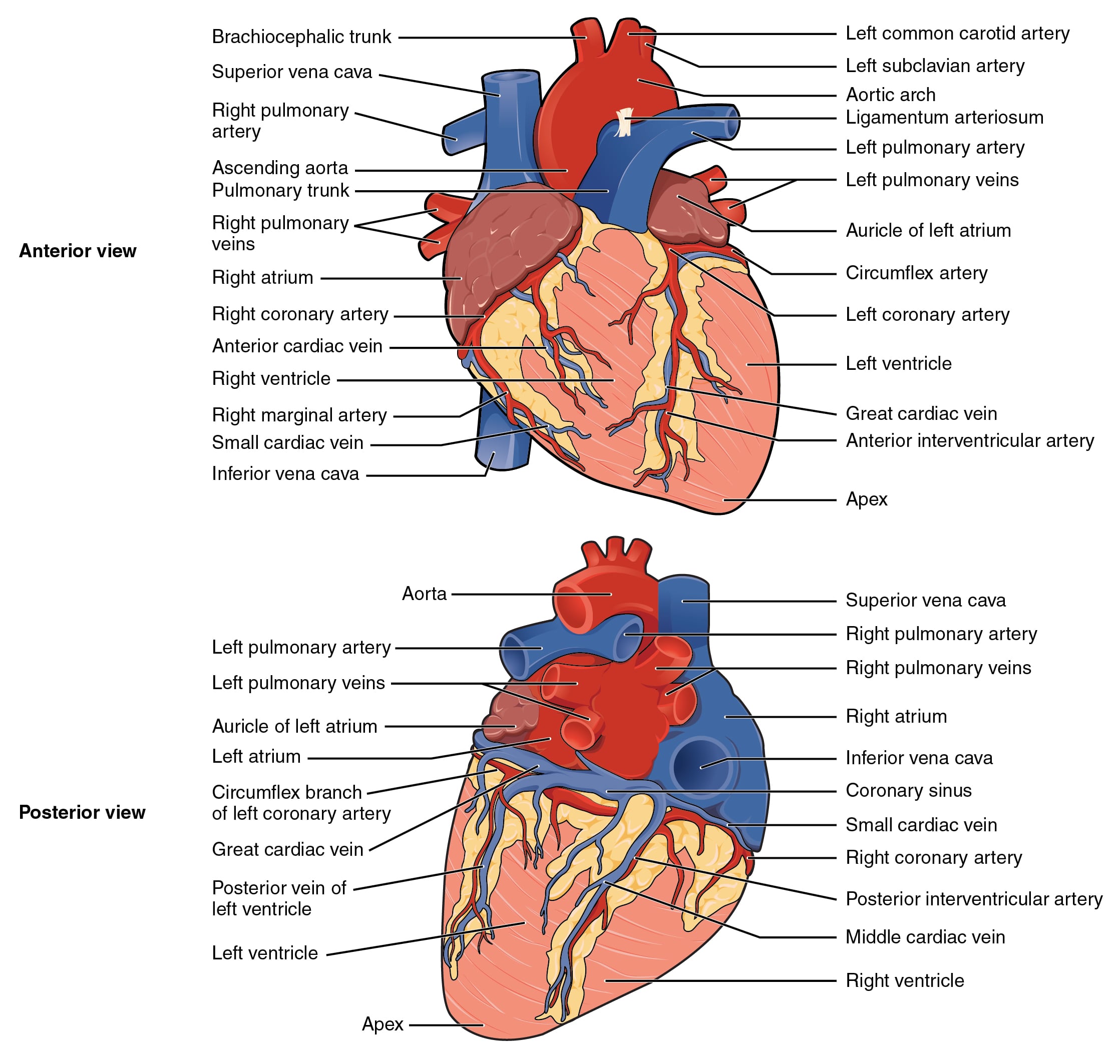
Blood supply of the heart
The heart receives its blood supply in diastole. The RCA (right coronary artery) and LCA (left coronary artery) arise from the right and left aortic sinuses of the ascending aorta, respectively.
-
RCA branches: courses in the coronary sulcus.
- SA nodal artery: supplies the SA node.
- AV nodal artery: supplies the AV node via the posterior interventricular artery.
- Posterior interventricular artery: lies in the posterior interventricular sulcus to supply parts of the right and left ventricles and the posterior interventricular septum.
-
LCA branches:
- LAD/ Left Anterior Descending / Anterior Interventricular Artery: most common site of coronary occlusion. Descends in the anterior interventricular sulcus and provides branches to the anterior part of the left ventricle, anterior two-thirds of the interventricular septum, Bundle of His, and apex of the heart.
- Left circumflex artery: via the marginal artery it supplies the left border of the heart. Also supplies the posterior inferior wall of the left ventricle.
Venous drainage of the heart
- Coronary sinus: lies in the posterior coronary sulcus and drains into the right atrium.
- Great cardiac vein: lies in the anterior interventricular sulcus with the LAD artery and drains into the coronary sinus.
- Middle cardiac vein: lies in the posterior interventricular sulcus and drains into the coronary sinus.
- Venae cordis minimae / Thebesian veins and anterior cardiac veins: open directly into the chambers of the heart.
Histology of the heart
The innermost layer of the heart is the endocardium, which is a lining of endothelium. The subendocardial space contains blood vessels. The endocardium is the last area of the heart to receive blood.
The middle layer is the myocardium, which is the muscle layer, and it also contains endocrine cells.
The epicardium is the outer layer and is also called the visceral pericardium. The coronary blood vessels lie on the epicardial layer.
Cardiac muscle cells are connected by intercalated discs. These cells branch and join to form a syncytium, which helps spread electrical impulses across the myocardium and allows the chambers to contract and relax as a single unit. Purkinje cells are modified myocytes specialized for conduction.
Intercalated discs comprise three special structures - fascia adherens, desmosomes, and gap junctions.
- The fascia adherens are bands of proteins that connect the actin filaments of the sarcomeres in each cardiac muscle fiber to the sarcomere in neighboring cells, producing a single unified chain of sarcomeres.
- Desmosomes help bind cardiac muscle cells together but form smaller, tighter junctions compared to the fascia adherens. Intermediate fibers inside each muscle fiber are connected by a series of proteins in the desmosome, which form an interlocking protein chain.
- Gap junctions form between cardiac muscle cells, allowing material to pass between cells. Each gap junction is made of connexin proteins that form a tunnel through the cell membranes of cardiac muscle cells, allowing small molecules, including ions, to pass through.