Blood vessels
Aorta and branches
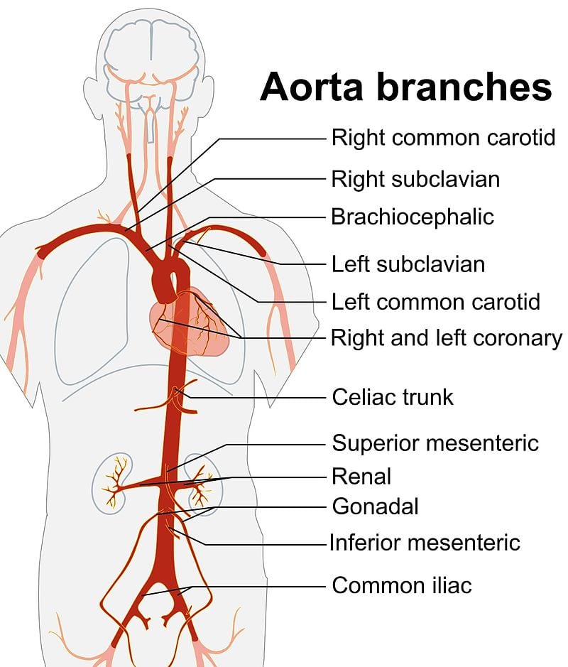
The aorta is the largest artery in the body. It has four major segments: the ascending aorta, the aortic arch, the thoracic (descending) aorta, and the abdominal aorta.
-
Ascending aorta: It arises from the left ventricle. The right and left coronary arteries originate from the right and left aortic sinuses, respectively.
-
Arch of aorta: The arch gives rise to several critical branches. There are three direct branches: the left common carotid artery, the left subclavian artery, and the brachiocephalic trunk (also called the innominate artery).
The brachiocephalic trunk divides into the right common carotid and right subclavian arteries.
The subclavian artery gives rise to four major branches: the vertebral artery, internal thoracic artery, thyrocervical trunk, and costocervical trunk. It then continues as the axillary artery.
The axillary artery continues as the brachial artery, which divides into the radial and ulnar arteries. These anastomose in the hand as the superficial and deep palmar arches.
The vertebral artery ascends to the base of the brain through the foramen magnum and joins the contralateral vertebral artery to form the basilar artery.
The common carotid artery divides into the external and internal carotid arteries. The external carotid artery ends by branching into the superficial temporal and maxillary arteries. The facial artery is a branch of the external carotid artery.
-
Thoracic aorta: It gives off visceral and parietal branches.
Visceral branches are pericardial, mediastinal, esophageal, and bronchial. There are two left bronchial arteries and one right bronchial artery. The left bronchial arteries arise from the thoracic aorta, while the right bronchial artery may arise from the thoracic aorta or the right third posterior intercostal artery.
Parietal branches are the posterior intercostal, subcostal, and superior phrenic arteries.
-
Abdominal aorta: The thoracic aorta becomes the abdominal aorta after passing through the aortic hiatus of the diaphragm. It gives rise to paired and unpaired branches as follows:
- Paired branches: Inferior phrenic, suprarenal, renal, gonadal, and lumbar arteries.
- Unpaired branches: Celiac trunk, superior mesenteric artery, inferior mesenteric artery, and median sacral artery.
The abdominal aorta bifurcates into two common iliac arteries, which further divide into internal and external iliac arteries. The femoral artery is a continuation of the external iliac artery.
Subclavian steal syndrome
Subclavian steal syndrome is characterized by retrograde flow in the vertebral artery due to subclavian artery stenosis or occlusion proximal to the origin of the vertebral artery. It is commonly caused by atherosclerosis and, more rarely, by Takayasu arteritis or giant cell arteritis.
It presents with upper limb claudication after exercising the upper limb and/or neurologic symptoms from relative cerebral ischemia, especially in the posterior circulation. Symptoms may include pre-syncope, dizziness, vertigo, blurry vision, and diplopia, characteristically following upper extremity exercise.
On examination:
- There is a blood pressure differential between both arms of more than 20 mmhg.
- The affected arm has weak or absent pulses.
- A bruit may be auscultated over the supraclavicular fossa.
Blood vessels histology
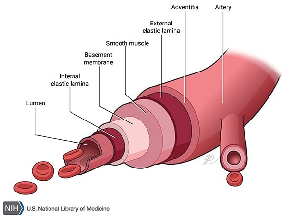
Blood vessel walls have three layers. From outside to inside, these are the tunica adventitia, tunica media, and tunica intima.
- The tunica adventitia contains type I collagen and is very tough.
- The tunica media contains smooth muscle cells and type III collagen.
- The tunica intima consists of the endothelial layer and secretes types I and IV collagen, among other things.
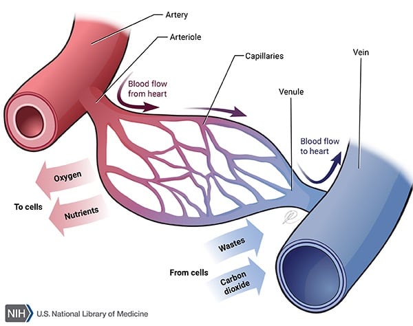
Capillaries are made of a single layer of endothelial cells surrounded by a basal lamina. They can be divided as follows:
- Continuous capillaries: They have zona occludens. Pores are absent. Seen in lung, muscle, brain (BB barrier).
- Fenestrated with diaphragms: They are made of endothelial cells joined together by fascia occludens, creating slit-like intercellular spaces, and contain fenestrae with diaphragms. Seen in endocrine glands, intestine, and kidney.
- Fenestrated without diaphragms: Seen in kidney glomerulus only.
- Discontinuous / sinusoids: They have a single layer of endothelial cells separated by wide gaps (no zona occludens). Seen in liver, spleen, bone marrow.