


This browser is too old to run Achievable.

Update it, or open this page in a current version of Chrome, Safari, Firefox or Edge. On an iPhone or iPad, update iOS: every browser there runs on the version of Safari that comes with it.

Achievable logoAchievable logo
NPTE-PTA
Sign in
Sign up
Purchase
Textbook
Practice exams
Support
How it works
Resources
Exam catalog
Mountain with a flag at the peak
Textbook
Introduction
1. Cardiovascular system
2. Pulmonary system
3. Neuromuscular system
4. Pediatrics
5. Musculoskeletal system
5.1 Anatomy of musculoskeletal system
5.2 Anatomical terminology and exercise training principles
5.3 Joint mechanics and phases of healing
5.4 Upper extremity anatomy
5.5 Special tests of upper extremity
5.6 Comparing clinical presentation and interventions for upper extremity
5.7 Lower extremity anatomy
5.8 Special tests of lower extremity
5.9 Comparing clinical presentation and interventions for lower extremity
5.9.1 Hip and knee pathologies
5.9.2 Lower leg, ankle, and foot pathologies
5.10 Spine and pelvis anatomy
5.11 Special tests of the spine, pelvis, and temporomandibular joint
5.12 Comparing clinical presentation and interventions for the spine, pelvis, and temporomandibular joint
5.13 Other musculoskeletal conditions
5.14 Gait
5.15 Prosthetics
5.16 Orthotics
5.17 Medications, imaging, and fractures
5.18 Surgical protocols
6. Other systems
7. Non-systems
Wrapping up
Achievable logoAchievable logo
5.9.1 Hip and knee pathologies
Achievable NPTE-PTA
5. Musculoskeletal system
5.9. Comparing clinical presentation and interventions for lower extremity
Our NPTE-PTA course is now in "early access" - get 50% off for a limited time.

Hip and knee pathologies

9 min read
Font
Discuss
Share
Feedback

Hip conditions

Acetaminophen and non-steroidal anti-inflammatory drugs (NSAIDs) are standard first-line analgesic/anti-inflammatory management across most of the conditions in this chapter. This is noted here rather than repeated in every condition’s medical management list; additional or different medical management is called out where it applies.

Avascular necrosis

  • Etiologies that result in impaired or loss of blood supply to the femoral head

  • Symptoms

    • Loss of range of motion in hip flexion, internal rotation, and abduction
    • Pain in the groin and/or thigh
    • Tenderness with palpation at the hip joint
    • Antalgic gait
  • Diagnosis

    • Clinical presentation
    • X-ray
    • Bone scans
    • CT or MRI imaging (MRI detects early bone changes that may not yet show on X-ray)
  • Medical management

    • Surgery: core decompression or bone grafting in early stages; total hip replacement once the femoral head has collapsed
  • Physical therapy management

    • Joint protection
    • Functional mobility training
    • Improve joint mechanics and mobility
    • Improve strength/coordination

Hip labral tear

  • A hip labral tear commonly causes anterior hip or groin pain along with mechanical symptoms such as clicking, locking, or catching of the hip, unlike the stiffness and capsular pattern of osteoarthritis

Trochanteric bursitis

  • Inflammation of the deep trochanteric bursa due to direct injury, irritation by the iliotibial band, or repetitive microtrauma

    • Can be associated with rheumatoid arthritis
  • Symptoms

    • Sharp or dull ache on the outside of the hip
    • Pain that radiates down the upper thigh
    • Pain that worsens with activities that involve the hip, such as walking, running, or climbing stairs
    • Tenderness at the lateral hip
  • Diagnosis

    • Clinical presentation
  • Medical management

    • Management of rheumatoid arthritis as indicated
    • A glucocorticoid (corticosteroid) injection may be considered for bursitis that is persistent when oral medications and rest are inadequate
  • Physical therapy management

    • Interventions matched to the phase of healing (acute vs subacute vs chronic), as set by the PT in the plan of care

Proximal hamstring tendinopathy

  • Proximal hamstring tendinopathy causes deep pain at the ischial tuberosity (sit bone) that is worse with running, prolonged sitting, and hip flexion with the knee extended

Iliotibial band tightness

  • Tight iliotibial band, abnormal gait pattern

    • Can lead to trochanteric bursitis
    • Can lead to increased lateral tracking of the patella
  • Symptoms

    • Pain that worsens with activity, especially running, cycling, or going up and down stairs
    • Lateral knee pain that is worst near 30 degrees of knee flexion, where the IT band slides over the lateral femoral condyle
    • Swelling on the outside of the knee
    • Tenderness to the touch on the outside of the knee
  • Diagnosis

    • Clinical presentation
      • Special tests- Ober’s and Noble’s tests
  • Medical management

    • Treatment of trochanteric bursitis as appropriate
  • Physical therapy management

    • Stretching of the IT band
    • Hip abductor strengthening is a core part of rehabilitation: stretching and activity modification address the early phase, and strengthening the gluteus medius and other abductors becomes the main long-term goal
    • Gait training- appropriate running shoes, orthotics may be prescribed
    • Reduction of pain/inflammation
    • Soft tissue and manual techniques
    • Joint mobility and functional training

Key distinction: Trochanteric bursitis causes lateral hip pain with point tenderness over the greater trochanter. IT band tightness causes lateral knee pain that’s reproduced by Ober’s or Noble’s test and worsens with running, cycling, or stairs.

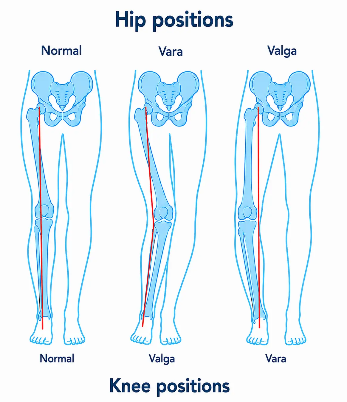
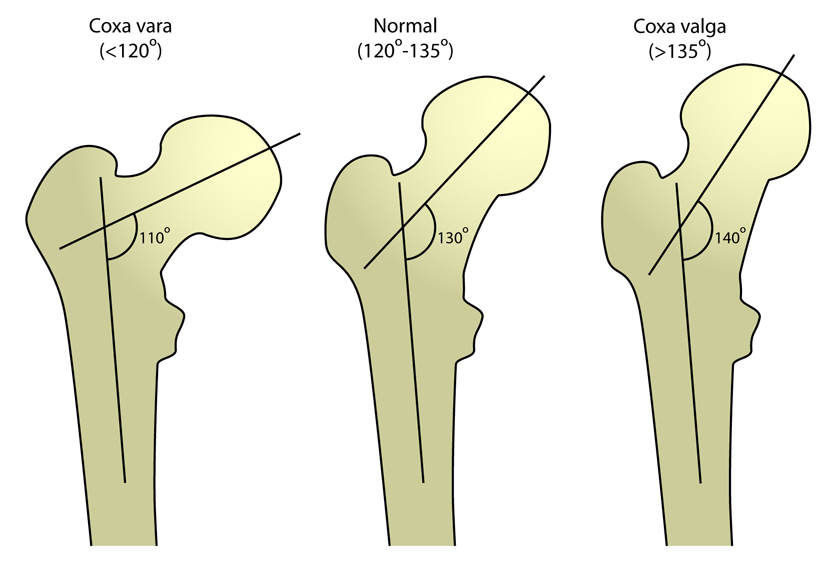
Coxa vara and coxa valga

  • Caused by a defect in the ossification (formation) of the femoral neck; it can also be a result of avascular necrosis due to septic arthritis

  • The position of the knee will be the direct opposite of the hip

    • Coxa vara- angle of femoral neck <115 degrees, knee in valgus
    • Coxa valga- angle of femoral neck >125 degrees, knee in varus
  • Symptoms

    • Coxa vara
      • Leg length discrepancy
      • Pain in the hip and/or leg
      • Stiffness with abduction
      • Prominent greater trochanter
      • Limited hip mobility
    • Coxa valga
      • Increased anterior pelvic tilt
  • Diagnosis

    • Clinical presentation
    • X-ray
  • Medical management

    • Surgery (corrective osteotomy) if the deformity is severe or progressive (e.g., Hilgenreiner-epiphyseal angle >60 degrees in developmental coxa vara)
    • Assistive devices as indicated
  • Physical therapy management

    • Improve joint mobility and mechanics
    • Use of orthotics for leg length discrepancy
    • Use of muscle energy techniques to correct pelvic tilt
Normal, vara, and valga hip and knee positions.
Hip and knee positions
Achievable
Angle of inclination of hip
Behrang Amini, MD/PhD
/
Wikimedia Commons
/
CC BY-SA 3.0

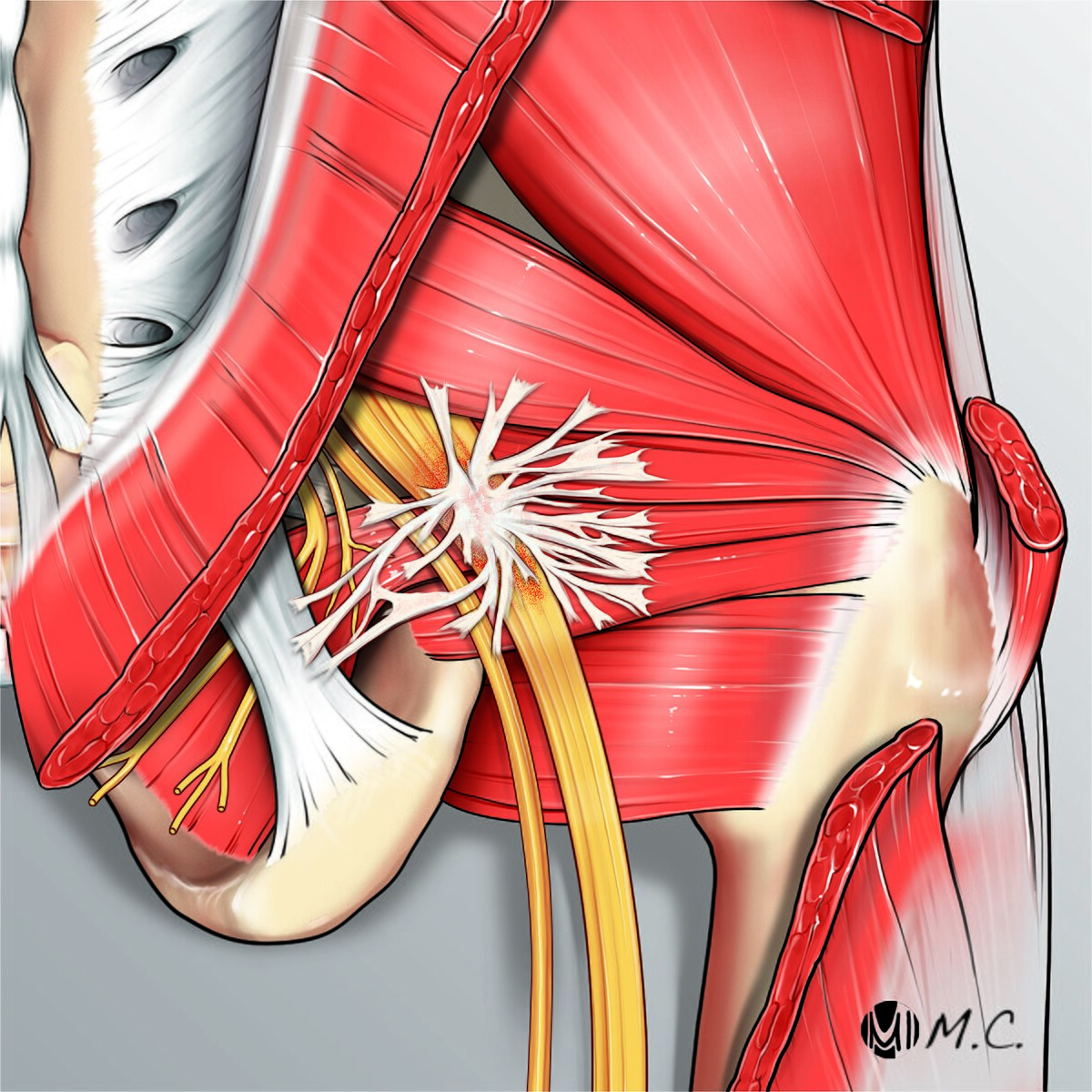
Piriformis syndrome

  • Tightness or spasm of the piriformis can result in compression of the sciatic nerve

    • Tightness can be a result of repetitive movement of the piriformis or overuse
    • Compression of the sciatic nerve can cause radiation of pain down the leg
  • Symptoms

    • Restriction in internal rotation
    • Pain with palpation of the piriformis muscle
    • Referred pain to the lower posterior leg
    • Weakness of external rotation
  • Diagnosis

    • Clinical presentation
      • Special test- piriformis test
      • Rule out lumbar spine involvement
    • Electrodiagnostics of the sciatic nerve
  • Physical therapy management

    • Pain management and reduction of pain from the posterior leg to only the piriformis (centralization of pain)
    • Muscle mobility and strengthening
    • Muscle balance restoration
    • Correction of biomechanical faults with orthoses
Sciatic nerve entrapment
Massimiliano Crespi, This work was commissioned privately to be used on wikipedia
/
Wikimedia Commons
/
CC0 1.0

Knee conditions

Ligament sprains

  • Can involve one (single plane) or multiple (rotary instability) ligaments of the knee

    • Anterior cruciate ligament
    • Posterior cruciate ligament
    • Medial collateral ligament
    • Lateral collateral ligament
  • Classification of sprains

    • First degree: minimal instability of the knee joint; ligament stretched but not torn
    • Second degree: minimal- moderate instability of the knee joint; ligament is partially torn
    • Third degree: extreme instability; ligament is completely torn and ruptured
  • Symptoms

    • First degree: minimal pain and swelling
    • Second degree: moderate pain, swelling, tenderness to the joint, and joint laxity noted with certain movements
    • Third degree: significant pain, swelling, and instability due to a complete tear
  • Diagnosis

    • Clinical presentation
      • Special tests based on ligament injury
        • Anterior cruciate ligament - Lachman, anterior drawer
        • Posterior cruciate ligament- posterior drawer test
        • Medial collateral ligament- valgus knee test
        • Lateral collateral ligament - varus knee test
      • MRI
  • Medical management

    • Surgery is indicated if functional instability is present, which can be for grade two or three sprains
  • Physical therapy management

    • If surgery is performed, interventions will be based on protocols post-surgery
    • Landing technique: to land safely from a jump, keep the knees bent, the chest high and the buttocks back, and land softly rather than stiff-legged
    • Reduction of pain and inflammation
    • Functional training
    • Joint mobility

Meniscus injuries

  • A combination of flexion, compression, and rotary forces on the knee, causing abnormal stress to the knee

  • Symptoms

    • Pain increases with twisting or weight-bearing activities
    • A sensation of the knee giving way or locking up, especially when squatting, standing up, or turning
    • A feeling that the knee is not stable or may give out
  • Diagnosis

    • Clinical presentation
      • Special tests- McMurray, Apley, Thessaly
    • MRI
  • Medical management

    • Surgery may be indicated
  • Physical therapy management

    • Reduction of pain and inflammation
    • Functional training
    • Joint mobility

Patellofemoral pain syndrome

  • Dysfunction of the knee due to trauma, muscle imbalance/tightness, improper loading of joints

  • Symptoms

    • Pain in the front of the knee, around or behind the kneecap
    • Pain when going up or down stairs
    • Pain when squatting or kneeling
    • Pain after prolonged sitting with bent knees
    • A grinding or catching sensation in the knee
  • Diagnosis

    • Clinical presentation- description of pain when increased load to the patella, such as stair climbing or squatting
  • Physical therapy management

    • Reduction of pain and inflammation
    • Taping; patellar mobilization (performed by the PT)
    • Functional training
    • Biofeedback for vastus medialis
    • Prefabricated foot orthoses (selected by the PT) for short-term pain relief in patients with excessive foot pronation, combined with exercise

Patellar tendinopathy

  • Degeneration of the patellar tendon due to overload and/or jumping-related activities

    • Can be related to patellofemoral pain syndrome
  • Symptoms

    • Pain that’s worse with activity, such as running, jumping, or walking
    • Pain that’s worse when going downhill or descending stairs
    • Pain that’s worse with knee flexion or extension
    • Tenderness on the front of the knee
    • Tenderness at the lower pole of the kneecap, where the patellar tendon attaches
  • Diagnosis

    • Clinical presentation
      • Pain localized to the patellar tendon at the lower pole of the kneecap, provoked by tendon loading such as a single-leg decline squat
    • Ultrasound or MRI, often to rule out other causes such as patellofemoral pain
  • Medical management

    • Exercise-based (tendon loading) rehabilitation first; injections or surgery (such as arthroscopic debridement) only for cases that don’t respond
  • Physical therapy management

    • Improve lower extremity strength- specifically quadriceps and hamstrings
    • Functional training
    • Joint mobility
    • Patella taping or bracing

Patellar instability

  • Patellar instability causes a feeling that the kneecap will dislocate (apprehension) or actual dislocations; a first-time dislocation is usually treated with exercises to strengthen the thigh muscles and a stabilizing brace

Pes anserine bursitis

  • Pes anserine bursitis causes tenderness over the proximal medial tibia, below the knee joint line, where the sartorius, gracilis, and semitendinosus tendons insert, and it is aggravated by stair climbing and rising from a seated position

Key distinction: Patellofemoral pain syndrome causes diffuse anterior knee pain that’s worse with stair climbing, squatting, or prolonged sitting with bent knees. Patellar tendinopathy causes localized tenderness at the patellar tendon (below the kneecap) that’s worse with jumping, running, or descending stairs.

Avascular necrosis (femoral head)

  • Impaired blood supply to femoral head; causes groin/thigh pain, antalgic gait, limited flexion/IR/abduction
  • Diagnosed via X-ray, bone scan, MRI (MRI catches early changes)
  • Early: core decompression/bone graft; late (collapsed head): total hip replacement
  • PT: joint protection, mobility, strength/coordination

Trochanteric bursitis vs. IT band tightness

  • Bursitis: inflammation of trochanteric bursa (direct injury, IT band irritation, overuse, or RA); lateral hip pain, radiates down thigh, point tenderness over greater trochanter
  • IT band tightness: can cause bursitis and lateral patellar tracking issues; lateral knee pain worsened by running/cycling/stairs, positive Ober’s/Noble’s tests
  • PT for IT band: stretching, gait retraining/orthotics, soft tissue/manual therapy
  • Key distinction: bursitis = lateral hip pain + trochanter tenderness; IT band = lateral knee pain + positive Ober’s/Noble’s

Coxa vara/valga

  • Femoral neck ossification defect or AVN; knee position opposite of hip deformity
    • Coxa vara: neck angle <115°, knee valgus; leg length discrepancy, abduction stiffness, prominent trochanter
    • Coxa valga: neck angle >125°, knee varus; increased anterior pelvic tilt
  • Diagnosis: X-ray; severe/progressive cases (HEA >60°) may need corrective osteotomy
  • PT: joint mobility, orthotics for leg length, muscle energy techniques for pelvic tilt

Piriformis syndrome

  • Piriformis tightness/spasm compresses sciatic nerve → referred posterior leg pain
  • Restricted IR, weak ER, tender piriformis; positive piriformis test; must rule out lumbar spine
  • PT goal: centralize pain to piriformis, restore muscle balance/strength, correct biomechanics with orthoses

Knee ligament sprains

  • Graded 1st (stretch) to 3rd degree (complete tear/instability)
  • Special tests by ligament: ACL (Lachman, anterior drawer), PCL (posterior drawer), MCL (valgus test), LCL (varus test); confirm with MRI
  • Surgery considered for grade 2–3 with functional instability; PT follows post-op protocols, focuses on pain control, mobility, functional training

Meniscus injuries

  • Caused by combined flexion/compression/rotation forces
  • Symptoms: pain with twisting/weight-bearing, locking/giving way sensation
  • Special tests: McMurray, Apley, Thessaly; confirm with MRI
  • Management: possible surgery; PT addresses pain, mobility, functional training

Patellofemoral pain syndrome vs. patellar tendinopathy

  • PFPS: muscle imbalance/overload causes diffuse anterior knee pain, worse with stairs/squatting/prolonged sitting; treat with taping, patellar mobilization (by the PT), VMO biofeedback, orthotics
  • Patellar tendinopathy: tendon degeneration from overload/jumping; localized tenderness at inferior patellar pole, worse with jumping/running/downhill/stairs; provoked by single-leg decline squat; ultrasound/MRI mainly to rule out other causes
    • Medical management: exercise-based tendon loading first-line; injections/surgery only if unresponsive
  • PT for tendinopathy: strengthen quads/hamstrings, functional training, taping/bracing
  • Key distinction: PFPS = diffuse pain worsened by sitting/squatting; tendinopathy = localized tendon tenderness worsened by jumping/running

Sign up for free to take 19 quiz questions on this topic

Previous
Next  | 5.9.2 Lower leg, ankle, and foot pathologies
All rights reserved ©2016 - 2026 Achievable, Inc.

Hip and knee pathologies

Hip conditions

Acetaminophen and non-steroidal anti-inflammatory drugs (NSAIDs) are standard first-line analgesic/anti-inflammatory management across most of the conditions in this chapter. This is noted here rather than repeated in every condition’s medical management list; additional or different medical management is called out where it applies.

Avascular necrosis

  • Etiologies that result in impaired or loss of blood supply to the femoral head

  • Symptoms

    • Loss of range of motion in hip flexion, internal rotation, and abduction
    • Pain in the groin and/or thigh
    • Tenderness with palpation at the hip joint
    • Antalgic gait
  • Diagnosis

    • Clinical presentation
    • X-ray
    • Bone scans
    • CT or MRI imaging (MRI detects early bone changes that may not yet show on X-ray)
  • Medical management

    • Surgery: core decompression or bone grafting in early stages; total hip replacement once the femoral head has collapsed
  • Physical therapy management

    • Joint protection
    • Functional mobility training
    • Improve joint mechanics and mobility
    • Improve strength/coordination

Hip labral tear

  • A hip labral tear commonly causes anterior hip or groin pain along with mechanical symptoms such as clicking, locking, or catching of the hip, unlike the stiffness and capsular pattern of osteoarthritis

Trochanteric bursitis

  • Inflammation of the deep trochanteric bursa due to direct injury, irritation by the iliotibial band, or repetitive microtrauma

    • Can be associated with rheumatoid arthritis
  • Symptoms

    • Sharp or dull ache on the outside of the hip
    • Pain that radiates down the upper thigh
    • Pain that worsens with activities that involve the hip, such as walking, running, or climbing stairs
    • Tenderness at the lateral hip
  • Diagnosis

    • Clinical presentation
  • Medical management

    • Management of rheumatoid arthritis as indicated
    • A glucocorticoid (corticosteroid) injection may be considered for bursitis that is persistent when oral medications and rest are inadequate
  • Physical therapy management

    • Interventions matched to the phase of healing (acute vs subacute vs chronic), as set by the PT in the plan of care

Proximal hamstring tendinopathy

  • Proximal hamstring tendinopathy causes deep pain at the ischial tuberosity (sit bone) that is worse with running, prolonged sitting, and hip flexion with the knee extended

Iliotibial band tightness

  • Tight iliotibial band, abnormal gait pattern

    • Can lead to trochanteric bursitis
    • Can lead to increased lateral tracking of the patella
  • Symptoms

    • Pain that worsens with activity, especially running, cycling, or going up and down stairs
    • Lateral knee pain that is worst near 30 degrees of knee flexion, where the IT band slides over the lateral femoral condyle
    • Swelling on the outside of the knee
    • Tenderness to the touch on the outside of the knee
  • Diagnosis

    • Clinical presentation
      • Special tests- Ober’s and Noble’s tests
  • Medical management

    • Treatment of trochanteric bursitis as appropriate
  • Physical therapy management

    • Stretching of the IT band
    • Hip abductor strengthening is a core part of rehabilitation: stretching and activity modification address the early phase, and strengthening the gluteus medius and other abductors becomes the main long-term goal
    • Gait training- appropriate running shoes, orthotics may be prescribed
    • Reduction of pain/inflammation
    • Soft tissue and manual techniques
    • Joint mobility and functional training

Key distinction: Trochanteric bursitis causes lateral hip pain with point tenderness over the greater trochanter. IT band tightness causes lateral knee pain that’s reproduced by Ober’s or Noble’s test and worsens with running, cycling, or stairs.

Coxa vara and coxa valga

  • Caused by a defect in the ossification (formation) of the femoral neck; it can also be a result of avascular necrosis due to septic arthritis

  • The position of the knee will be the direct opposite of the hip

    • Coxa vara- angle of femoral neck <115 degrees, knee in valgus
    • Coxa valga- angle of femoral neck >125 degrees, knee in varus
  • Symptoms

    • Coxa vara
      • Leg length discrepancy
      • Pain in the hip and/or leg
      • Stiffness with abduction
      • Prominent greater trochanter
      • Limited hip mobility
    • Coxa valga
      • Increased anterior pelvic tilt
  • Diagnosis

    • Clinical presentation
    • X-ray
  • Medical management

    • Surgery (corrective osteotomy) if the deformity is severe or progressive (e.g., Hilgenreiner-epiphyseal angle >60 degrees in developmental coxa vara)
    • Assistive devices as indicated
  • Physical therapy management

    • Improve joint mobility and mechanics
    • Use of orthotics for leg length discrepancy
    • Use of muscle energy techniques to correct pelvic tilt

Piriformis syndrome

  • Tightness or spasm of the piriformis can result in compression of the sciatic nerve

    • Tightness can be a result of repetitive movement of the piriformis or overuse
    • Compression of the sciatic nerve can cause radiation of pain down the leg
  • Symptoms

    • Restriction in internal rotation
    • Pain with palpation of the piriformis muscle
    • Referred pain to the lower posterior leg
    • Weakness of external rotation
  • Diagnosis

    • Clinical presentation
      • Special test- piriformis test
      • Rule out lumbar spine involvement
    • Electrodiagnostics of the sciatic nerve
  • Physical therapy management

    • Pain management and reduction of pain from the posterior leg to only the piriformis (centralization of pain)
    • Muscle mobility and strengthening
    • Muscle balance restoration
    • Correction of biomechanical faults with orthoses

Knee conditions

Ligament sprains

  • Can involve one (single plane) or multiple (rotary instability) ligaments of the knee

    • Anterior cruciate ligament
    • Posterior cruciate ligament
    • Medial collateral ligament
    • Lateral collateral ligament
  • Classification of sprains

    • First degree: minimal instability of the knee joint; ligament stretched but not torn
    • Second degree: minimal- moderate instability of the knee joint; ligament is partially torn
    • Third degree: extreme instability; ligament is completely torn and ruptured
  • Symptoms

    • First degree: minimal pain and swelling
    • Second degree: moderate pain, swelling, tenderness to the joint, and joint laxity noted with certain movements
    • Third degree: significant pain, swelling, and instability due to a complete tear
  • Diagnosis

    • Clinical presentation
      • Special tests based on ligament injury
        • Anterior cruciate ligament - Lachman, anterior drawer
        • Posterior cruciate ligament- posterior drawer test
        • Medial collateral ligament- valgus knee test
        • Lateral collateral ligament - varus knee test
      • MRI
  • Medical management

    • Surgery is indicated if functional instability is present, which can be for grade two or three sprains
  • Physical therapy management

    • If surgery is performed, interventions will be based on protocols post-surgery
    • Landing technique: to land safely from a jump, keep the knees bent, the chest high and the buttocks back, and land softly rather than stiff-legged
    • Reduction of pain and inflammation
    • Functional training
    • Joint mobility

Meniscus injuries

  • A combination of flexion, compression, and rotary forces on the knee, causing abnormal stress to the knee

  • Symptoms

    • Pain increases with twisting or weight-bearing activities
    • A sensation of the knee giving way or locking up, especially when squatting, standing up, or turning
    • A feeling that the knee is not stable or may give out
  • Diagnosis

    • Clinical presentation
      • Special tests- McMurray, Apley, Thessaly
    • MRI
  • Medical management

    • Surgery may be indicated
  • Physical therapy management

    • Reduction of pain and inflammation
    • Functional training
    • Joint mobility

Patellofemoral pain syndrome

  • Dysfunction of the knee due to trauma, muscle imbalance/tightness, improper loading of joints

  • Symptoms

    • Pain in the front of the knee, around or behind the kneecap
    • Pain when going up or down stairs
    • Pain when squatting or kneeling
    • Pain after prolonged sitting with bent knees
    • A grinding or catching sensation in the knee
  • Diagnosis

    • Clinical presentation- description of pain when increased load to the patella, such as stair climbing or squatting
  • Physical therapy management

    • Reduction of pain and inflammation
    • Taping; patellar mobilization (performed by the PT)
    • Functional training
    • Biofeedback for vastus medialis
    • Prefabricated foot orthoses (selected by the PT) for short-term pain relief in patients with excessive foot pronation, combined with exercise

Patellar tendinopathy

  • Degeneration of the patellar tendon due to overload and/or jumping-related activities

    • Can be related to patellofemoral pain syndrome
  • Symptoms

    • Pain that’s worse with activity, such as running, jumping, or walking
    • Pain that’s worse when going downhill or descending stairs
    • Pain that’s worse with knee flexion or extension
    • Tenderness on the front of the knee
    • Tenderness at the lower pole of the kneecap, where the patellar tendon attaches
  • Diagnosis

    • Clinical presentation
      • Pain localized to the patellar tendon at the lower pole of the kneecap, provoked by tendon loading such as a single-leg decline squat
    • Ultrasound or MRI, often to rule out other causes such as patellofemoral pain
  • Medical management

    • Exercise-based (tendon loading) rehabilitation first; injections or surgery (such as arthroscopic debridement) only for cases that don’t respond
  • Physical therapy management

    • Improve lower extremity strength- specifically quadriceps and hamstrings
    • Functional training
    • Joint mobility
    • Patella taping or bracing

Patellar instability

  • Patellar instability causes a feeling that the kneecap will dislocate (apprehension) or actual dislocations; a first-time dislocation is usually treated with exercises to strengthen the thigh muscles and a stabilizing brace

Pes anserine bursitis

  • Pes anserine bursitis causes tenderness over the proximal medial tibia, below the knee joint line, where the sartorius, gracilis, and semitendinosus tendons insert, and it is aggravated by stair climbing and rising from a seated position

Key distinction: Patellofemoral pain syndrome causes diffuse anterior knee pain that’s worse with stair climbing, squatting, or prolonged sitting with bent knees. Patellar tendinopathy causes localized tenderness at the patellar tendon (below the kneecap) that’s worse with jumping, running, or descending stairs.

Key points

Avascular necrosis (femoral head)

  • Impaired blood supply to femoral head; causes groin/thigh pain, antalgic gait, limited flexion/IR/abduction
  • Diagnosed via X-ray, bone scan, MRI (MRI catches early changes)
  • Early: core decompression/bone graft; late (collapsed head): total hip replacement
  • PT: joint protection, mobility, strength/coordination

Trochanteric bursitis vs. IT band tightness

  • Bursitis: inflammation of trochanteric bursa (direct injury, IT band irritation, overuse, or RA); lateral hip pain, radiates down thigh, point tenderness over greater trochanter
  • IT band tightness: can cause bursitis and lateral patellar tracking issues; lateral knee pain worsened by running/cycling/stairs, positive Ober’s/Noble’s tests
  • PT for IT band: stretching, gait retraining/orthotics, soft tissue/manual therapy
  • Key distinction: bursitis = lateral hip pain + trochanter tenderness; IT band = lateral knee pain + positive Ober’s/Noble’s

Coxa vara/valga

  • Femoral neck ossification defect or AVN; knee position opposite of hip deformity
    • Coxa vara: neck angle <115°, knee valgus; leg length discrepancy, abduction stiffness, prominent trochanter
    • Coxa valga: neck angle >125°, knee varus; increased anterior pelvic tilt
  • Diagnosis: X-ray; severe/progressive cases (HEA >60°) may need corrective osteotomy
  • PT: joint mobility, orthotics for leg length, muscle energy techniques for pelvic tilt

Piriformis syndrome

  • Piriformis tightness/spasm compresses sciatic nerve → referred posterior leg pain
  • Restricted IR, weak ER, tender piriformis; positive piriformis test; must rule out lumbar spine
  • PT goal: centralize pain to piriformis, restore muscle balance/strength, correct biomechanics with orthoses

Knee ligament sprains

  • Graded 1st (stretch) to 3rd degree (complete tear/instability)
  • Special tests by ligament: ACL (Lachman, anterior drawer), PCL (posterior drawer), MCL (valgus test), LCL (varus test); confirm with MRI
  • Surgery considered for grade 2–3 with functional instability; PT follows post-op protocols, focuses on pain control, mobility, functional training

Meniscus injuries

  • Caused by combined flexion/compression/rotation forces
  • Symptoms: pain with twisting/weight-bearing, locking/giving way sensation
  • Special tests: McMurray, Apley, Thessaly; confirm with MRI
  • Management: possible surgery; PT addresses pain, mobility, functional training

Patellofemoral pain syndrome vs. patellar tendinopathy

  • PFPS: muscle imbalance/overload causes diffuse anterior knee pain, worse with stairs/squatting/prolonged sitting; treat with taping, patellar mobilization (by the PT), VMO biofeedback, orthotics
  • Patellar tendinopathy: tendon degeneration from overload/jumping; localized tenderness at inferior patellar pole, worse with jumping/running/downhill/stairs; provoked by single-leg decline squat; ultrasound/MRI mainly to rule out other causes
    • Medical management: exercise-based tendon loading first-line; injections/surgery only if unresponsive
  • PT for tendinopathy: strengthen quads/hamstrings, functional training, taping/bracing
  • Key distinction: PFPS = diffuse pain worsened by sitting/squatting; tendinopathy = localized tendon tenderness worsened by jumping/running

More from Comparing clinical presentation and interventions for lower extremity

  • Lower leg, ankle, and foot pathologies