



Shoulder soft tissue conditions
Glenohumeral dislocation and instability
- Dislocations of the glenohumeral joint caused by traumatic or atraumatic reasons
- Trauma is due to direct injury, most commonly a fall on an outstretched hand (FOOSH) mechanism of injury
- Atraumatic can be due to repetitive injury, causing hypermobility
- Types of dislocations:
- Anterior-inferior dislocations: Most common type (95%). Mechanism is a combination of motions including: excessive horizontal abduction, abduction, external (lateral) rotation, and extension/hyperextension of the upper extremity.
- If traumatic can lead to: Disruption anterior glenohumeral/capsular ligament, subscapularis, and anterior/inferior glenoid labrum
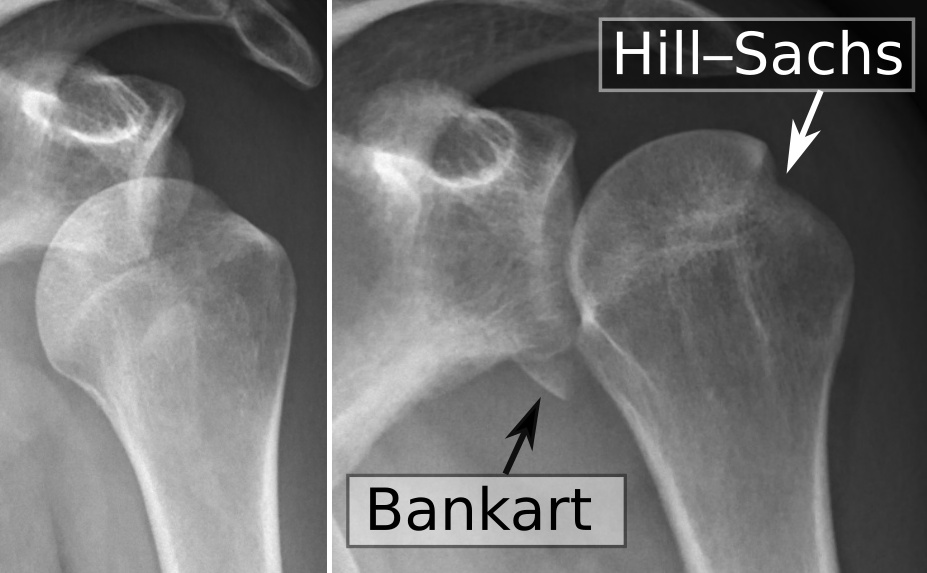
- Hill-Sachs lesion: compression fracture of the posterior humeral head
- Bankart lesion: avulsion of the anterior-inferior glenoid labrum
- Axillary nerve injury: numbness, tingling, and weakness in the deltoid
- If traumatic can lead to: Disruption anterior glenohumeral/capsular ligament, subscapularis, and anterior/inferior glenoid labrum
- Posterior dislocations: rare, caused by horizontal adduction and internal rotation
- Anterior-inferior dislocations: Most common type (95%). Mechanism is a combination of motions including: excessive horizontal abduction, abduction, external (lateral) rotation, and extension/hyperextension of the upper extremity.
- Symptoms
- Popping during movements
- Repeated dislocations or subluxations of the glenohumeral joint
- Diagnosis
- Clinical presentation
- Special tests - apprehension test (highly specific for anterior instability); sulcus sign (positive for inferior instability)
- X-ray
- Physical therapy interventions
- Strengthening the rotator cuff and scapular stabilizers (dynamic stabilizers) to prevent recurrent dislocation
- Functional training
- Avoidance of apprehension position
- Avoid combined abduction/external rotation after an anterior stabilization procedure, per protocol
- Surgical protocols if indicated
- With anticipated return to function in 3-4 months
Labral tears
-
Tear in the cartilage ring that surrounds the shoulder joint; divided into above the middle of the socket and below the middle of the socket
- Above the middle of the socket is called a SLAP (superior labral anterior-posterior) tear; can also involve the biceps tendon
- Below the middle of the socket: Bankart lesion (see glenohumeral dislocation and instability above); may also include a bony Bankart (avulsion fracture of the anterior-inferior glenoid rim)
-
Labral tears are associated with traumatic injury or repetitive shoulder dislocations
-
Symptoms:
- Pain increased with overhead movement or behind the back
- Shoulder weakness
- Shoulder instability/dislocation
- Pain with resisted flexion of the biceps
- Tenderness over the anterior shoulder
-
Diagnosis
- Clinical presentation
- MRI
- Arthroscopic surgery - gold standard for diagnosis
-
Physical therapy interventions
- Focus on return to function without pain
- Correction of muscle imbalances
- Address underlying causes of labral tears
- Surgical protocols if indicated
- With anticipated return to function in 3-4 months
Rotator cuff tendonitis and impingement syndrome
- Caused by mechanical impingement of the distal attachment of the rotator cuff, causing inflammation of the tendons
- Increased risk for development of tendonitis due to poor vascularity at attachment sites
- Inadequate scapular upward rotation and posterior tilt during arm elevation reduce the subacromial space and contribute to impingement
- Symptoms
- Pain in the anterior and superior aspect of the shoulder
- Pain and weakness that worsen with overhead movements, such as reaching, throwing, or lifting objects
- Pain that may radiate down the arm
- Pain that is worse at night
- Diagnosis
- Clinical presentation
- Special tests
- Neer test - forced forward flexion with the scapula stabilized compresses the supraspinatus against the anteroinferior acromion
- Hawkins-Kennedy test - shoulder flexed to 90 degrees with internal rotation compresses the supraspinatus under the coracoacromial ligament
- Painful arc
- Shoulder pain that occurs when abducting an arm between 60 and 120 degrees.
- MRI
- Medical management
- Surgical (subacromial) decompression if indicated
- Physical therapy intervention
- Avoidance of shoulder elevation greater than 90 degrees to avoid subacromial compression - acute phase
- Postural re-education - improve forward head, rounded shoulders, and kyphotic posture (upper cross syndrome)
- Correction of muscle imbalances
- Improve joint mobility
- Return to previous functional status
- Follow the post-operative protocol after subacromial decompression if indicated
Rotator cuff tear
-
Acute tears are caused by a fall on an outstretched hand (FOOSH), sudden heavy lifting, or loading the shoulder in an awkward position
-
Degenerative or partial tears develop over time from chronic tendonitis and poor blood supply at the tendon insertion
-
Symptoms
- Pain when raising or lowering the arm
- Weakness when lifting or rotating the arm
- Pain when reaching behind the back
- Pain that worsens after lying down on the affected side
- Pain that is worse at night
-
Diagnosis
- Clinical presentation
- Special tests - empty can test, drop arm test, manual muscle testing of infraspinatus and supraspinatus
- MRI
-
Physical therapy intervention
- Conservative management: scapular stabilization and graded rotator cuff strengthening in pain-free range
- Surgical protocols if indicated, avoiding active motion that breaks protocol in the early phase after a repair
- After a rotator cuff repair, the arm is kept in a sling with passive motion only for the first 4 to 6 weeks while the tendon heals back to bone; active exercises follow, and the PT introduces strengthening at about 8 to 12 weeks
Subacromial/subdeltoid bursitis
-
Subacromial and subdeltoid bursae become inflamed (close relationship with rotator cuff tendonitis)
- The bursa becomes trapped (impinged) beneath the acromion arch
-
Symptoms
- Pain may worsen with overhead movements, such as reaching for objects or lifting the arm.
- The shoulder may be tender to the touch, especially around the acromion process.
- The bursa may become inflamed and swollen, causing a visible bulge or lump in the shoulder.
- The shoulder may feel stiff and difficult to move
- When moving the shoulder, there may be clicking or popping sounds as the bursa rubs against the acromion bone.
- Empty end-feel: passive movement stops because of the patient’s pain, not a physical block.
-
Diagnosis:
- Clinical examination - ruling in and out other diagnoses based on symptomatology, as well as assessment of range of motion and muscle strength
-
Physical therapy intervention
- Avoidance of provoking overhead activity in the acute phase, then postural correction and progressive strengthening
Bicipital tendonitis
-
Inflammation of the long head of the biceps
- Cases can involve mechanical impingement of the long head of the biceps under the coracoacromial arch, or irritation as it glides within the bicipital groove
-
Symptoms
- Sharp, throbbing, or dull pain in the front of the shoulder that may radiate down the upper arm - pain worsens with overhead movements, such as reaching, lifting, or throwing.
- Localized tenderness over the biceps tendon, particularly where it passes over the shoulder joint.
- A feeling or sound of snapping or popping in the shoulder when moving the arm.
- Weakness in the biceps muscle, making it difficult to lift or bend the arm.
-
Diagnosis
- Clinical presentation, positive Speed’s and Yergason’s tests.
- MRI
-
Physical therapy intervention
- Avoidance of provoking overhead activity in the acute phase, then postural correction and progressive strengthening
Adhesive capsulitis
-
Restriction in shoulder motion due to inflammation of the joint capsule
- Restrictions are in external rotation (greatest), then abduction, then internal rotation (capsular pattern of the shoulder)
- Progresses through three stages: freezing (painful, progressively restricted motion), frozen (stiff but less painful), and thawing (gradual return of motion)
- Risk factors include diabetes, thyroid disease, cardiovascular disease, and prolonged immobilization (for example, after injury or surgery)
-
Symptoms
- Restriction in external rotation, abduction, and internal rotation, causing functional deficits
-
Diagnosis
- Clinical presentation
- Functional limitations
-
Medical management
- Management of diabetes, cardiovascular, or thyroid disease if applicable
-
Physical therapy management
- Improve joint mobility in the capsular pattern - gentle mobilization in the freezing stage, more aggressive stretching/mobilization in the frozen and thawing stages
- Return to prior functional status
- Improve muscle imbalances
Use this table to tell apart conditions that present similarly:
| Condition | Key distinguishing finding | Positive tests | PT emphasis |
|---|---|---|---|
| Impingement syndrome | Painful arc, 60-120 degrees of abduction | Neer, Hawkins-Kennedy | Postural correction; avoid elevation above 90 degrees acutely |
| Rotator cuff tear | Weakness with overhead lifting | Drop arm, empty can | Scapular stabilization, graded cuff strengthening |
| Bursitis | Point tenderness over the acromion; empty end-feel | Clinical exam | Relative rest, then postural correction/strengthening |
| Bicipital tendonitis | Tenderness over the bicipital groove | Speed’s, Yergason’s | Relative rest, then postural correction/strengthening |
| Adhesive capsulitis | Capsular pattern: ER most limited, then abduction, then IR | Clinical presentation | Gentle mobility (freezing), more aggressive stretching/mobilization (frozen/thawing) |