


This browser is too old to run Achievable.

Update it, or open this page in a current version of Chrome, Safari, Firefox or Edge. On an iPhone or iPad, update iOS: every browser there runs on the version of Safari that comes with it.

Achievable logoAchievable logo
NPTE-PTA
Sign in
Sign up
Purchase
Textbook
Practice exams
Support
How it works
Resources
Exam catalog
Mountain with a flag at the peak
Textbook
Introduction
1. Cardiovascular system
2. Pulmonary system
3. Neuromuscular system
4. Pediatrics
5. Musculoskeletal system
5.1 Anatomy of musculoskeletal system
5.2 Anatomical terminology and exercise training principles
5.3 Joint mechanics and phases of healing
5.4 Upper extremity anatomy
5.5 Special tests of upper extremity
5.6 Comparing clinical presentation and interventions for upper extremity
5.7 Lower extremity anatomy
5.8 Special tests of lower extremity
5.9 Comparing clinical presentation and interventions for lower extremity
5.10 Spine and pelvis anatomy
5.11 Special tests of the spine, pelvis, and temporomandibular joint
5.12 Comparing clinical presentation and interventions for the spine, pelvis, and temporomandibular joint
5.13 Other musculoskeletal conditions
5.14 Gait
5.15 Prosthetics
5.16 Orthotics
5.17 Medications, imaging, and fractures
5.18 Surgical protocols
6. Other systems
7. Non-systems
Wrapping up
Achievable logoAchievable logo
5.17 Medications, imaging, and fractures
Achievable NPTE-PTA
5. Musculoskeletal system
Our NPTE-PTA course is now in "early access" - get 50% off for a limited time.

Medications, imaging, and fractures

11 min read
Font
Discuss
Share
Feedback

Imaging

There are several types of medical imaging used to assess different tissues and structures in the body. Below are the main types.

PTA role: You don’t order or interpret imaging to make a diagnosis - that’s the role of the supervising PT or physician. You use imaging findings already documented in the chart as background for the established plan of care, and you report any new or changed symptoms (for example, signs suggesting a new fracture) back to the PT.

X-ray (radiography)

  • Best for: Bones, fractures, joint alignment, lung conditions
  • How it works: Uses ionizing radiation to create images of dense structures
  • Common uses:
    • Fractures & dislocations
    • Arthritis
    • Lung infections (e.g., pneumonia)
    • Foreign objects
  • Limitations: limited soft tissue visibility, radiation exposure
  • Interpreting an X-ray
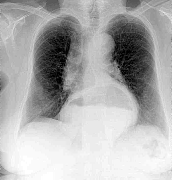
    • High-density tissue (e.g., bone) - absorbs x-rays to a greater degree, and appears white on the film.
    • Low-density tissue (e.g., the lungs) - absorbs X-rays to a lesser degree, and appears black on the film
    • Intermediate density tissue (e.g., muscle and fat) - appears as shades of grey on the x-ray film
Radiograph of lungs
Radiograph of lungs
Diego Grez
/
Wikimedia Commons
/
CC BY-SA 3.0

Computed tomography (CT scan)

  • Best for: Bones, soft tissues, internal bleeding, internal organs, and the brain.
  • How it works: Combines multiple X-rays to create cross-sectional images
  • Common uses:
    • Bone fractures and complex injuries
    • Internal bleeding (trauma)
    • Stroke
    • Tumors
    • Lung and abdominal conditions
  • Limitations: higher radiation exposure than X-rays
  • Interpreting CT scan
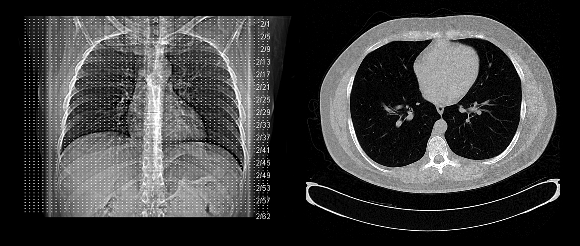
    • Dense structures (like bone and calcifications) appear lighter (white).
    • Lucent structures (like air and fat) appear darker (black).
Examples of CT scan
Examples of CT scan
Ptrump16
/
Wikimedia Commons
/
CC BY-SA 4.0

Magnetic resonance imaging (MRI)

  • Best for: Soft tissues, brain, muscles, ligaments, and nerves
  • How it works: Uses strong magnets and radio waves to generate detailed images
  • Common uses:
    • Ligament and tendon injuries
    • Brain and spinal cord conditions
    • Tumors
    • Disc herniations
  • Limitations: expensive, time-consuming, may be contraindicated with certain ferromagnetic metal implants and devices (e.g., some pacemakers, aneurysm clips) - safety depends on the specific implant
  • Interpreting MRI
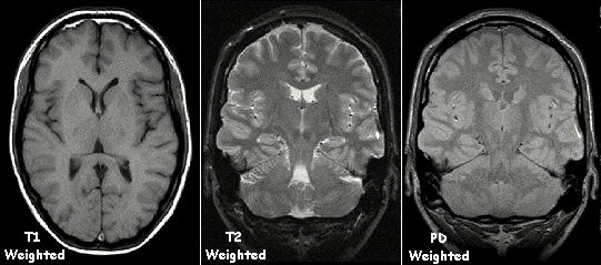
    • T1 MRI highlights anatomy, provides crisp images, and shows fluids as dark
    • T2 MRI focuses on pathology, making fluids bright, which is ideal for visualizing inflammation, edema
Examples of MRI
Examples of MRI
KieranMaher at English Wikibooks, Adapted with permission by Kieran Maher using Graphic Converter from Applied Imaging Technology by Heggie, Liddell & Maher (2000).
/
Wikimedia Commons
/
Public domain

Ultrasound

  • Best for: Soft tissues, pregnancy, blood flow
  • How it works: Uses high-frequency sound waves to create real-time images.
  • Common uses:
    • Pregnancy monitoring
    • Soft tissue injuries (e.g., muscle tears)
    • Blood clots (Doppler ultrasound)
    • Organ imaging (e.g., liver, kidneys)
  • Limitations: poor image quality for bones and deep structures

Nuclear medicine imaging (e.g., PET scan, bone scan)

  • Best for: Organ function, cancer detection, metabolic activity
  • How it works: Uses radioactive tracers to highlight metabolic activity
  • Common uses:
    • Cancer detection (PET scan)
    • Bone metastases (bone scan)
    • Stress fracture/microfracture (bone scan)
    • Thyroid and kidney function
  • Limitations: radiation exposure, high cost

Fluoroscopy

  • Best for: Real-time imaging of movement (e.g., swallowing, joint motion)
  • How it works: Continuous X-ray imaging allows real-time assessment
  • Common uses:
    • Barium swallow for digestive tract
    • Cardiac catheterization
    • Joint injections
  • Limitations: higher radiation exposure than standard X-rays

Example: choosing the right imaging study

A patient reports a popping sensation and knee instability after twisting the joint during sports. The supervising PT suspects a ligament tear. Which imaging modality is best suited to visualize this soft-tissue injury?

Answer: MRI - it provides the soft-tissue detail needed to visualize ligaments, tendons, and cartilage. X-ray is best for bone, and CT is best for bone and internal bleeding, so neither shows a ligament tear as clearly.

Lab values and bone density testing

Besides imaging, the chart may include lab work and bone density results relevant to musculoskeletal conditions:

  • DEXA scan (bone density): a T-score of −2.5 or lower indicates osteoporosis; a T-score below −1.0 but above −2.5 indicates osteopenia.
  • ESR and CRP: elevated levels suggest inflammation or infection.
  • Rheumatoid factor and anti-CCP: elevated levels support a diagnosis of rheumatoid arthritis.
  • Uric acid: elevated levels are associated with gout.
  • Alkaline phosphatase, calcium, and vitamin D: reflect bone metabolism and healing.

Medications

Medications for musculoskeletal conditions target pain, inflammation, and muscle spasm or spasticity; the main classes are NSAIDs, opioids, corticosteroids, and antispasticity/muscle-relaxant agents.

PTA role: You don’t prescribe or adjust these medications, but you do monitor patients for the adverse effects below during each session and report changes to the supervising PT. Stop the intervention and notify the PT if you notice signs such as labored or slowed breathing in a patient on opioids, new bone pain or a fall in a patient on long-term corticosteroids (increased fracture risk), or unexplained bruising, black stools, or GI complaints in a patient on NSAIDs.

  • Non-steroidal anti-inflammatory drugs (NSAIDs)
    • Mechanism of action: inhibit cyclooxygenase (COX) enzymes, which reduces prostaglandin production and decreases inflammation, pain, and fever
    • Key adverse effects to monitor: GI ulcers, indigestion, and increased bleeding risk
  • Opioids
    • Mechanism of action: bind to opioid receptors in the central nervous system, altering the perception of pain
    • Key adverse effects to monitor: decreased respiration rate (respiratory depression), constipation, and addiction risk with long-term use
  • Corticosteroids
    • Mechanism of action: suppress the immune and inflammatory response throughout the body when taken systemically (for example, by mouth), or mainly at the site when injected locally
    • Key adverse effects to monitor: osteoporosis (increased fracture risk with long-term use), high blood pressure, and weight gain/diabetes
  • Baclofen (antispasticity agent)
    • Mechanism of action: a GABA-B receptor agonist that acts mainly at the spinal cord to suppress spinal reflexes and reduce spasticity - distinct from the centrally acting muscle relaxants below, which target acute muscle spasm rather than spasticity
    • Key adverse effects to monitor: drowsiness, dizziness, fatigue, and muscle weakness; abrupt withdrawal can cause seizures, hallucinations, and rebound spasticity
  • Muscle relaxants (for acute muscle spasm)
    • Mechanism of action: acts on the central nervous system (CNS) to interfere with the transmission of nerve impulses to muscles, reducing muscle spasms and tension
    • Key adverse effects to monitor: drowsiness, dizziness, muscle weakness, and nausea

PT implications:

  • Analgesics and NSAIDs can mask pain, so the patient may push past safe limits - rely on objective signs and RPE rather than self-reported pain alone.
  • When the PT’s plan allows, time sessions to coincide with the medication’s peak effect so the patient can tolerate the intervention.
  • Drowsiness and dizziness from opioids, baclofen, and muscle relaxants raise fall risk - guard closely during transfers and gait.
  • After a local corticosteroid injection, the tendon is temporarily weaker, so follow the PT’s plan about avoiding heavy loading in that area.

Fractures

A fracture is a partial or complete break in the bone. There are many different types of fractures. Bone fractures are often caused by falls, injury, or a direct hit or kick to the body. Overuse or repetitive motions can cause stress fractures. Bone healing moves through four stages: inflammatory → soft callus → hard callus → remodeling.

Types of fractures

  • Open (compound): The bone breaks through the skin, exposing it to the environment
  • Closed (simple): The bone breaks but does not penetrate the skin.
  • Transverse: A straight break across the bone
  • Oblique: A diagonal break at an angle to the bone
  • Spiral: A twisting break that spirals around the bone
  • Greenstick: A partial break that occurs in children’s flexible bones
  • Comminuted: The bone breaks into multiple fragments
  • Stress fracture: A small, hairline crack caused by repetitive stress
  • Impacted fracture: The broken ends of the bone are driven into each other
  • Avulsion fracture: A small piece of bone is pulled away by a tendon or ligament

An acute fracture typically causes intense pain with swelling, bruising, and tenderness at the injury, trouble moving the part, and sometimes a deformity such as a limb that looks out of place.

PTA role - fracture complications requiring immediate notification:

  • Compartment syndrome: pain out of proportion to the injury, pain with passive stretch, paresthesia, pallor, pulselessness
  • Deep vein thrombosis (DVT): calf pain, swelling, warmth
  • Fat embolism syndrome: see its own section below
  • Delayed union or nonunion: healing that’s slower than expected or fails to progress

Salter-Harris fracture

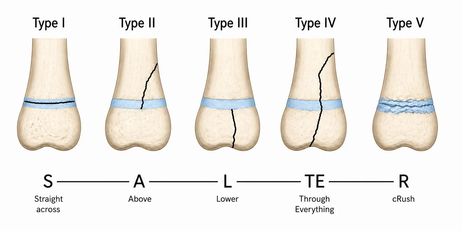
A Salter-Harris fracture is a type of bone fracture that occurs in children and adolescents, involving the growth plate (physis). Salter-Harris fractures are classified into five types based on the location and extent of the fracture, using the mnemonic SALTR:

Type Mnemonic Structures involved Key point
I S - straight across Physis only No bone involvement
II A - above Physis and metaphysis Most common type
III L - lower Physis and epiphysis Involves joint surface
IV TE - through everything Metaphysis, physis, and epiphysis Crosses entire bone
V R - rammed (crush) Crush injury to the growth plate Often not visible initially, worst prognosis
Five Salter-Harris classification types of pediatric growth plate fractures.
Salter-Harris Fractures
Achievable

Fat embolism syndrome

After a long-bone fracture, fat droplets from the marrow enter the circulation. A few patients develop fat embolism syndrome, most often after fractures of the femur, pelvis or tibia, and after intramedullary nailing or arthroplasty. Signs usually appear 24 to 72 hours after the injury:

  • Respiratory: tachypnea, dyspnea and cyanosis; oxygen saturation can fall before the patient complains
  • Neurological: confusion, drowsiness, seizures or coma
  • Skin: a flat petechial rash on the chest, axilla, neck and conjunctiva
  • Other: tachycardia and fever

PTA role: During early mobilization after a femur fracture or intramedullary nailing, check oxygen saturation, heart rate, respiratory rate and orientation. New shortness of breath, falling saturation, new confusion or a petechial rash is a medical emergency: stop the session, keep the patient at rest with the head raised, call for emergency help and stay with the patient.

Medical Imaging Modalities

  • PTA role: use imaging findings already in the chart for context; never order/interpret, but report new or changed symptoms to the PT
  • X-ray: bones/joints/lungs; ionizing radiation; dense tissue (bone) = white, air = black, soft tissue = gray; poor soft-tissue detail
  • CT: cross-sectional X-rays; best for bone, soft tissue, internal bleeding, brain; dense structures white, air/fat dark; higher radiation than X-ray
  • MRI: best for soft tissue (ligaments, nerves, discs); magnets/radio waves, no radiation; T1 = anatomy (fluid dark), T2 = pathology (fluid bright, shows edema/inflammation); caution with ferromagnetic implants
  • Ultrasound: real-time soft tissue/blood flow/pregnancy imaging; no radiation; poor for bone/deep structures
  • Nuclear medicine (PET, bone scan): shows metabolic activity via radioactive tracers; used for cancer, bone metastases, stress fractures
  • Fluoroscopy: continuous real-time X-ray for movement (swallowing, joint motion, injections); higher radiation exposure

Lab Values & Bone Density

  • DEXA T-score: −2.5 or lower = osteoporosis; −1.0 to −2.5 = osteopenia
  • ESR/CRP elevated = inflammation/infection; RF/anti-CCP elevated = rheumatoid arthritis; uric acid elevated = gout
  • Alkaline phosphatase, calcium, vitamin D reflect bone metabolism/healing

Medications for Musculoskeletal Conditions

  • NSAIDs: inhibit COX → reduce inflammation/pain; watch for GI ulcers, bleeding
  • Opioids: bind CNS opioid receptors; watch for respiratory depression, constipation, addiction
  • Corticosteroids: suppress immune/inflammatory response body-wide when systemic (mainly local when injected); watch for osteoporosis/fracture risk, hypertension, weight gain
  • Baclofen: GABA-B agonist, spinal-level antispasticity agent; watch for drowsiness, weakness, withdrawal seizures
  • Muscle relaxants: CNS-acting for acute spasm; watch for drowsiness, weakness, nausea
  • PTA implications: pain meds can mask symptoms (use objective signs/RPE); time therapy to peak drug effect; guard against fall risk from sedating drugs; avoid heavy loading after corticosteroid injections

Fracture Types & Complications

  • Open (through skin) vs closed; patterns include transverse, oblique, spiral, comminuted, greenstick (children), stress, impacted, avulsion
  • Watch for and report: compartment syndrome (pain out of proportion, pain with passive stretch, paresthesia, pallor, pulselessness), DVT (calf pain/swelling/warmth), fat embolism (dyspnea, confusion, petechiae), delayed/nonunion
  • Healing stages: inflammatory → soft callus → hard callus → remodeling

Salter-Harris Fractures (Pediatric Growth Plate)

  • Mnemonic SALTR: Straight across (I, physis only) → Above (II, physis+metaphysis, most common) → Lower (III, physis+epiphysis, joint involved) → Through everything (IV, all three) → Rammed/crush (V, worst prognosis, often not visible initially)

Fat Embolism Syndrome

  • Occurs 24-72 hours after long-bone fracture (femur, pelvis, tibia) or IM nailing/arthroplasty
  • Classic triad: respiratory distress (tachypnea, dyspnea, cyanosis), neuro changes (confusion, seizures), petechial rash (chest/axilla/neck)
  • PTA role: monitor vitals/orientation during mobilization; new dyspnea, falling O2 sat, confusion, or rash = medical emergency — stop session, position with head raised, call for help, stay with patient

Sign up for free to take 21 quiz questions on this topic

Previous
Next  | 5.18 Surgical protocols
All rights reserved ©2016 - 2026 Achievable, Inc.

Medications, imaging, and fractures

Imaging

There are several types of medical imaging used to assess different tissues and structures in the body. Below are the main types.

PTA role: You don’t order or interpret imaging to make a diagnosis - that’s the role of the supervising PT or physician. You use imaging findings already documented in the chart as background for the established plan of care, and you report any new or changed symptoms (for example, signs suggesting a new fracture) back to the PT.

X-ray (radiography)

  • Best for: Bones, fractures, joint alignment, lung conditions
  • How it works: Uses ionizing radiation to create images of dense structures
  • Common uses:
    • Fractures & dislocations
    • Arthritis
    • Lung infections (e.g., pneumonia)
    • Foreign objects
  • Limitations: limited soft tissue visibility, radiation exposure
  • Interpreting an X-ray
    • High-density tissue (e.g., bone) - absorbs x-rays to a greater degree, and appears white on the film.
    • Low-density tissue (e.g., the lungs) - absorbs X-rays to a lesser degree, and appears black on the film
    • Intermediate density tissue (e.g., muscle and fat) - appears as shades of grey on the x-ray film

Computed tomography (CT scan)

  • Best for: Bones, soft tissues, internal bleeding, internal organs, and the brain.
  • How it works: Combines multiple X-rays to create cross-sectional images
  • Common uses:
    • Bone fractures and complex injuries
    • Internal bleeding (trauma)
    • Stroke
    • Tumors
    • Lung and abdominal conditions
  • Limitations: higher radiation exposure than X-rays
  • Interpreting CT scan
    • Dense structures (like bone and calcifications) appear lighter (white).
    • Lucent structures (like air and fat) appear darker (black).

Magnetic resonance imaging (MRI)

  • Best for: Soft tissues, brain, muscles, ligaments, and nerves
  • How it works: Uses strong magnets and radio waves to generate detailed images
  • Common uses:
    • Ligament and tendon injuries
    • Brain and spinal cord conditions
    • Tumors
    • Disc herniations
  • Limitations: expensive, time-consuming, may be contraindicated with certain ferromagnetic metal implants and devices (e.g., some pacemakers, aneurysm clips) - safety depends on the specific implant
  • Interpreting MRI
    • T1 MRI highlights anatomy, provides crisp images, and shows fluids as dark
    • T2 MRI focuses on pathology, making fluids bright, which is ideal for visualizing inflammation, edema

Ultrasound

  • Best for: Soft tissues, pregnancy, blood flow
  • How it works: Uses high-frequency sound waves to create real-time images.
  • Common uses:
    • Pregnancy monitoring
    • Soft tissue injuries (e.g., muscle tears)
    • Blood clots (Doppler ultrasound)
    • Organ imaging (e.g., liver, kidneys)
  • Limitations: poor image quality for bones and deep structures

Nuclear medicine imaging (e.g., PET scan, bone scan)

  • Best for: Organ function, cancer detection, metabolic activity
  • How it works: Uses radioactive tracers to highlight metabolic activity
  • Common uses:
    • Cancer detection (PET scan)
    • Bone metastases (bone scan)
    • Stress fracture/microfracture (bone scan)
    • Thyroid and kidney function
  • Limitations: radiation exposure, high cost

Fluoroscopy

  • Best for: Real-time imaging of movement (e.g., swallowing, joint motion)
  • How it works: Continuous X-ray imaging allows real-time assessment
  • Common uses:
    • Barium swallow for digestive tract
    • Cardiac catheterization
    • Joint injections
  • Limitations: higher radiation exposure than standard X-rays

Example: choosing the right imaging study

A patient reports a popping sensation and knee instability after twisting the joint during sports. The supervising PT suspects a ligament tear. Which imaging modality is best suited to visualize this soft-tissue injury?

Answer: MRI - it provides the soft-tissue detail needed to visualize ligaments, tendons, and cartilage. X-ray is best for bone, and CT is best for bone and internal bleeding, so neither shows a ligament tear as clearly.

Lab values and bone density testing

Besides imaging, the chart may include lab work and bone density results relevant to musculoskeletal conditions:

  • DEXA scan (bone density): a T-score of −2.5 or lower indicates osteoporosis; a T-score below −1.0 but above −2.5 indicates osteopenia.
  • ESR and CRP: elevated levels suggest inflammation or infection.
  • Rheumatoid factor and anti-CCP: elevated levels support a diagnosis of rheumatoid arthritis.
  • Uric acid: elevated levels are associated with gout.
  • Alkaline phosphatase, calcium, and vitamin D: reflect bone metabolism and healing.

Medications

Medications for musculoskeletal conditions target pain, inflammation, and muscle spasm or spasticity; the main classes are NSAIDs, opioids, corticosteroids, and antispasticity/muscle-relaxant agents.

PTA role: You don’t prescribe or adjust these medications, but you do monitor patients for the adverse effects below during each session and report changes to the supervising PT. Stop the intervention and notify the PT if you notice signs such as labored or slowed breathing in a patient on opioids, new bone pain or a fall in a patient on long-term corticosteroids (increased fracture risk), or unexplained bruising, black stools, or GI complaints in a patient on NSAIDs.

  • Non-steroidal anti-inflammatory drugs (NSAIDs)
    • Mechanism of action: inhibit cyclooxygenase (COX) enzymes, which reduces prostaglandin production and decreases inflammation, pain, and fever
    • Key adverse effects to monitor: GI ulcers, indigestion, and increased bleeding risk
  • Opioids
    • Mechanism of action: bind to opioid receptors in the central nervous system, altering the perception of pain
    • Key adverse effects to monitor: decreased respiration rate (respiratory depression), constipation, and addiction risk with long-term use
  • Corticosteroids
    • Mechanism of action: suppress the immune and inflammatory response throughout the body when taken systemically (for example, by mouth), or mainly at the site when injected locally
    • Key adverse effects to monitor: osteoporosis (increased fracture risk with long-term use), high blood pressure, and weight gain/diabetes
  • Baclofen (antispasticity agent)
    • Mechanism of action: a GABA-B receptor agonist that acts mainly at the spinal cord to suppress spinal reflexes and reduce spasticity - distinct from the centrally acting muscle relaxants below, which target acute muscle spasm rather than spasticity
    • Key adverse effects to monitor: drowsiness, dizziness, fatigue, and muscle weakness; abrupt withdrawal can cause seizures, hallucinations, and rebound spasticity
  • Muscle relaxants (for acute muscle spasm)
    • Mechanism of action: acts on the central nervous system (CNS) to interfere with the transmission of nerve impulses to muscles, reducing muscle spasms and tension
    • Key adverse effects to monitor: drowsiness, dizziness, muscle weakness, and nausea

PT implications:

  • Analgesics and NSAIDs can mask pain, so the patient may push past safe limits - rely on objective signs and RPE rather than self-reported pain alone.
  • When the PT’s plan allows, time sessions to coincide with the medication’s peak effect so the patient can tolerate the intervention.
  • Drowsiness and dizziness from opioids, baclofen, and muscle relaxants raise fall risk - guard closely during transfers and gait.
  • After a local corticosteroid injection, the tendon is temporarily weaker, so follow the PT’s plan about avoiding heavy loading in that area.

Fractures

A fracture is a partial or complete break in the bone. There are many different types of fractures. Bone fractures are often caused by falls, injury, or a direct hit or kick to the body. Overuse or repetitive motions can cause stress fractures. Bone healing moves through four stages: inflammatory → soft callus → hard callus → remodeling.

Types of fractures

  • Open (compound): The bone breaks through the skin, exposing it to the environment
  • Closed (simple): The bone breaks but does not penetrate the skin.
  • Transverse: A straight break across the bone
  • Oblique: A diagonal break at an angle to the bone
  • Spiral: A twisting break that spirals around the bone
  • Greenstick: A partial break that occurs in children’s flexible bones
  • Comminuted: The bone breaks into multiple fragments
  • Stress fracture: A small, hairline crack caused by repetitive stress
  • Impacted fracture: The broken ends of the bone are driven into each other
  • Avulsion fracture: A small piece of bone is pulled away by a tendon or ligament

An acute fracture typically causes intense pain with swelling, bruising, and tenderness at the injury, trouble moving the part, and sometimes a deformity such as a limb that looks out of place.

PTA role - fracture complications requiring immediate notification:

  • Compartment syndrome: pain out of proportion to the injury, pain with passive stretch, paresthesia, pallor, pulselessness
  • Deep vein thrombosis (DVT): calf pain, swelling, warmth
  • Fat embolism syndrome: see its own section below
  • Delayed union or nonunion: healing that’s slower than expected or fails to progress

Salter-Harris fracture

A Salter-Harris fracture is a type of bone fracture that occurs in children and adolescents, involving the growth plate (physis). Salter-Harris fractures are classified into five types based on the location and extent of the fracture, using the mnemonic SALTR:

Type Mnemonic Structures involved Key point
I S - straight across Physis only No bone involvement
II A - above Physis and metaphysis Most common type
III L - lower Physis and epiphysis Involves joint surface
IV TE - through everything Metaphysis, physis, and epiphysis Crosses entire bone
V R - rammed (crush) Crush injury to the growth plate Often not visible initially, worst prognosis

Fat embolism syndrome

After a long-bone fracture, fat droplets from the marrow enter the circulation. A few patients develop fat embolism syndrome, most often after fractures of the femur, pelvis or tibia, and after intramedullary nailing or arthroplasty. Signs usually appear 24 to 72 hours after the injury:

  • Respiratory: tachypnea, dyspnea and cyanosis; oxygen saturation can fall before the patient complains
  • Neurological: confusion, drowsiness, seizures or coma
  • Skin: a flat petechial rash on the chest, axilla, neck and conjunctiva
  • Other: tachycardia and fever

PTA role: During early mobilization after a femur fracture or intramedullary nailing, check oxygen saturation, heart rate, respiratory rate and orientation. New shortness of breath, falling saturation, new confusion or a petechial rash is a medical emergency: stop the session, keep the patient at rest with the head raised, call for emergency help and stay with the patient.

Key points

Medical Imaging Modalities

  • PTA role: use imaging findings already in the chart for context; never order/interpret, but report new or changed symptoms to the PT
  • X-ray: bones/joints/lungs; ionizing radiation; dense tissue (bone) = white, air = black, soft tissue = gray; poor soft-tissue detail
  • CT: cross-sectional X-rays; best for bone, soft tissue, internal bleeding, brain; dense structures white, air/fat dark; higher radiation than X-ray
  • MRI: best for soft tissue (ligaments, nerves, discs); magnets/radio waves, no radiation; T1 = anatomy (fluid dark), T2 = pathology (fluid bright, shows edema/inflammation); caution with ferromagnetic implants
  • Ultrasound: real-time soft tissue/blood flow/pregnancy imaging; no radiation; poor for bone/deep structures
  • Nuclear medicine (PET, bone scan): shows metabolic activity via radioactive tracers; used for cancer, bone metastases, stress fractures
  • Fluoroscopy: continuous real-time X-ray for movement (swallowing, joint motion, injections); higher radiation exposure

Lab Values & Bone Density

  • DEXA T-score: −2.5 or lower = osteoporosis; −1.0 to −2.5 = osteopenia
  • ESR/CRP elevated = inflammation/infection; RF/anti-CCP elevated = rheumatoid arthritis; uric acid elevated = gout
  • Alkaline phosphatase, calcium, vitamin D reflect bone metabolism/healing

Medications for Musculoskeletal Conditions

  • NSAIDs: inhibit COX → reduce inflammation/pain; watch for GI ulcers, bleeding
  • Opioids: bind CNS opioid receptors; watch for respiratory depression, constipation, addiction
  • Corticosteroids: suppress immune/inflammatory response body-wide when systemic (mainly local when injected); watch for osteoporosis/fracture risk, hypertension, weight gain
  • Baclofen: GABA-B agonist, spinal-level antispasticity agent; watch for drowsiness, weakness, withdrawal seizures
  • Muscle relaxants: CNS-acting for acute spasm; watch for drowsiness, weakness, nausea
  • PTA implications: pain meds can mask symptoms (use objective signs/RPE); time therapy to peak drug effect; guard against fall risk from sedating drugs; avoid heavy loading after corticosteroid injections

Fracture Types & Complications

  • Open (through skin) vs closed; patterns include transverse, oblique, spiral, comminuted, greenstick (children), stress, impacted, avulsion
  • Watch for and report: compartment syndrome (pain out of proportion, pain with passive stretch, paresthesia, pallor, pulselessness), DVT (calf pain/swelling/warmth), fat embolism (dyspnea, confusion, petechiae), delayed/nonunion
  • Healing stages: inflammatory → soft callus → hard callus → remodeling

Salter-Harris Fractures (Pediatric Growth Plate)

  • Mnemonic SALTR: Straight across (I, physis only) → Above (II, physis+metaphysis, most common) → Lower (III, physis+epiphysis, joint involved) → Through everything (IV, all three) → Rammed/crush (V, worst prognosis, often not visible initially)

Fat Embolism Syndrome

  • Occurs 24-72 hours after long-bone fracture (femur, pelvis, tibia) or IM nailing/arthroplasty
  • Classic triad: respiratory distress (tachypnea, dyspnea, cyanosis), neuro changes (confusion, seizures), petechial rash (chest/axilla/neck)
  • PTA role: monitor vitals/orientation during mobilization; new dyspnea, falling O2 sat, confusion, or rash = medical emergency — stop session, position with head raised, call for help, stay with patient

More from Musculoskeletal system

  • Anatomy of musculoskeletal system
  • Anatomical terminology and exercise training principles
  • Joint mechanics and phases of healing
  • Upper extremity anatomy
  • Lower extremity anatomy