Major lymph node groups and drainage areas
It’s important to know the major lymph node groups and the areas they drain. This information helps you localize infections and understand how cancers spread.
A sentinel node is the first lymph node (within a regional group) that receives lymph from a specific area. Because it’s the first “stop,” it’s often the first node involved in cancer spread. A sentinel lymph node biopsy helps detect early metastasis. It’s performed by injecting a blue dye or radioactive tracer into the drainage area; the first node to take up the dye/tracer is identified as the sentinel node.
Inguinal lymph nodes
There are 2 major groups: superficial and deep.
Superficial nodes
These form a T-shaped arrangement, located just below the inguinal ligament and along the great saphenous vein. They drain superficial structures (i.e., structures above the deep fascia). Drainage areas include:
- Abdominal wall below the umbilicus
- Lower limb except the posterolateral area of the calf and the dorsolateral foot
- External (NOT internal) genitalia
- Lower part of vagina
- Vulva, scrotum
- Anal canal
- Perineum
- Cornu of uterus
Deep nodes
These are located medial to the femoral vein within the femoral sheath. Cloquet’s node is the most superior node in this group, just under the inguinal ligament. Deep inguinal nodes drain the deep lymphatics of the distal lower extremity and perineum, including the glans penis and clitoris. They drain into the external iliac lymph nodes and finally into paraaortic nodes.
Lymphatic drainage of female internal genitalia
Lymphatic drainage depends on the specific structure.
- Upper ⅔ of the vagina drains into external and internal iliac lymph nodes.
- Lower ⅓ of the vagina drains into superficial inguinal lymph nodes.
- Most of the fundus of the uterus drains into pre and paraaortic nodes.
- The part of the fundus along the round ligament drains into the superficial inguinal nodes.
- Cornu drains to superficial inguinal nodes.
- Body drains mainly into the external iliac and lumbar nodes.
- Cervix drains to the external and internal iliac nodes.
Lymphatic drainage of male internal genitalia
- Lymph from the testes and epididymis drains into para aortic / lumbar lymph nodes.
- Seminal vesicles drain into external and internal iliac lymph nodes.
- Lymphatics from the prostate drain mainly to internal iliac and some end in external iliac lymph nodes.
Lymphatic drainage of the face
- Forehead and anterior part of face drain mainly to submandibular and a few to buccal lymph nodes.
- Lateral part of face and lateral parts of eyelids drain to parotid nodes.
- Lower lip and chin drain to submental lymph nodes.
Important lymph node groups in the head and neck region
Following is a list of them:
- Anterior cervical: (includes jugulodigastric) drains posterior pharynx, tonsils, thyroid gland, throat. Classically enlarged in CMV infections, infectious mononucleosis and toxoplasmosis.
- Posterior cervical: drains scalp and neck, thorax, cervical and axillary nodes.
- Tonsillar: drains tonsils and posterior pharynx.
- Submental: drains lower lip, floor of mouth, tip of tongue and cheeks.
- Submandibular: drains floor of mouth, submandibular gland, tongue, lips, conjunctivae.
- Supraclavicular: drains mediastinum, lungs, esophagus, abdomen via thoracic duct.
Lymphatic drainage of the gastrointestinal tract
| Part of GIT | Draining lymph nodes |
|---|---|
| Esophagus | Mediastinal lymph nodes |
| Stomach, pancreas, liver, spleen, upper duodenum | Celiac lymph nodes |
| Lower part of duodenum (distal to major duodenal papilla), jejunum, ileum, colon up to the proximal ⅔ of transverse colon | Superior mesenteric nodes |
| Distal ⅓ of transverse colon to upper rectum | Inferior mesenteric lymph nodes |
| Lower part of rectum, anal canal above pectinate line | Internal iliac nodes* |
| Anal canal below pectinate line | Superficial inguinal nodes |
*The bladder drains to internal iliac nodes (finally into para aortic) while kidneys, adrenals and upper part of renal collecting system drain directly into para aortic nodes.
Lymphatic drainage of the breast
Breast lymph drains primarily to two node groups:
- Axillary lymph nodes: drain the lateral parts of the breast, including the superior, lateral, and inferior quadrants and the nipple. They receive close to 75% of breast lymph drainage.
- Parasternal/internal mammary lymph nodes: drain the medial quadrants.
Miscellaneous (but important) nodes and drainage areas
- Popliteal node: posterior calf and dorsolateral foot
- Epitrochlear node: medial side of arm below the elbow
- Supraclavicular node: On the right think of lungs, mediastinum. On the left think of the stomach, abdomen.
- Deltopectoral node: arm
- Axillary node: arm, breast, thorax, neck.
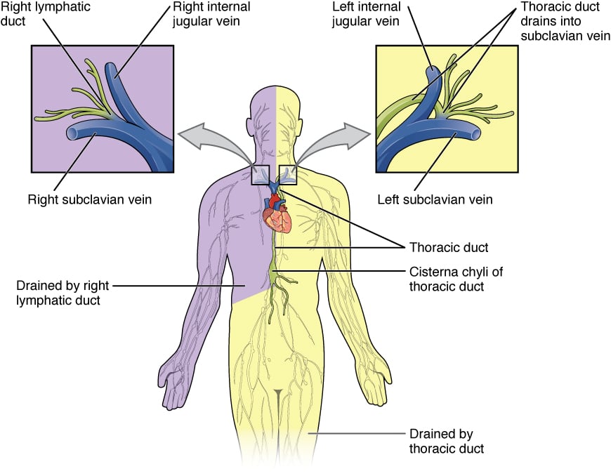
Thoracic duct
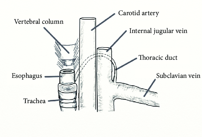
The thoracic duct begins at the level of the second lumbar vertebrae as the dilated cisterna chyli. It enters the thoracic cavity through the aortic opening of the diaphragm. It then passes through the posterior and superior mediastinum and finally drains into the junction of the left subclavian vein and the internal jugular vein at the base of the neck.
The abdominal and thoracic parts of the thoracic duct show active peristalsis. It carries lymph (chyle) from all areas of the body except those drained by the right lymphatic duct (see below).
Cervical course of the thoracic duct. The thoracic duct enters the neck lateral to the esophagus, ascending superiorly and laterally behind to the carotid and internal jugular vein before turning inferiorly and anteriorly to join the venous circulation at the confluence of the internal jugular vein and subclavian vein.
Right lymphatic duct
The right lymphatic duct drains lymph from the right side of the head and neck, right upper extremity, right side of the thorax, right lung, right side of the heart, and a part of the liver. It opens into the right subclavian vein.