



Skin structure, wound healing, and dressings
Foundational skin information
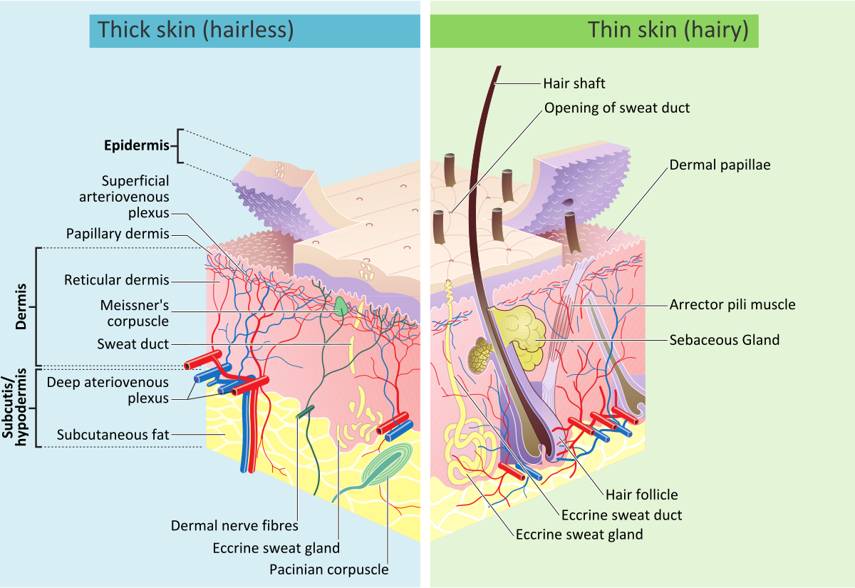
The integumentary system consists of the skin, hair, nails, glands, and sensory receptors. It serves as the first line of defense against injury, infection, and dehydration while regulating temperature and sensation. Below are the layers of skin and their descriptions:
Epidermis (outermost layer)
- Avascular (no blood vessels)
- Composed of keratinized stratified squamous epithelium
- Key cells:
- Keratinocytes - strength and waterproofing (keratin production)
- Melanocytes - pigmentation and ultraviolet light protection
- Langerhans cells - immune defense
- Merkel cells - touch sensation
- The epidermis is organized into several layers, from the deep stratum basale (site of active cell division) to the superficial stratum corneum, a barrier layer of dead, keratinized cells
Dermis (middle layer)
- Vascularized, supplies nutrients to the epidermis
- Contains:
- Blood vessels
- Nerve endings
- Hair follicles
- Sweat and sebaceous glands
- Collagen and elastin fibers (structural support)
Hypodermis (subcutaneous tissue - deepest layer)
- Composed of: fat (adipose tissue), connective tissue
- Function: insulation, energy storage, shock absorption
Related to skin
- Hair - protects against UV, insulates scalp
- Nails - keratinized structures for protection and dexterity
- Sebaceous (oil) glands - secrete sebum (moisturizes and protects skin)
- Sweat glands - regulate temperature and remove waste
- Eccrine glands - watery sweat, found over most of the body
- Apocrine glands - thicker secretion, found in the axillae and groin
Wound healing phases
Inflammatory phase
- Initiated immediately and lasts 3-4 days
- Marked by hemostasis (stopping bleeding) and phagocytosis (removal of debris and pathogens)
Proliferative phase
- Lasts until day 21 post-injury
- Collagen formation: A whitish protein substance that adds to the tensile strength of the wound
- Granulation tissue (beefy red, moist) fills the wound bed, new capillaries form (angiogenesis), myofibroblasts pull the wound edges together (wound contraction), and epithelial cells migrate across the surface (epithelialization)
- New epithelial tissue appears light pink and translucent as it grows over the granulation tissue from the wound margins
Maturation phase
- Begins at day 21 and lasts up to 1-2 years
- Leads to scar formation
- Collagen is remodeled, but the scar reaches only about 70-80% of the original tensile strength
Types of healing
Primary intention (first intention)
- Tissue surfaces are closed with minimal tissue loss
- Example: Surgical incision
Secondary intention
- Extensive tissue loss prevents edges from being approximated
- Example: Pressure ulcer
- Longer repair time, increased scarring, and higher infection risk
Tertiary intention healing (delayed closure)
- Wound is left open for a period before closure
- Example: Abdominal wound left open for drainage before later closure
Factors that impair wound healing
- Local factors:
- Infection - bacteria compete with healing cells for oxygen and nutrients
- Poor perfusion - reduces oxygen and nutrient delivery to the wound bed
- Pressure - compresses vessels and restricts local blood flow
- Too much or too little moisture - excess moisture causes maceration, while dryness slows cell migration
- Necrotic tissue - acts as a physical barrier to granulation
- Systemic factors:
- Diabetes - impairs circulation and immune response
- Malnutrition or low protein intake - limits collagen synthesis
- Smoking - causes vasoconstriction, reducing oxygen delivery
- Corticosteroids or chemotherapy - suppress the inflammatory response needed to start healing
- Advanced age - slower cell turnover and reduced skin elasticity
- Obesity - reduces vascularity of adipose tissue and increases tension on the wound
Classification of wound drainage
- Serous - Clear or pale yellow, thin and watery fluid from the serum portion of blood
- Sanguineous - Bright red, bloody drainage
- Serosanguineous - Pinkish drainage, a mix of blood and serum
- Purulent - Thick, yellow or yellow-green pus, indicating infection
How to measure wounds accurately
Measuring wounds is essential for assessing healing progress and treatment effectiveness. Proper wound measurement includes length, width, depth, and any tunneling or undermining. Below are the tools and steps for how to measure a wound.
Tools needed for wound measurement
- Disposable ruler (centimeters or millimeters)
- Sterile cotton-tipped applicator (for depth)
- Wound probe (for tunneling or undermining)
- Gloves (to maintain infection control)
- Transparent film grid (optional for tracing irregular shapes)
Step-by-step wound measurement
Measuring length and width
- Patient positioning: Ensure the patient is in the same position each time for consistency
- Use a ruler: Place a disposable ruler over the wound
- Length: measure head-to-toe (12 to 6 o’clock), with the patient’s head at 12 o’clock
- Width: measure side-to-side (3 to 9 o’clock), perpendicular to length
- Use the same method at every visit so measurements stay comparable over time
- Documentation format: Length × Width (e.g., 5.2 cm × 3.4 cm)
Measuring wound depth
- Hold a sterile cotton-tipped applicator perpendicular to the skin surface at the deepest part of the wound
- Grasp or mark the applicator where it is level with the wound edge
- Remove and measure against a ruler
- Example documentation: Depth = 0.8 cm
Measuring wound volume
- Measuring wound volume can be done by multiplying length, width, and depth
- Alternatively, measure the amount of hydrogel required to fill the wound
Steps for measuring tunneling and undermining
- Insert a sterile probe into the suspected tunnel/undermined area
- Measure using a ruler
- Use the clock method to describe location (e.g., “Tunneling at 3 o’clock position, depth 1.5 cm”)
Additional documentation details
- Shape and edges: Irregular, round, oval, jagged
- Tissue type: Granulation, slough, eschar
- Exudate: amount (none, scant, moderate, heavy) and type (serous, purulent, sanguineous)
- Periwound skin: Erythema, edema, maceration, induration
Types of dressing
Dressing selection follows the principle of moist wound healing: the wound bed should stay moist enough to support cell migration and granulation, but not so wet that the surrounding skin breaks down (maceration). Match the dressing’s absorbency to the wound’s exudate level - add moisture to a dry wound, absorb moisture from a heavily draining one, and choose a middle-ground option for light-to-moderate drainage.
Pressure injury stages (I-IV, unstageable, and deep tissue injury) are introduced in the later chapter on debridement and wound classification by etiology. Arterial, venous, and diabetic/neuropathic ulcers are covered in that same chapter, and burns in burns and other common skin conditions.
| Dressing | Exudate level | Best for | Avoid |
|---|---|---|---|
| Gauze | Moderate-heavy | Packing deep or surgical wounds; deeper pressure injuries | Caution: dry wounds (can adhere to the wound bed and delay healing) |
| Transparent film | None-minimal | Superficial wounds, abrasions, IV sites; shallow pressure injuries | Contraindicated: infected or heavily exudating wounds |
| Hydrocolloid | Light-moderate | Shallow pressure injuries, venous ulcers; keeps the wound bed moist | Contraindicated: infected wounds or heavy exudate |
| Hydrogel | None-minimal | Dry wounds, burns, painful wounds, necrotic tissue (autolytic debridement) | Caution: heavily exudating wounds (risk of maceration) |
| Foam | Moderate-heavy | Venous ulcers, diabetic foot ulcers; cushions deeper pressure injuries | Caution: dry wounds or minimal exudate |
| Alginate | Heavy | Infected or tunneling wounds; seaweed-derived, absorbs large fluid volumes | Caution: dry wounds or minimal exudate |
| Collagen | Light-moderate | Chronic non-healing or granulating wounds | Contraindicated: collagen (often bovine-derived) allergy. Caution: dry wounds or wounds with eschar |
| Antimicrobial (silver or iodine) | Moderate-heavy | Infected wounds, burns, surgical wounds at risk of infection | Contraindicated: silver or iodine allergy; iodine is also contraindicated with thyroid disorder or pregnancy/lactation. Caution: long-term use (cytotoxicity risk) |
When an alginate dressing is removed, it can look yellowish-tan or brown and have a mild, unpleasant odor - this is normal and comes from the interaction between the seaweed-based fibers and the wound’s exudate, not necessarily a sign of infection.
If any of the warning signs listed in the PTA scope-of-practice note above appear during a dressing change, stop and notify the supervising PT before proceeding.
Wound cleansing: normal saline is an isotonic wound cleanser that does not interfere with normal wound repair, while hydrogen peroxide is cytotoxic and can delay wound healing.