


This browser is too old to run Achievable.

Update it, or open this page in a current version of Chrome, Safari, Firefox or Edge. On an iPhone or iPad, update iOS: every browser there runs on the version of Safari that comes with it.

Achievable logoAchievable logo
NPTE-PTA
Sign in
Sign up
Purchase
Textbook
Practice exams
Support
How it works
Resources
Exam catalog
Mountain with a flag at the peak
Textbook
Introduction
1. Cardiovascular system
2. Pulmonary system
3. Neuromuscular system
4. Pediatrics
5. Musculoskeletal system
6. Other systems
6.1 Integumentary system
6.2 Lymphedema
6.3 Gastrointestinal and reproductive systems
6.4 Urinary incontinence and renal disorders
6.5 Endocrine and metabolic systems
6.6 Cancer and psychological conditions
6.7 Infectious disease, immune disorders, and hematological disorders
7. Non-systems
Wrapping up
Achievable logoAchievable logo
6.2 Lymphedema
Achievable NPTE-PTA
6. Other systems
Our NPTE-PTA course is now in "early access" - get 50% off for a limited time.

Lymphedema

8 min read
Font
Discuss
Share
Feedback

Anatomy and physiology

Lymphatic physiology

The lymphatic system works with the venous system to return fluid to the bloodstream, removes plasma proteins from tissue, and filters antigens, bacteria, and waste products. It also produces lymphocytes, a type of white blood cell that supports immune function; all white blood cells originate in the bone marrow, but organs like the thymus, spleen, and lymph nodes are crucial for lymphocyte development and function.

Lymph nodes

Lymph nodes are a series of sinuses that filter antigens and bacteria out of lymph fluid, which forms when plasma seeps out of blood capillaries into the surrounding tissue. This filtering is key to the body’s immune response.

The body contains roughly 600 lymph nodes, with about 300 of them in the head and neck. The axillary region has the largest concentration in the upper body and filters lymph from the arms and breasts, while the inguinal, or groin, region has the largest concentration in the lower body and filters lymph from the legs and lower abdomen.

Two primary lymphatic ducts of the body

  • Right lymphatic duct
  • Thoracic duct

Lymph fluid composition

Lymph fluid is a transparent, yellowish fluid containing water, proteins, lipids, minerals, and hormones. The body produces roughly 2-3 L of this fluid daily, which the lymphatic vessels return to the bloodstream.

Pathology

Lymphedema

  • Excessive accumulation of lymph fluid due to dysfunction in the lymphatic system, leading to increased edema
  • Lymphedema is a chronic condition that cannot be cured, and it may develop years after treatment has ended. Protecting the skin prevents infection, and there are ways to keep lymphedema from getting worse.
  • Primary lymphedema: a malformation of the lymphatic system, either congenital (Milroy disease), appearing around puberty (Meige disease, also called lymphedema praecox), or appearing after age 35 (lymphedema tarda)
  • Secondary lymphedema: acquired from lymph node removal or surgery, radiation, trauma, infection (including filariasis, the most common cause worldwide), or obesity
  • Compared with other causes of limb swelling: lymphedema is usually one-sided, affects the feet or hands, and is Stemmer-positive in later stages, while venous or cardiac edema is usually bilateral and pitting, and lipedema is symmetric, spares the feet, and is Stemmer-negative

Effects of lymphedema

  • Chronic swelling
  • Excessive tissue proteins
  • Fibrotic changes
  • Chronic inflammation
  • Infections - cellulitis
  • Skin changes, such as peau d’orange (dimpled skin resembling orange peel)

Related impairments

  • Functional deficits
  • Loss of mobility
  • Difficulty wearing normal clothing
  • Psychological issues

Stages of lymphedema

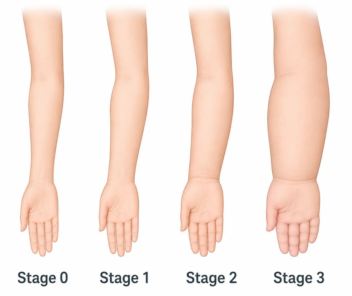
Lymphedema is staged by how reversible the swelling is and how much fibrosis has developed, ranging from stage 0 (no visible swelling) to stage 3 (irreversible hardening).

The following table compares the four stages of lymphedema on reversibility, pitting, fibrosis, and Stemmer sign.


Stage Reversible? Pitting Fibrosis Stemmer sign
0 (latent) No visible swelling yet None None Negative
1 Yes, spontaneously (subsides with elevation) Soft, pitting Little to none Negative
2 Not spontaneously; reduced with treatment Only with strong pressure, or none Usually present Present
3 (elephantiasis) No None Significant, with skin hardening Present
Lymphedema progression from Stage 0 to Stage 3, showing increasing arm and hand swelling.
Stages of lymphedema
Achievable
Definitions
Stemmer sign
Inability to lift a fold of skin on the dorsal surface of the second toe or finger; typically associated with stage 2 and stage 3 lymphedema.
Lymphotome
A specific region of the skin that drains lymph to a particular group of lymph nodes.

Goals of manual lymph drainage

Manual lymph drainage (MLD) is one part of complete decongestive therapy (CDT), the standard treatment approach for lymphedema, which also includes compression, exercise, and skin care. CDT has two phases: an intensive phase of daily treatment to reduce limb volume, and a maintenance phase where the patient self-manages with garments and exercise. The PTA carries out the PT’s plan of care, collects data such as limb girth, and reports changes back to the PT rather than modifying the plan independently.

  • Peristalsis of lymph fluid
  • Increase lymph volume in lymph vessels
  • Decrease congestion in the interstitium

Basic principles

Decongestion always begins proximally, clearing the nodes closest to the trunk before moving to the affected lymphotome, since clearing proximal nodes first opens a path for fluid to drain. Treatment starts with deep breathing, then AROM exercises in a proximal-to-distal progression. Massage is then directed toward the cleared nodes in a distal-to-proximal direction, using very light pressure so the lymphatic vessels aren’t collapsed.

Data collection

  • Measure circumferential girth at standardized points (for example, every 4 cm from a bony landmark) and compare with the unaffected limb
  • Water-displacement volumetry gives a more precise measure of limb volume when it’s available
  • Assess pitting depth and rebound time, and check for the Stemmer sign
  • Inspect the skin and tissue for fibrosis, peau d’orange, and open wounds
  • After axillary lymph node surgery, measure shoulder flexion and abduction against the other side, and note any compensation the patient uses to reach overhead
  • Look for axillary web syndrome (cording): tight, rope-like cords from the axilla down the medial arm, sometimes to the forearm or base of the thumb, that limit shoulder abduction. It usually appears within 8 weeks of surgery and often resolves within 12 weeks; cords that aren’t visible may appear when the shoulder is taken into full abduction. Report new cording or lost shoulder motion to the PT

Contraindications

Contraindications fall into two categories: absolute contraindications mean treatment stops and the PT is notified, while relative contraindications mean treatment continues only with PT-directed modification.

Absolute contraindications

  • Acute infection or cellulitis
  • Acute or decompensated congestive heart failure
  • Acute deep vein thrombosis
  • Acute renal failure
  • Acute pulmonary edema

Relative contraindications

  • Malignancy
  • Arterial disease (compression is contraindicated in severe arterial insufficiency)
  • Sensation deficits
  • Low blood counts
  • Areas of local inflammation

Precautions

Precautions require caution, not a treatment stop:

  • Avoid direct treatment over open wounds; adjust technique as directed by the PT
  • Perform exercise only with compression in place
  • Avoid heat sources, such as saunas and hot tubs, on the affected limb
  • Avoid blood pressure readings, injections, and blood draws on the affected limb
  • Monitor the affected limb closely and treat any skin breaks promptly to prevent infection
  • Protect an at-risk limb (after lymph node removal or radiation, even with no swelling yet) when the patient is treated for something else: no saunas, hot tubs, or heat modalities on that limb; nothing constricting it, such as tight clothing or a compression garment or bandage that rolls or pinches; and care to avoid burns and other skin injury, since heat, constriction, and injury can each overload the lymphatic system

Adverse responses: stop and report to the PT

  • New redness, warmth, or fever suggest cellulitis - stop treatment and notify the PT or physician
  • New calf pain or swelling may indicate a DVT
  • Numbness, tingling, pain, or color change distal to a bandage or garment means remove the compression
  • Shortness of breath during MLD may signal fluid overload
  • Increasing girth or heaviness after exercise should be reported to the PT

Example: Recognizing an adverse response

You’re treating a patient six weeks after a mastectomy with axillary lymph node dissection. During today’s visit, you notice the patient’s forearm is newly red and warm, and the patient mentions feeling feverish.

Answer: These findings point to cellulitis, not ordinary lymphedema swelling. You stop MLD and compression for the session, document the skin changes, and notify the PT so the patient can be evaluated before treatment continues.

Bandaging techniques

Short-stretch bandaging

  • Use short-stretch (low-stretch) bandages rather than long-stretch elastic wraps: they provide low pressure at rest and a high working pressure during muscle contraction
  • Apply in multiple layers with padding, using a pressure gradient that is firmest distally and decreases moving proximally
  • Worn nearly 24 hours a day during the intensive phase of CDT
  • Check distal circulation and sensation after application

Compression garments

  • Maintains the reduction gained during therapy
  • Transition to garments when reduction plateaus
  • Garments for daytime wear, options for nighttime
  • Insurance coverage varies greatly
  • In stage 0 or 1 lymphedema, self-care education, infection prevention and compression garments may be enough; in stage 2 and 3, intensive clinical care (manual lymph drainage with multilayer short-stretch bandaging) is essential, and garments are used in the maintenance phase

Therapeutic exercise

  • Exercises facilitate muscle pumping
  • Should be done with compression
  • Progress from proximally to distally
  • Resistance starts light and progresses slowly, with the compression garment on; slowly progressive resistance training does not worsen lymphedema and reduced flare-ups in breast cancer-related lymphedema

Patient education

  • Home exercise
  • Self-massage
  • Skin care
  • Precautions
  • Compression garments

Lymphatic system basics

  • Returns fluid to bloodstream, filters proteins/antigens/bacteria, produces lymphocytes
  • ~600 lymph nodes; largest clusters axillary (arms/breasts) and inguinal (legs/lower abdomen)
  • Two main ducts: right lymphatic duct, thoracic duct; ~2-3 L lymph fluid/day

Lymphedema overview

  • Excess lymph fluid from lymphatic dysfunction
  • Primary (congenital/Milroy, puberty/Meige, after 35/tarda) vs secondary (surgery, radiation, trauma, infection/filariasis most common worldwide, obesity)
  • Usually unilateral, affects hands/feet, Stemmer-positive late (vs. bilateral pitting edema or symmetric Stemmer-negative lipedema)
  • Effects: chronic swelling, fibrosis, inflammation, cellulitis risk, peau d’orange skin; causes mobility loss, clothing difficulty, psychological impact

Staging

  • Stage 0 (latent, no swelling, Stemmer-negative) → 1 (reversible w/ elevation, soft pitting) → 2 (not spontaneously reversible, fibrosis, Stemmer-positive) → 3/elephantiasis (irreversible, severe fibrosis)
  • Stemmer sign: can’t lift skin fold on dorsal toe/finger, marks stage 2-3
  • Lymphotome: skin region draining to specific node group

MLD & CDT

  • MLD is part of complete decongestive therapy (CDT): also compression, exercise, skin care
  • CDT phases: intensive (daily, reduce volume) and maintenance (self-management)
  • PTA collects data (e.g., girth), reports to PT, doesn’t alter plan
  • Goals: promote lymph peristalsis, increase vessel volume, reduce interstitial congestion
  • Technique: clear proximal nodes first; deep breathing → proximal-to-distal AROM → light massage distal-to-proximal

Data collection

  • Girth measured at standardized points vs. unaffected limb; water-displacement volumetry if available
  • Assess pitting, rebound, Stemmer sign, skin/tissue fibrosis or wounds
  • After axillary surgery: compare shoulder flexion/abduction with the other side; watch for axillary web syndrome (cords from axilla down medial arm limiting abduction, usually within 8 weeks) and report it

Contraindications & precautions

  • Absolute (stop, notify PT): acute infection/cellulitis, acute or decompensated CHF, acute DVT, acute renal failure, acute pulmonary edema
  • Relative (modify per PT): malignancy, arterial disease, sensation deficits, low blood counts, local inflammation
  • Precautions: avoid wounds, heat, BP cuffs/injections on affected limb; exercise only with compression on; same heat/constriction/injury limits on an at-risk limb treated for other problems
  • Stop/report: redness/fever (cellulitis), calf pain (DVT), numbness/color change (remove compression), shortness of breath (fluid overload), increased girth post-exercise

Bandaging & garments

  • Short-stretch bandages: low resting, high working pressure; layered, distal-to-proximal gradient; worn ~24 hrs/day intensive phase; check circulation after
  • Garments maintain reduction once plateaued; day/night options; variable insurance coverage

Exercise & education

  • Exercise with compression on, proximal-to-distal, light/slow resistance progression—safe, reduces flare-ups
  • Education: home exercise, self-massage, skin care, precautions, garment use

Sign up for free to take 23 quiz questions on this topic

Previous
Next  | 6.3 Gastrointestinal and reproductive systems
All rights reserved ©2016 - 2026 Achievable, Inc.

Lymphedema

Anatomy and physiology

Lymphatic physiology

The lymphatic system works with the venous system to return fluid to the bloodstream, removes plasma proteins from tissue, and filters antigens, bacteria, and waste products. It also produces lymphocytes, a type of white blood cell that supports immune function; all white blood cells originate in the bone marrow, but organs like the thymus, spleen, and lymph nodes are crucial for lymphocyte development and function.

Lymph nodes

Lymph nodes are a series of sinuses that filter antigens and bacteria out of lymph fluid, which forms when plasma seeps out of blood capillaries into the surrounding tissue. This filtering is key to the body’s immune response.

The body contains roughly 600 lymph nodes, with about 300 of them in the head and neck. The axillary region has the largest concentration in the upper body and filters lymph from the arms and breasts, while the inguinal, or groin, region has the largest concentration in the lower body and filters lymph from the legs and lower abdomen.

Two primary lymphatic ducts of the body

  • Right lymphatic duct
  • Thoracic duct

Lymph fluid composition

Lymph fluid is a transparent, yellowish fluid containing water, proteins, lipids, minerals, and hormones. The body produces roughly 2-3 L of this fluid daily, which the lymphatic vessels return to the bloodstream.

Pathology

Lymphedema

  • Excessive accumulation of lymph fluid due to dysfunction in the lymphatic system, leading to increased edema
  • Lymphedema is a chronic condition that cannot be cured, and it may develop years after treatment has ended. Protecting the skin prevents infection, and there are ways to keep lymphedema from getting worse.
  • Primary lymphedema: a malformation of the lymphatic system, either congenital (Milroy disease), appearing around puberty (Meige disease, also called lymphedema praecox), or appearing after age 35 (lymphedema tarda)
  • Secondary lymphedema: acquired from lymph node removal or surgery, radiation, trauma, infection (including filariasis, the most common cause worldwide), or obesity
  • Compared with other causes of limb swelling: lymphedema is usually one-sided, affects the feet or hands, and is Stemmer-positive in later stages, while venous or cardiac edema is usually bilateral and pitting, and lipedema is symmetric, spares the feet, and is Stemmer-negative

Effects of lymphedema

  • Chronic swelling
  • Excessive tissue proteins
  • Fibrotic changes
  • Chronic inflammation
  • Infections - cellulitis
  • Skin changes, such as peau d’orange (dimpled skin resembling orange peel)

Related impairments

  • Functional deficits
  • Loss of mobility
  • Difficulty wearing normal clothing
  • Psychological issues

Stages of lymphedema

Lymphedema is staged by how reversible the swelling is and how much fibrosis has developed, ranging from stage 0 (no visible swelling) to stage 3 (irreversible hardening).

The following table compares the four stages of lymphedema on reversibility, pitting, fibrosis, and Stemmer sign.


Stage Reversible? Pitting Fibrosis Stemmer sign
0 (latent) No visible swelling yet None None Negative
1 Yes, spontaneously (subsides with elevation) Soft, pitting Little to none Negative
2 Not spontaneously; reduced with treatment Only with strong pressure, or none Usually present Present
3 (elephantiasis) No None Significant, with skin hardening Present
Definitions
Stemmer sign
Inability to lift a fold of skin on the dorsal surface of the second toe or finger; typically associated with stage 2 and stage 3 lymphedema.
Lymphotome
A specific region of the skin that drains lymph to a particular group of lymph nodes.

Goals of manual lymph drainage

Manual lymph drainage (MLD) is one part of complete decongestive therapy (CDT), the standard treatment approach for lymphedema, which also includes compression, exercise, and skin care. CDT has two phases: an intensive phase of daily treatment to reduce limb volume, and a maintenance phase where the patient self-manages with garments and exercise. The PTA carries out the PT’s plan of care, collects data such as limb girth, and reports changes back to the PT rather than modifying the plan independently.

  • Peristalsis of lymph fluid
  • Increase lymph volume in lymph vessels
  • Decrease congestion in the interstitium

Basic principles

Decongestion always begins proximally, clearing the nodes closest to the trunk before moving to the affected lymphotome, since clearing proximal nodes first opens a path for fluid to drain. Treatment starts with deep breathing, then AROM exercises in a proximal-to-distal progression. Massage is then directed toward the cleared nodes in a distal-to-proximal direction, using very light pressure so the lymphatic vessels aren’t collapsed.

Data collection

  • Measure circumferential girth at standardized points (for example, every 4 cm from a bony landmark) and compare with the unaffected limb
  • Water-displacement volumetry gives a more precise measure of limb volume when it’s available
  • Assess pitting depth and rebound time, and check for the Stemmer sign
  • Inspect the skin and tissue for fibrosis, peau d’orange, and open wounds
  • After axillary lymph node surgery, measure shoulder flexion and abduction against the other side, and note any compensation the patient uses to reach overhead
  • Look for axillary web syndrome (cording): tight, rope-like cords from the axilla down the medial arm, sometimes to the forearm or base of the thumb, that limit shoulder abduction. It usually appears within 8 weeks of surgery and often resolves within 12 weeks; cords that aren’t visible may appear when the shoulder is taken into full abduction. Report new cording or lost shoulder motion to the PT

Contraindications

Contraindications fall into two categories: absolute contraindications mean treatment stops and the PT is notified, while relative contraindications mean treatment continues only with PT-directed modification.

Absolute contraindications

  • Acute infection or cellulitis
  • Acute or decompensated congestive heart failure
  • Acute deep vein thrombosis
  • Acute renal failure
  • Acute pulmonary edema

Relative contraindications

  • Malignancy
  • Arterial disease (compression is contraindicated in severe arterial insufficiency)
  • Sensation deficits
  • Low blood counts
  • Areas of local inflammation

Precautions

Precautions require caution, not a treatment stop:

  • Avoid direct treatment over open wounds; adjust technique as directed by the PT
  • Perform exercise only with compression in place
  • Avoid heat sources, such as saunas and hot tubs, on the affected limb
  • Avoid blood pressure readings, injections, and blood draws on the affected limb
  • Monitor the affected limb closely and treat any skin breaks promptly to prevent infection
  • Protect an at-risk limb (after lymph node removal or radiation, even with no swelling yet) when the patient is treated for something else: no saunas, hot tubs, or heat modalities on that limb; nothing constricting it, such as tight clothing or a compression garment or bandage that rolls or pinches; and care to avoid burns and other skin injury, since heat, constriction, and injury can each overload the lymphatic system

Adverse responses: stop and report to the PT

  • New redness, warmth, or fever suggest cellulitis - stop treatment and notify the PT or physician
  • New calf pain or swelling may indicate a DVT
  • Numbness, tingling, pain, or color change distal to a bandage or garment means remove the compression
  • Shortness of breath during MLD may signal fluid overload
  • Increasing girth or heaviness after exercise should be reported to the PT

Example: Recognizing an adverse response

You’re treating a patient six weeks after a mastectomy with axillary lymph node dissection. During today’s visit, you notice the patient’s forearm is newly red and warm, and the patient mentions feeling feverish.

Answer: These findings point to cellulitis, not ordinary lymphedema swelling. You stop MLD and compression for the session, document the skin changes, and notify the PT so the patient can be evaluated before treatment continues.

Bandaging techniques

Short-stretch bandaging

  • Use short-stretch (low-stretch) bandages rather than long-stretch elastic wraps: they provide low pressure at rest and a high working pressure during muscle contraction
  • Apply in multiple layers with padding, using a pressure gradient that is firmest distally and decreases moving proximally
  • Worn nearly 24 hours a day during the intensive phase of CDT
  • Check distal circulation and sensation after application

Compression garments

  • Maintains the reduction gained during therapy
  • Transition to garments when reduction plateaus
  • Garments for daytime wear, options for nighttime
  • Insurance coverage varies greatly
  • In stage 0 or 1 lymphedema, self-care education, infection prevention and compression garments may be enough; in stage 2 and 3, intensive clinical care (manual lymph drainage with multilayer short-stretch bandaging) is essential, and garments are used in the maintenance phase

Therapeutic exercise

  • Exercises facilitate muscle pumping
  • Should be done with compression
  • Progress from proximally to distally
  • Resistance starts light and progresses slowly, with the compression garment on; slowly progressive resistance training does not worsen lymphedema and reduced flare-ups in breast cancer-related lymphedema

Patient education

  • Home exercise
  • Self-massage
  • Skin care
  • Precautions
  • Compression garments
Key points

Lymphatic system basics

  • Returns fluid to bloodstream, filters proteins/antigens/bacteria, produces lymphocytes
  • ~600 lymph nodes; largest clusters axillary (arms/breasts) and inguinal (legs/lower abdomen)
  • Two main ducts: right lymphatic duct, thoracic duct; ~2-3 L lymph fluid/day

Lymphedema overview

  • Excess lymph fluid from lymphatic dysfunction
  • Primary (congenital/Milroy, puberty/Meige, after 35/tarda) vs secondary (surgery, radiation, trauma, infection/filariasis most common worldwide, obesity)
  • Usually unilateral, affects hands/feet, Stemmer-positive late (vs. bilateral pitting edema or symmetric Stemmer-negative lipedema)
  • Effects: chronic swelling, fibrosis, inflammation, cellulitis risk, peau d’orange skin; causes mobility loss, clothing difficulty, psychological impact

Staging

  • Stage 0 (latent, no swelling, Stemmer-negative) → 1 (reversible w/ elevation, soft pitting) → 2 (not spontaneously reversible, fibrosis, Stemmer-positive) → 3/elephantiasis (irreversible, severe fibrosis)
  • Stemmer sign: can’t lift skin fold on dorsal toe/finger, marks stage 2-3
  • Lymphotome: skin region draining to specific node group

MLD & CDT

  • MLD is part of complete decongestive therapy (CDT): also compression, exercise, skin care
  • CDT phases: intensive (daily, reduce volume) and maintenance (self-management)
  • PTA collects data (e.g., girth), reports to PT, doesn’t alter plan
  • Goals: promote lymph peristalsis, increase vessel volume, reduce interstitial congestion
  • Technique: clear proximal nodes first; deep breathing → proximal-to-distal AROM → light massage distal-to-proximal

Data collection

  • Girth measured at standardized points vs. unaffected limb; water-displacement volumetry if available
  • Assess pitting, rebound, Stemmer sign, skin/tissue fibrosis or wounds
  • After axillary surgery: compare shoulder flexion/abduction with the other side; watch for axillary web syndrome (cords from axilla down medial arm limiting abduction, usually within 8 weeks) and report it

Contraindications & precautions

  • Absolute (stop, notify PT): acute infection/cellulitis, acute or decompensated CHF, acute DVT, acute renal failure, acute pulmonary edema
  • Relative (modify per PT): malignancy, arterial disease, sensation deficits, low blood counts, local inflammation
  • Precautions: avoid wounds, heat, BP cuffs/injections on affected limb; exercise only with compression on; same heat/constriction/injury limits on an at-risk limb treated for other problems
  • Stop/report: redness/fever (cellulitis), calf pain (DVT), numbness/color change (remove compression), shortness of breath (fluid overload), increased girth post-exercise

Bandaging & garments

  • Short-stretch bandages: low resting, high working pressure; layered, distal-to-proximal gradient; worn ~24 hrs/day intensive phase; check circulation after
  • Garments maintain reduction once plateaued; day/night options; variable insurance coverage

Exercise & education

  • Exercise with compression on, proximal-to-distal, light/slow resistance progression—safe, reduces flare-ups
  • Education: home exercise, self-massage, skin care, precautions, garment use

More from Other systems

  • Gastrointestinal and reproductive systems
  • Urinary incontinence and renal disorders
  • Endocrine and metabolic systems
  • Cancer and psychological conditions
  • Infectious disease, immune disorders, and hematological disorders