



Debridement and wound classification by etiology
Types of debridement
Debridement is the removal of slough or eschar/necrotic tissue from a wound base. The goal is to remove these unhealthy wound products and leave only granulation tissue.
Debridement methods are either selective or non-selective. Selective methods (sharp, enzymatic, autolytic, biological) remove only nonviable tissue, while non-selective methods (wet-to-dry gauze, whirlpool) remove viable and nonviable tissue alike, so they’re avoided once a wound bed is mostly granulating.
Autolytic debridement (selective)
- Definition: Uses the body’s own enzymes and moisture to break down necrotic tissue.
- Methods: Hydrocolloid, hydrogel, or transparent film dressings.
- Indications:
- Wounds with necrotic tissue (eschar or slough).
- Patients who cannot tolerate more aggressive debridement.
- Minimal to moderate exudating wounds.
- Contraindications:
- Infected wounds.
- Heavily exudating wounds.
Mechanical debridement (non-selective)
- Definition: Uses physical forces to remove necrotic tissue.
- Methods:
- Wet-to-dry dressings (gauze applied wet and removed dry).
- Hydrotherapy (whirlpool).
- Pulsed lavage (pressurized irrigation).
- Indications:
- Moderate to heavily necrotic wounds.
- Wounds with thick, adherent slough or debris.
- Contraindications:
- Granulating wounds (removes healthy tissue along with necrotic tissue).
- Precautions:
- Painful wounds (premedicate or adjust per the PT’s plan of care).
Enzymatic debridement (selective)
- Definition: Uses topical enzymatic agents to break down necrotic tissue.
- Methods: Application of collagenase (e.g., Santyl).
- Indications:
- Wounds with thick, adherent necrotic tissue.
- Patients unable to tolerate surgical or sharp debridement.
- Contraindications:
- Infected wounds without antibiotic coverage.
- Clean wounds with no necrotic tissue.
Sharp debridement (selective)
- Definition: Uses scalpels, scissors, or forceps to remove necrotic tissue manually.
- Who performs it: Performed by the PT (or physician); APTA lists sharp selective debridement as an intervention only the physical therapist may perform, so the PTA does not perform it.
- Indications:
- Wounds with extensive necrosis or thick eschar.
- Infected wounds requiring immediate removal of necrotic tissue.
- Contraindications:
- Bleeding disorders or uncontrolled coagulopathy.
- Poor vascular supply (risk of delayed healing).
- Precautions:
- Anticoagulated patients (proceed per the PT’s plan of care).
Surgical debridement
- Definition: Performed by a surgeon in an operating room to remove large amounts of necrotic tissue.
- Indications:
- Deep wounds with extensive necrosis.
- Life-threatening infections (e.g., necrotizing fasciitis).
- Chronic non-healing wounds requiring aggressive intervention.
- Contraindications:
- Patients who are medically unstable for surgery.
Biological (maggot) debridement (selective)
- Definition: Uses sterile maggots (larvae of Lucilia sericata) to digest necrotic tissue and bacteria.
- Indications:
- Chronic, non-healing wounds with necrotic tissue.
- Infected wounds (maggots secrete antimicrobial enzymes).
- Contraindications:
- Patient refusal.
- Allergies to fly larvae or materials used in dressing containment.
Wound classifications by etiology
Wounds are also classified by their underlying cause, which shapes their location, appearance, and treatment. Wounds without their own staging system (such as arterial and venous wounds) are described by depth: partial thickness extends through the epidermis into part of the dermis, while full thickness extends through the dermis into subcutaneous tissue or deeper.
Arterial wounds
Cause: Reduced arterial blood supply leading to ischemia and necrosis.
Characteristics
- Location: Toes, lateral malleolus, dorsum of the foot, distal lower extremities.
- Appearance: punched-out, deep with well-defined edges.
- Wound bed: Pale, necrotic, minimal granulation tissue.
- Drainage: minimal (often dry).
- Surrounding skin: Cool, shiny, thin, hair loss, pallor with elevation, dependent rubor.
- Pain: severe, worsens with elevation, relieved in a dependent position.
- Pulses: Diminished or absent.
- Risk factors: Peripheral arterial disease (PAD), smoking, diabetes, and hypertension.
- Key management: No compression and no elevation; protect the limb; stable dry eschar is usually left intact rather than debrided.
Venous wounds
Cause: Venous insufficiency leading to fluid retention, inflammation, and skin breakdown.
Characteristics
- Location: Medial malleolus, lower leg (gaiter region).
- Appearance: Shallow, irregular borders, large wound area.
- Wound bed: Granular, red with yellow fibrinous tissue.
- Drainage: Moderate to heavy (wet wound)
- Surrounding skin: Edema, hemosiderin staining (brown discoloration), lipodermatosclerosis (thickened skin).
- Pain: Mild to moderate, relieved by elevation.
- Pulses: Present but may be difficult to palpate due to edema.
- Risk factors: Chronic venous insufficiency, varicose veins, obesity, prolonged standing.
- Key management: Compression and elevation, once the ankle-brachial index (ABI) confirms adequate arterial flow.
Pressure wounds
Cause: Prolonged pressure, shear, or friction leading to ischemic damage.
Characteristics
- Location: Bony prominences (sacrum, heels, ischial tuberosities, greater trochanter).
- Appearance: Varies by stage, from non-blanchable redness to deep tissue damage.
- Drainage: Variable (depends on stage and infection presence).
- Surrounding skin: May show maceration, erythema, or induration.
- Pain: Variable (more in early stages, less in advanced necrotic ulcers).
- Risk factors: Immobility, malnutrition, incontinence, neuropathy.
- Key management: Pressure relief, frequent repositioning, and appropriate support surfaces.
Friction, shear, and pressure during movement: watching bed mobility, transfers, and sitting shows where these forces load the skin.
- Friction: skin rubbed against a surface, as when a patient is dragged across a sheet or wears an ill-fitting prosthesis or shoe, causes blisters and superficial erosions. Lift with a draw sheet or lifting device rather than dragging.
- Shear: bone and deep tissue slide while the skin stays in place, as when a seated patient slides down a chair or the head of the bed is raised more than 30°, which can cause deep tissue injury. Keep the head of the bed below 30° unless the patient needs it higher (for example, during tube feeding, for reflux, or for breathing), and limit the time spent there.
- Pressure: turn a patient confined to bed at least every 2 hours, and have a seated patient shift position every 15-30 minutes.
Interventions for other systems also stress the skin: pressure from orthoses, splints, and casts (see orthotics), compression garments and bandages, friction and shear during transfers and bed mobility, and burns from thermal modalities or irritation under electrodes (see modalities). Inspect the skin under and after every device and modality, and report redness that doesn’t fade, blisters, or burns to the PT.
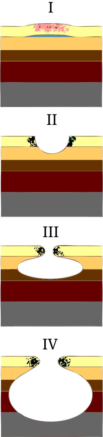
Grading: NPIAP pressure injury staging
The National Pressure Injury Advisory Panel (NPIAP) describes the following categories:
- Stage 1: Non-blanchable erythema, intact skin.
- Stage 2: Partial-thickness skin loss (blister or shallow ulcer).
- Stage 3: Full-thickness skin loss with visible fat, but no bone/muscle.
- Stage 4: Full-thickness with exposed bone, muscle, or tendon.
- Unstageable: Wound covered by necrotic tissue, depth unclear.
- Deep tissue injury: Intact or non-intact skin with persistent non-blanchable deep red, maroon, or purple discoloration.
Diabetic wounds
Cause: Peripheral neuropathy, pressure, and poor circulation due to diabetes.
Characteristics
- Location: plantar surface of foot, metatarsal heads, heels, toes.
- Appearance: punched-out, round, deep, with well-defined edges.
- Wound bed: Pale or granulating tissue, may have necrosis.
- Drainage: minimal unless infected.
- Surrounding skin: Calloused, dry, cracked, loss of protective sensation (neuropathy).
- Pain: typically painless due to neuropathy.
- Pulses: May be present or diminished (if arterial disease is also present).
- Risk factors: Diabetes, poor glycemic control, loss of sensation, and foot deformities.
- Key management: Offloading (total contact cast or therapeutic footwear), 5.07 monofilament testing for protective sensation, and glucose control.
Grading: Wagner classification for diabetic foot ulcers
- Grade 0: Intact skin with high risk (calluses, foot deformities).
- Grade 1: Superficial ulcer (partial/full-thickness skin loss).
- Grade 2: Ulcer extending to tendon, bone, or capsule.
- Grade 3: Deep ulcer with abscess, osteomyelitis.
- Grade 4: Localized gangrene.
- Grade 5: Extensive gangrene of the foot requiring amputation.
Comparing arterial, venous, and diabetic wounds
The following table compares arterial, venous, and diabetic/neuropathic wounds by location, depth and edges, drainage, pain and position, pulses, and key management.
| Feature | Arterial | Venous | Diabetic/neuropathic |
|---|---|---|---|
| Location | Toes, lateral malleolus, dorsum of foot | Medial malleolus, gaiter region | Plantar surface, metatarsal heads |
| Depth/edges | Punched-out, deep, well-defined | Shallow, irregular | Punched-out, deep, well-defined |
| Drainage | Minimal (dry) | Moderate to heavy | Minimal unless infected |
| Pain and position | Severe; worse with elevation | Mild; relieved by elevation | Typically painless (neuropathy) |
| Pulses | Diminished or absent | Present, hard to palpate | Variable |
| Key management | No compression/elevation; protect limb | Compression and elevation (after ABI) | Offloading, monofilament testing, glucose control |