



Burns and other common skin conditions
Burns
Causes: Thermal, friction, electrical, chemical, radiation
Electrical injury can cause cardiac arrhythmias, so cardiac monitoring is indicated for arrhythmias, chest pain, or any suggestion of cardiac damage; muscle destruction can also cause rhabdomyolysis with electrolyte disturbances. A burn can cause hypovolemic shock because of fluid lost from the skin.
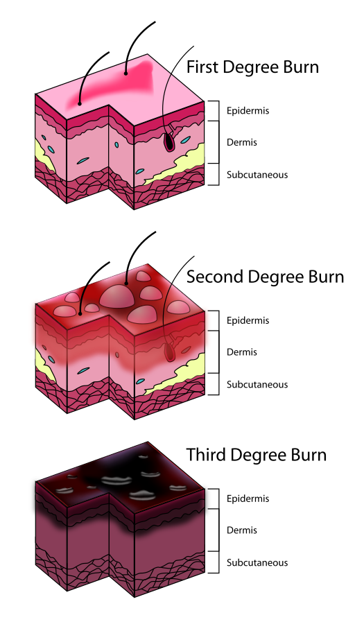
The presentation of burns is determined by the classification. See below:
Superficial (first-degree burn)
- Layers affected: Epidermis only
- Appearance: Red, dry, no blisters
- Pain level: Mild to moderate
- Healing time: 3-7 days without scarring
- Example: Sunburn
Partial-thickness (second-degree burn)
A. Superficial partial-thickness
- Layers affected: Epidermis + upper dermis
- Appearance: Red, moist, blisters present
- Pain level: Very painful (nerve endings intact)
- Healing time: 10-21 days, minimal to no scarring
B. Deep partial-thickness
- Layers affected: Epidermis + deeper dermis
- Appearance: Mottled red/white, sluggish blanching
- Pain level: Less painful (nerve endings damaged)
- Healing time: 3-6 weeks, scarring likely
Full-thickness (third-degree burn)
- Layers affected: Epidermis + entire dermis (may extend to subcutaneous tissue)
- Appearance: White, leathery, charred, dry
- Pain level: Painless (nerve endings destroyed)
- Healing time: Requires skin grafting, significant scarring
Subdermal (fourth-degree burn)
- Layers affected: Epidermis, dermis, fat, muscle, and bone
- Appearance: Black, charred, necrotic tissue
- Pain level: Painless (complete nerve destruction)
- Healing time: Requires surgical intervention; amputation is possible
Use of grafts for burns
Full-thickness burns typically require grafting to aid in re-epithelialization, and deep partial-thickness burns often (but not always) benefit from grafting to speed healing and reduce scarring. Grafts can be taken from different areas of the body, another person, or another species. Below are the different types of grafts:
- Autografts: taken from the individual’s own body
- Allografts: taken from another human, frequently a cadaver
- Xenografts: taken from another species (e.g., pig, cow)
Interventions for burns
The specific interventions for burns are determined by the depth of the wound and/or the level of tissue damage. See below for specifics regarding interventions:
Data collection is a key part of burn care: the PTA measures wound dimensions (length × width, and depth where relevant) and documents wound bed appearance (e.g., granulation, slough, eschar) and drainage, reporting changes to the supervising PT. Reclassifying burn depth is the responsibility of the PT or physician, not the PTA.
Medical management of burns
First-degree burns
- Typically managed with cool water, over-the-counter pain relievers (NSAIDs or acetaminophen), and soothing agents such as aloe vera gel or petroleum jelly.
Second-degree burns
- In addition to the above, the medical team may apply a topical antibiotic cream (such as bacitracin or neomycin), cover the burn with a non-stick dressing, and use silver sulfadiazine cream to prevent infection.
Third-degree burns
- Managed with IV fluids and pain relievers, antibiotic therapy, and topical dressings such as mafenide acetate or silver sulfadiazine.
- Surgery may be necessary to remove the burned skin and graft new skin.
- After a severe burn the body is hypermetabolic and breaks down protein rapidly, so calorie and protein needs rise; burned adults need an estimated 1.5 to 2.0 g/kg/day of protein.
Other medications
- Corticosteroids may be used to reduce inflammation.
- Antihistamines may be used to relieve itching.
- Sedatives may be used to manage pain and anxiety.
The PTA carries out wound care and exercise according to the PT’s plan of care and does not select or change medications or topical agents. Because corticosteroids can slow wound healing and topical antimicrobials such as silver can be cytotoxic with prolonged use, the PTA should report use of these medications to the supervising PT if wound progress stalls or worsens.
PT interventions for burns
- Position in the anti-contracture position - opposite the position of comfort (e.g., neck extension, shoulder abduction around 90°, elbow and knee extension, ankle dorsiflexion).
- Begin AROM/PROM and early mobility as soon as the PT’s plan allows, to prevent contracture and preserve function.
- Apply splints as prescribed by the PT to maintain anti-contracture positioning between exercise sessions.
- Perform scar massage and apply pressure garments (worn roughly 23 hours per day) to manage hypertrophic scarring once the wound has closed.
- Watch the scar during movement: tight scar tissue over a joint blanches (turns white) under stretch and limits soft tissue glide, and stretching is commonly dosed by scar blanching and the patient’s tolerance. Massaging the scar while stretching helps increase range of motion.
- Hold range of motion to a new graft site for the period specified by the surgeon or PT (commonly 3-5 days) before resuming mobilization.
- Dry skin and itching can last for months after a burn has healed, so regular use of a water-based moisturizing cream is recommended once the wound has closed.
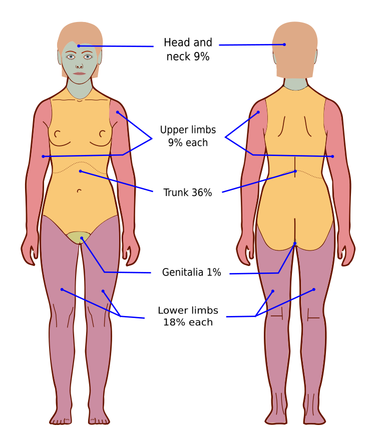
Rule of nines for burn assessment
The rule of nines is a quick method to estimate the total body surface area (TBSA) affected by burns. It differs for adults and children due to variations in body proportions.
Rule of nines for adults
In adults, the body is divided into sections, each representing 9% or a multiple of 9% of the total body surface area:
| Body part | Percentage (%) of TBSA |
| Head & neck | 9% (4.5% front, 4.5% back) |
| Each arm | 9% (4.5% front, 4.5% back) |
| Each leg | 18% (9% front, 9% back) |
| Anterior torso | 18% |
| Posterior torso | 18% |
| Perineum (genital area) | 1% |
Example: A burn covering the entire front of one leg and half the anterior torso would be 9% + 9% = 18% TBSA.
Rule of nines for children
Children have a larger head-to-body ratio, so the rule of nines is adjusted:
| Body part | Percentage (%) of TBSA |
| Head & neck | 18% (9% front, 9% back) |
| Each arm | 9% (4.5% front, 4.5% back) |
| Each leg | 14% (7% front, 7% back) |
| Anterior torso | 18% |
| Posterior torso | 18% |
Example: A child with burns covering the entire head and one arm would have 18% + 9% = 27% TBSA.
Other common skin conditions
Impetigo
Impetigo is a highly contagious bacterial skin infection that commonly affects children, marked by honey-colored, crusted lesions around the mouth, nose, and hands. It is superficial and easily treatable, without deep tissue damage.
Cellulitis
Cellulitis is a bacterial infection of the deeper skin and subcutaneous tissue. Unlike impetigo, it presents with diffuse redness, warmth, swelling, and pain, and it can spread quickly and cause systemic symptoms.
Herpes zoster (shingles)
Herpes zoster (shingles) is a reactivation of the varicella-zoster virus that causes a painful, blistering rash following a dermatomal pattern - limited to one side of the body along a nerve path. A significant complication is post-herpetic neuralgia, chronic nerve pain that can persist after the rash resolves.
The following table compares impetigo, cellulitis, and herpes zoster by cause, hallmark presentation, contagion status, treatment, and the PTA’s role.
| Condition | Cause | Hallmark presentation | Contagious? | Treatment | PTA implication |
|---|---|---|---|---|---|
| Impetigo | Bacterial (Staphylococcus aureus or Streptococcus pyogenes) | Honey-colored, crusted lesions around mouth, nose, hands | Yes | Antibiotics | Contact precautions |
| Cellulitis | Bacterial (Staphylococcus aureus or Streptococcus pyogenes) | Diffuse redness, warmth, swelling, pain; possible fever/chills | Not directly contagious | Antibiotics, possibly hospitalization | Report spreading redness, warmth, or fever to the PT |
| Herpes zoster (shingles) | Viral (reactivated varicella-zoster virus) | Painful, blistering, dermatomal rash on one side of the body | Yes, until lesions crust | Antiviral medication | Standard precautions with lesions covered if localized in an immunocompetent patient; airborne + contact precautions if disseminated or the patient is immunocompromised |
Dermatitis (eczema)
Dermatitis is a non-infectious inflammatory skin condition that presents as red, dry, scaly, itchy skin, often triggered by allergens, irritants, or stress. Treatment involves moisturizers, corticosteroids, and avoiding triggers. PT implication: avoid harsh soaps or hot water during skin or wound care, and monitor skin integrity under dressings or orthoses. Eczema (atopic dermatitis) usually appears on the inner folds of the elbows and behind the knees, and intense itching is the main symptom.
Psoriasis
Psoriasis is a chronic autoimmune condition causing rapid skin cell turnover and thick, scaly plaques, typically on the scalp, elbows, knees, and lower back; it can be linked to psoriatic arthritis. Treatment includes topical steroids, phototherapy, and systemic immunosuppressants. PT implication: watch for joint involvement from psoriatic arthritis and avoid friction or trauma over active plaques.
Scleroderma
Scleroderma is a chronic autoimmune connective tissue disease characterized by abnormal collagen deposition, leading to skin thickening, fibrosis, and vascular dysfunction. It can affect the skin, blood vessels, and internal organs, including the lungs, heart, kidneys, and gastrointestinal tract.
There are two main types:
1. Localized scleroderma - Primarily affects the skin and underlying tissues, often in morphea or linear forms.
2. Systemic sclerosis (systemic scleroderma) - Involves widespread fibrosis affecting the skin and internal organs.
Common symptoms include tight, hard skin, Raynaud’s phenomenon, joint pain, and digestive issues. There is no cure, but treatment focuses on managing symptoms and slowing disease progression with immunosuppressants, vasodilators, and physical therapy. PT implication: cold modalities are contraindicated with Raynaud’s phenomenon, and other thermal modalities need caution, and skin fibrosis should be monitored for loss of joint range of motion.