


Achievable logoAchievable logo
NPTE-PTA
Sign in
Sign up
Purchase
Textbook
Practice exams
Support
How it works
Resources
Exam catalog
Mountain with a flag at the peak
Textbook
Introduction
1. Cardiopulmonary system
2. Pulmonary system
3. Neuromuscular system
4. Pediatrics
5. Musculoskeletal system
6. Other system
6.1 Integumentary system
6.1.1 Skin structure, wound healing, and dressings
6.1.2 Debridement and wound classification by etiology
6.1.3 Burns and other common skin conditions
6.2 Lymphedema
6.3 Gastrointestinal and reproductive systems
6.4 Urinary incontinence and renal disorders
6.5 Endocrine and metabolic systems
6.6 Cancer and psychological conditions
6.7 Infectious disease, immune disorders, and hematological disorders
7. Non systems
Wrapping up
Achievable logoAchievable logo
6.1.1 Skin structure, wound healing, and dressings
Achievable NPTE-PTA
6. Other system
6.1. Integumentary system
Our NPTE-PTA course is now in "early access" - get 50% off for a limited time.

Skin structure, wound healing, and dressings

7 min read
Font
Discuss
Share
Feedback

Foundational skin information

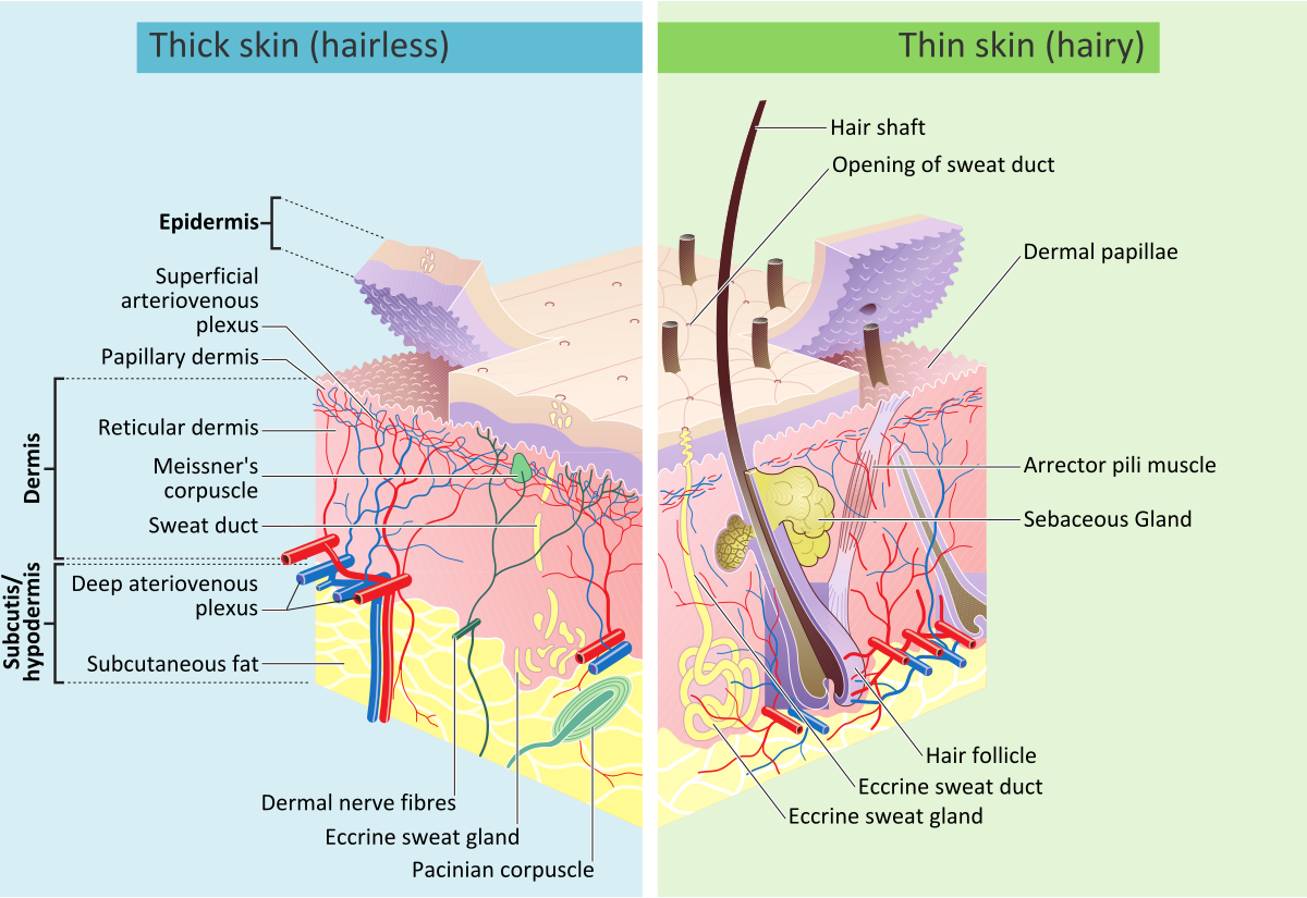
The integumentary system consists of the skin, hair, nails, glands, and sensory receptors. It serves as the first line of defense against injury, infection, and dehydration while regulating temperature and sensation. Below are the layers of skin and their descriptions:

Epidermis (outermost layer)

  • Avascular (no blood vessels)
  • Composed of keratinized stratified squamous epithelium
  • Key cells:
    • Keratinocytes - strength and waterproofing (keratin production)
    • Melanocytes - pigmentation and ultraviolet light protection
    • Langerhans cells - immune defense
    • Merkel cells - touch sensation
  • The epidermis is organized into several layers, from the deep stratum basale (site of active cell division) to the superficial stratum corneum, a barrier layer of dead, keratinized cells

Dermis (middle layer)

  • Vascularized, supplies nutrients to the epidermis
  • Contains:
    • Blood vessels
    • Nerve endings
    • Hair follicles
    • Sweat and sebaceous glands
    • Collagen and elastin fibers (structural support)

Hypodermis (subcutaneous tissue - deepest layer)

  • Composed of: fat (adipose tissue), connective tissue
  • Function: insulation, energy storage, shock absorption

Related to skin

  • Hair - protects against UV, insulates scalp
  • Nails - keratinized structures for protection and dexterity
  • Sebaceous (oil) glands - secrete sebum (moisturizes and protects skin)
  • Sweat glands - regulate temperature and remove waste
  • Eccrine glands - watery sweat, found everywhere
Layers of the skin
Layers of the skin
By - Madhero88 and M.Komorniczak, https://en.wikipedia.org/wiki/File:Skin_layers.png, CC BY-SA 3.0
/
Wikimedia Commons
/
CC BY-SA 3.0

Wound healing phases

Inflammatory phase

  • Initiated immediately and lasts 3-4 days
  • Marked by hemostasis (stopping bleeding) and phagocytosis (removal of debris and pathogens)

Proliferative phase

  • Lasts until day 21 post-injury
  • Collagen formation: A whitish protein substance that adds to the tensile strength of the wound
  • Eschar formation may occur

Maturation phase

  • Begins at day 21 and lasts up to 1-2 years
  • Leads to scar formation

Types of healing

Primary intention (first intention)

  • Tissue surfaces are closed with minimal tissue loss
  • Example: Surgical incision

Secondary intention

  • Extensive tissue loss prevents edges from being approximated
  • Example: Pressure ulcer
  • Longer repair time, increased scarring, and higher infection risk

Tertiary intention healing (delayed closure)

  • Wound is left open for a period before closure
  • Example: Abdominal wound left open for drainage before later closure

Classification of wound drainage

  • Serous - Clear to brownish fluid from the serum portion of blood
  • Sanguineous - Bright red, bloody drainage
  • Serosanguineous - Pinkish drainage, a mix of blood and serum
  • Purulent - Thick, yellow or yellow-green pus, indicating infection

How to measure wounds accurately

Measuring wounds is essential for assessing healing progress and treatment effectiveness. Proper wound measurement includes length, width, depth, and any tunneling or undermining. Below are the tools and steps for how to measure a wound.

PTA scope of practice: The PTA collects wound data (measurements, drainage, tissue type) and recognizes changes such as signs of infection or deterioration, then reports these findings to the supervising PT. Selecting a different dressing type or debridement method, or modifying the overall plan of care, is outside the PTA’s scope (performing dressing changes as directed in the plan of care is within it) - that responsibility belongs to the supervising PT.

Tools needed for wound measurement

  • Disposable ruler (centimeters or millimeters)
  • Sterile cotton-tipped applicator (for depth)
  • Wound probe (for tunneling or undermining)
  • Gloves (to maintain infection control)
  • Transparent film grid (optional for tracing irregular shapes)

Step-by-step wound measurement

Measuring length and width

  • Patient positioning: Ensure the patient is in the same position each time for consistency
  • Use a ruler: Place a disposable ruler over the wound
  • Length measurement: Measure from the longest point head-to-toe (cephalad to caudal)
  • Width measurement: Measure perpendicular to the length, at the widest point from side to side
  • Documentation format: Length × Width (e.g., 5.2 cm × 3.4 cm)

Measuring wound depth

  • Insert a sterile cotton-tipped applicator into the deepest part of the wound
  • Mark the level where it meets the wound edge (this should be perpendicular to the cotton applicator)
  • Remove and measure against a ruler
  • Example documentation: Depth = 0.8 cm

Measuring wound volume

  • Measuring wound volume can be done by multiplying length, width, and depth
  • Alternatively, measure the amount of hydrogel required to fill the wound
Definitions
Tunneling
Tract extending deeper into tissue
Undermining
Cavity or space under wound edges

Steps for measuring tunneling and undermining

  • Insert a sterile probe into the suspected tunnel/undermined area
  • Measure using a ruler
  • Use the clock method to describe location (e.g., “Tunneling at 3 o’clock position, depth 1.5 cm”)

Additional documentation details

  • Shape and edges: Irregular, round, oval, jagged
  • Tissue type: Granulation, slough, eschar
  • Exudate: amount (none, scant, moderate, heavy) and type (serous, purulent, sanguineous)
  • Periwound skin: Erythema, edema, maceration, induration

Types of dressing

Gauze dressings

  • Names: Sterile gauze, non-sterile gauze, impregnated gauze
  • Indications: Used for wounds with moderate to heavy exudate, surgical wounds, and packing deep wounds, and pressure injuries stages III, IV.
  • Contraindications: Not ideal for dry wounds (can cause adherence and delay healing)

Transparent film dressings

  • Names: Tegaderm, Opsite, Bioclusive
  • Indications: Superficial wounds, abrasions, and partial-thickness wounds with minimal exudate; also used for IV sites and pressure injuries stages I, II.
  • Contraindications: Should not be used on heavily exudating wounds or infected wounds

Hydrocolloid dressings

  • Names: Duoderm, Comfeel, Tegasorb
  • Indications: Light to moderate exudating wounds, pressure injuries stage II and shallow stage III, and venous leg ulcers. Helps maintain a moist wound environment.
  • Contraindications: Not suitable for infected wounds or wounds with heavy exudate.

Hydrogel dressings

  • Names: IntraSite Gel, Aquaform, Vigilon
  • Types: Gel, sheet, or beads (for deep wounds)
  • Indications: Dry wounds, burns, painful wounds, and wounds with necrotic tissue (autolytic debridement), and pressure injuries stages II, III, IV.
  • Contraindications: Should not be used on heavily exudating wounds as it can cause maceration.

Foam dressings

  • Names: Allevyn, Mepilex, Lyofoam
  • Indications: Moderate to heavily exudating wounds, venous ulcers, pressure ulcers, and diabetic foot ulcers and pressure injuries stages II, III, IV. Provides cushioning.
  • Contraindications: Not ideal for dry wounds or wounds with minimal exudate.

Alginate dressings

  • Names: Kaltostat, Sorbsan, Algicell
  • Indications: Heavy exudating wounds, infected wounds, and wounds with tunneling. Derived from seaweed, absorbs large amounts of fluid, and pressure injuries stages III, IV.
  • Contraindications: Not recommended for dry wounds or wounds with minimal exudate.
  • Characteristics: When an alginate dressing is removed, it can have a yellowish, tan, or brown color and may have a mild, but unpleasant odor. While the appearance and smell can be alarming, they are often normal and result from the interaction between the seaweed-based dressing and the wound’s exudate.

Collagen dressings

  • Names: Promogran, Fibracol, Biostep
  • Indications: Chronic non-healing wounds, pressure ulcers, wounds with granulation tissue, and pressure injuries stages II, III, IV.
  • Encourages new tissue growth.
  • Contraindications: Not for dry wounds or wounds with eschar. Avoid in patients with collagen allergies.

Antimicrobial dressings

  • Names: Silvercel, Acticoat, Iodosorb
  • Indications: Infected wounds, burns, surgical wounds at risk of infection, and pressure injuries stages II, III, IV. Contains silver or iodine to reduce bacterial load.
  • Contraindications: Avoid in patients allergic to silver or iodine. Not recommended for long-term use due to potential cytotoxicity.

Foundational skin information

  • Integumentary system: skin, hair, nails, glands, sensory receptors
  • Functions: defense against injury/infection/dehydration, temperature regulation, sensation

Epidermis (outermost layer)

  • Avascular; keratinized stratified squamous epithelium
  • Key cells:
    • Keratinocytes - strength/waterproofing
    • Melanocytes - pigmentation/UV protection
    • Langerhans cells - immune defense
    • Merkel cells - touch sensation
  • Layers: stratum basale (deep, active division) to stratum corneum (superficial, dead cells)

Dermis (middle layer)

  • Vascularized; nourishes epidermis
  • Contains blood vessels, nerve endings, hair follicles, sweat/sebaceous glands
  • Collagen and elastin provide structural support

Hypodermis (deepest layer)

  • Composed of adipose and connective tissue
  • Function: insulation, energy storage, shock absorption

Related to skin

  • Hair - UV protection, insulation
  • Nails - protection, dexterity
  • Sebaceous glands - secrete sebum (moisturize/protect)
  • Sweat/eccrine glands - temperature regulation, waste removal

Wound healing phases

  • Inflammatory phase: immediate, lasts 3-4 days; hemostasis and phagocytosis
  • Proliferative phase: until day 21; collagen formation (tensile strength), possible eschar
  • Maturation phase: day 21 to 1-2 years; results in scar formation

Types of healing

  • Primary intention: minimal tissue loss, edges closed (e.g., surgical incision)
  • Secondary intention: extensive tissue loss, edges not approximated (e.g., pressure ulcer); longer healing, more scarring/infection risk
  • Tertiary intention: delayed closure, wound left open then closed (e.g., abdominal wound for drainage)

Classification of wound drainage

  • Serous - clear/brownish fluid
  • Sanguineous - bright red, bloody
  • Serosanguineous - pink (blood + serum mix)
  • Purulent - thick yellow/green pus (infection sign)

How to measure wounds accurately

  • Measure length, width, depth, tunneling/undermining for healing assessment
  • PTA scope: collects data, reports changes/infection signs to PT
    • Cannot change dressing type, debridement method, or plan of care (outside scope)
    • Can perform dressing changes as directed

Tools for wound measurement

  • Disposable ruler, sterile cotton-tipped applicator, wound probe, gloves, transparent film grid (optional)

Step-by-step measurement

  • Consistent patient positioning each time
  • Length: longest point head-to-toe (cephalad-caudal)
  • Width: perpendicular to length, widest point
  • Documentation: Length × Width (e.g., 5.2 cm × 3.4 cm)
  • Depth: insert applicator into deepest point, mark, measure against ruler

Measuring wound volume

  • Calculate: length × width × depth
  • Alternative: measure hydrogel needed to fill wound

Tunneling vs undermining

  • Tunneling: tract extending deeper into tissue
  • Undermining: cavity/space under wound edges
  • Measure with sterile probe; document using clock method (e.g., “3 o’clock position, depth 1.5 cm”)

Additional documentation details

  • Shape/edges: irregular, round, oval, jagged
  • Tissue type: granulation, slough, eschar
  • Exudate: amount (none/scant/moderate/heavy) and type
  • Periwound skin: erythema, edema, maceration, induration

Gauze dressings

  • Names: sterile/non-sterile/impregnated gauze
  • Use: moderate-heavy exudate, surgical wounds, deep packing, stages III-IV
  • Avoid: dry wounds (causes adherence)

Transparent film dressings

  • Names: Tegaderm, Opsite, Bioclusive
  • Use: superficial/minimal exudate wounds, IV sites, stages I-II
  • Avoid: heavy exudate or infected wounds

Hydrocolloid dressings

  • Names: Duoderm, Comfeel, Tegasorb
  • Use: light-moderate exudate, stage II/shallow III, venous ulcers; maintains moisture
  • Avoid: infected or heavily exudating wounds

Hydrogel dressings

  • Names: IntraSite Gel, Aquaform, Vigilon
  • Forms: gel, sheet, beads
  • Use: dry wounds, burns, necrotic tissue (autolytic debridement), stages II-IV
  • Avoid: heavy exudate (causes maceration)

Foam dressings

  • Names: Allevyn, Mepilex, Lyofoam
  • Use: moderate-heavy exudate, venous/diabetic/pressure ulcers, stages II-IV; provides cushioning
  • Avoid: dry or minimal exudate wounds

Alginate dressings

  • Names: Kaltostat, Sorbsan, Algicell
  • Use: heavy exudate, infected wounds, tunneling; seaweed-derived, high absorption, stages III-IV
  • Avoid: dry/minimal exudate wounds
  • Note: removal may show yellow/tan/brown color, mild odor - often normal

Collagen dressings

  • Names: Promogran, Fibracol, Biostep
  • Use: chronic non-healing wounds, granulation tissue, stages II-IV; promotes tissue growth
  • Avoid: dry wounds, eschar, collagen allergies

Antimicrobial dressings

  • Names: Silvercel, Acticoat, Iodosorb
  • Use: infected wounds, burns, at-risk surgical wounds, stages II-IV; contains silver/iodine
  • Avoid: silver/iodine allergies; not for long-term use (cytotoxicity risk)

Sign up for free to take 10 quiz questions on this topic

Previous
Next  | 6.1.2 Debridement and wound classification by etiology
All rights reserved ©2016 - 2026 Achievable, Inc.

Skin structure, wound healing, and dressings

Foundational skin information

The integumentary system consists of the skin, hair, nails, glands, and sensory receptors. It serves as the first line of defense against injury, infection, and dehydration while regulating temperature and sensation. Below are the layers of skin and their descriptions:

Epidermis (outermost layer)

  • Avascular (no blood vessels)
  • Composed of keratinized stratified squamous epithelium
  • Key cells:
    • Keratinocytes - strength and waterproofing (keratin production)
    • Melanocytes - pigmentation and ultraviolet light protection
    • Langerhans cells - immune defense
    • Merkel cells - touch sensation
  • The epidermis is organized into several layers, from the deep stratum basale (site of active cell division) to the superficial stratum corneum, a barrier layer of dead, keratinized cells

Dermis (middle layer)

  • Vascularized, supplies nutrients to the epidermis
  • Contains:
    • Blood vessels
    • Nerve endings
    • Hair follicles
    • Sweat and sebaceous glands
    • Collagen and elastin fibers (structural support)

Hypodermis (subcutaneous tissue - deepest layer)

  • Composed of: fat (adipose tissue), connective tissue
  • Function: insulation, energy storage, shock absorption

Related to skin

  • Hair - protects against UV, insulates scalp
  • Nails - keratinized structures for protection and dexterity
  • Sebaceous (oil) glands - secrete sebum (moisturizes and protects skin)
  • Sweat glands - regulate temperature and remove waste
  • Eccrine glands - watery sweat, found everywhere

Wound healing phases

Inflammatory phase

  • Initiated immediately and lasts 3-4 days
  • Marked by hemostasis (stopping bleeding) and phagocytosis (removal of debris and pathogens)

Proliferative phase

  • Lasts until day 21 post-injury
  • Collagen formation: A whitish protein substance that adds to the tensile strength of the wound
  • Eschar formation may occur

Maturation phase

  • Begins at day 21 and lasts up to 1-2 years
  • Leads to scar formation

Types of healing

Primary intention (first intention)

  • Tissue surfaces are closed with minimal tissue loss
  • Example: Surgical incision

Secondary intention

  • Extensive tissue loss prevents edges from being approximated
  • Example: Pressure ulcer
  • Longer repair time, increased scarring, and higher infection risk

Tertiary intention healing (delayed closure)

  • Wound is left open for a period before closure
  • Example: Abdominal wound left open for drainage before later closure

Classification of wound drainage

  • Serous - Clear to brownish fluid from the serum portion of blood
  • Sanguineous - Bright red, bloody drainage
  • Serosanguineous - Pinkish drainage, a mix of blood and serum
  • Purulent - Thick, yellow or yellow-green pus, indicating infection

How to measure wounds accurately

Measuring wounds is essential for assessing healing progress and treatment effectiveness. Proper wound measurement includes length, width, depth, and any tunneling or undermining. Below are the tools and steps for how to measure a wound.

PTA scope of practice: The PTA collects wound data (measurements, drainage, tissue type) and recognizes changes such as signs of infection or deterioration, then reports these findings to the supervising PT. Selecting a different dressing type or debridement method, or modifying the overall plan of care, is outside the PTA’s scope (performing dressing changes as directed in the plan of care is within it) - that responsibility belongs to the supervising PT.

Tools needed for wound measurement

  • Disposable ruler (centimeters or millimeters)
  • Sterile cotton-tipped applicator (for depth)
  • Wound probe (for tunneling or undermining)
  • Gloves (to maintain infection control)
  • Transparent film grid (optional for tracing irregular shapes)

Step-by-step wound measurement

Measuring length and width

  • Patient positioning: Ensure the patient is in the same position each time for consistency
  • Use a ruler: Place a disposable ruler over the wound
  • Length measurement: Measure from the longest point head-to-toe (cephalad to caudal)
  • Width measurement: Measure perpendicular to the length, at the widest point from side to side
  • Documentation format: Length × Width (e.g., 5.2 cm × 3.4 cm)

Measuring wound depth

  • Insert a sterile cotton-tipped applicator into the deepest part of the wound
  • Mark the level where it meets the wound edge (this should be perpendicular to the cotton applicator)
  • Remove and measure against a ruler
  • Example documentation: Depth = 0.8 cm

Measuring wound volume

  • Measuring wound volume can be done by multiplying length, width, and depth
  • Alternatively, measure the amount of hydrogel required to fill the wound
Definitions
Tunneling
Tract extending deeper into tissue
Undermining
Cavity or space under wound edges

Steps for measuring tunneling and undermining

  • Insert a sterile probe into the suspected tunnel/undermined area
  • Measure using a ruler
  • Use the clock method to describe location (e.g., “Tunneling at 3 o’clock position, depth 1.5 cm”)

Additional documentation details

  • Shape and edges: Irregular, round, oval, jagged
  • Tissue type: Granulation, slough, eschar
  • Exudate: amount (none, scant, moderate, heavy) and type (serous, purulent, sanguineous)
  • Periwound skin: Erythema, edema, maceration, induration

Types of dressing

Gauze dressings

  • Names: Sterile gauze, non-sterile gauze, impregnated gauze
  • Indications: Used for wounds with moderate to heavy exudate, surgical wounds, and packing deep wounds, and pressure injuries stages III, IV.
  • Contraindications: Not ideal for dry wounds (can cause adherence and delay healing)

Transparent film dressings

  • Names: Tegaderm, Opsite, Bioclusive
  • Indications: Superficial wounds, abrasions, and partial-thickness wounds with minimal exudate; also used for IV sites and pressure injuries stages I, II.
  • Contraindications: Should not be used on heavily exudating wounds or infected wounds

Hydrocolloid dressings

  • Names: Duoderm, Comfeel, Tegasorb
  • Indications: Light to moderate exudating wounds, pressure injuries stage II and shallow stage III, and venous leg ulcers. Helps maintain a moist wound environment.
  • Contraindications: Not suitable for infected wounds or wounds with heavy exudate.

Hydrogel dressings

  • Names: IntraSite Gel, Aquaform, Vigilon
  • Types: Gel, sheet, or beads (for deep wounds)
  • Indications: Dry wounds, burns, painful wounds, and wounds with necrotic tissue (autolytic debridement), and pressure injuries stages II, III, IV.
  • Contraindications: Should not be used on heavily exudating wounds as it can cause maceration.

Foam dressings

  • Names: Allevyn, Mepilex, Lyofoam
  • Indications: Moderate to heavily exudating wounds, venous ulcers, pressure ulcers, and diabetic foot ulcers and pressure injuries stages II, III, IV. Provides cushioning.
  • Contraindications: Not ideal for dry wounds or wounds with minimal exudate.

Alginate dressings

  • Names: Kaltostat, Sorbsan, Algicell
  • Indications: Heavy exudating wounds, infected wounds, and wounds with tunneling. Derived from seaweed, absorbs large amounts of fluid, and pressure injuries stages III, IV.
  • Contraindications: Not recommended for dry wounds or wounds with minimal exudate.
  • Characteristics: When an alginate dressing is removed, it can have a yellowish, tan, or brown color and may have a mild, but unpleasant odor. While the appearance and smell can be alarming, they are often normal and result from the interaction between the seaweed-based dressing and the wound’s exudate.

Collagen dressings

  • Names: Promogran, Fibracol, Biostep
  • Indications: Chronic non-healing wounds, pressure ulcers, wounds with granulation tissue, and pressure injuries stages II, III, IV.
  • Encourages new tissue growth.
  • Contraindications: Not for dry wounds or wounds with eschar. Avoid in patients with collagen allergies.

Antimicrobial dressings

  • Names: Silvercel, Acticoat, Iodosorb
  • Indications: Infected wounds, burns, surgical wounds at risk of infection, and pressure injuries stages II, III, IV. Contains silver or iodine to reduce bacterial load.
  • Contraindications: Avoid in patients allergic to silver or iodine. Not recommended for long-term use due to potential cytotoxicity.
Key points

Foundational skin information

  • Integumentary system: skin, hair, nails, glands, sensory receptors
  • Functions: defense against injury/infection/dehydration, temperature regulation, sensation

Epidermis (outermost layer)

  • Avascular; keratinized stratified squamous epithelium
  • Key cells:
    • Keratinocytes - strength/waterproofing
    • Melanocytes - pigmentation/UV protection
    • Langerhans cells - immune defense
    • Merkel cells - touch sensation
  • Layers: stratum basale (deep, active division) to stratum corneum (superficial, dead cells)

Dermis (middle layer)

  • Vascularized; nourishes epidermis
  • Contains blood vessels, nerve endings, hair follicles, sweat/sebaceous glands
  • Collagen and elastin provide structural support

Hypodermis (deepest layer)

  • Composed of adipose and connective tissue
  • Function: insulation, energy storage, shock absorption

Related to skin

  • Hair - UV protection, insulation
  • Nails - protection, dexterity
  • Sebaceous glands - secrete sebum (moisturize/protect)
  • Sweat/eccrine glands - temperature regulation, waste removal

Wound healing phases

  • Inflammatory phase: immediate, lasts 3-4 days; hemostasis and phagocytosis
  • Proliferative phase: until day 21; collagen formation (tensile strength), possible eschar
  • Maturation phase: day 21 to 1-2 years; results in scar formation

Types of healing

  • Primary intention: minimal tissue loss, edges closed (e.g., surgical incision)
  • Secondary intention: extensive tissue loss, edges not approximated (e.g., pressure ulcer); longer healing, more scarring/infection risk
  • Tertiary intention: delayed closure, wound left open then closed (e.g., abdominal wound for drainage)

Classification of wound drainage

  • Serous - clear/brownish fluid
  • Sanguineous - bright red, bloody
  • Serosanguineous - pink (blood + serum mix)
  • Purulent - thick yellow/green pus (infection sign)

How to measure wounds accurately

  • Measure length, width, depth, tunneling/undermining for healing assessment
  • PTA scope: collects data, reports changes/infection signs to PT
    • Cannot change dressing type, debridement method, or plan of care (outside scope)
    • Can perform dressing changes as directed

Tools for wound measurement

  • Disposable ruler, sterile cotton-tipped applicator, wound probe, gloves, transparent film grid (optional)

Step-by-step measurement

  • Consistent patient positioning each time
  • Length: longest point head-to-toe (cephalad-caudal)
  • Width: perpendicular to length, widest point
  • Documentation: Length × Width (e.g., 5.2 cm × 3.4 cm)
  • Depth: insert applicator into deepest point, mark, measure against ruler

Measuring wound volume

  • Calculate: length × width × depth
  • Alternative: measure hydrogel needed to fill wound

Tunneling vs undermining

  • Tunneling: tract extending deeper into tissue
  • Undermining: cavity/space under wound edges
  • Measure with sterile probe; document using clock method (e.g., “3 o’clock position, depth 1.5 cm”)

Additional documentation details

  • Shape/edges: irregular, round, oval, jagged
  • Tissue type: granulation, slough, eschar
  • Exudate: amount (none/scant/moderate/heavy) and type
  • Periwound skin: erythema, edema, maceration, induration

Gauze dressings

  • Names: sterile/non-sterile/impregnated gauze
  • Use: moderate-heavy exudate, surgical wounds, deep packing, stages III-IV
  • Avoid: dry wounds (causes adherence)

Transparent film dressings

  • Names: Tegaderm, Opsite, Bioclusive
  • Use: superficial/minimal exudate wounds, IV sites, stages I-II
  • Avoid: heavy exudate or infected wounds

Hydrocolloid dressings

  • Names: Duoderm, Comfeel, Tegasorb
  • Use: light-moderate exudate, stage II/shallow III, venous ulcers; maintains moisture
  • Avoid: infected or heavily exudating wounds

Hydrogel dressings

  • Names: IntraSite Gel, Aquaform, Vigilon
  • Forms: gel, sheet, beads
  • Use: dry wounds, burns, necrotic tissue (autolytic debridement), stages II-IV
  • Avoid: heavy exudate (causes maceration)

Foam dressings

  • Names: Allevyn, Mepilex, Lyofoam
  • Use: moderate-heavy exudate, venous/diabetic/pressure ulcers, stages II-IV; provides cushioning
  • Avoid: dry or minimal exudate wounds

Alginate dressings

  • Names: Kaltostat, Sorbsan, Algicell
  • Use: heavy exudate, infected wounds, tunneling; seaweed-derived, high absorption, stages III-IV
  • Avoid: dry/minimal exudate wounds
  • Note: removal may show yellow/tan/brown color, mild odor - often normal

Collagen dressings

  • Names: Promogran, Fibracol, Biostep
  • Use: chronic non-healing wounds, granulation tissue, stages II-IV; promotes tissue growth
  • Avoid: dry wounds, eschar, collagen allergies

Antimicrobial dressings

  • Names: Silvercel, Acticoat, Iodosorb
  • Use: infected wounds, burns, at-risk surgical wounds, stages II-IV; contains silver/iodine
  • Avoid: silver/iodine allergies; not for long-term use (cytotoxicity risk)

More from Integumentary system

  • Debridement and wound classification by etiology
  • Burns and other common skin conditions