



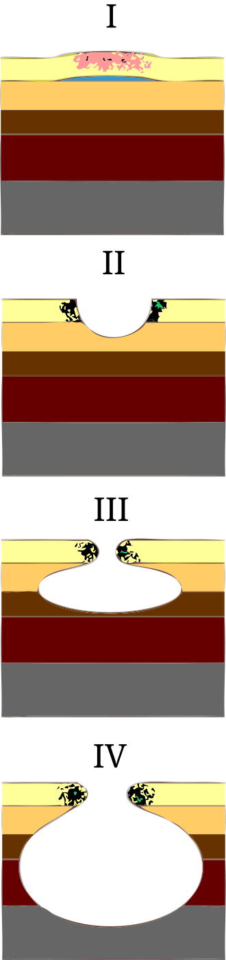
Debridement and wound classification by etiology
Types of debridement
Debridement is the removal of slough or eschar/necrotic tissue from a wound base. The goal is to remove these unhealthy wound products and leave only granulation tissue.
Autolytic debridement
- Definition: Uses the body’s own enzymes and moisture to break down necrotic tissue.
- Methods: Hydrocolloid, hydrogel, or transparent film dressings.
- Indications:
- Wounds with necrotic tissue (eschar or slough).
- Patients who cannot tolerate more aggressive debridement.
- Minimal to moderate exudating wounds.
- Contraindications:
- Infected wounds.
- Heavily exudating wounds.
Mechanical debridement
- Definition: Uses physical forces to remove necrotic tissue.
- Methods:
- Wet-to-dry dressings (gauze applied wet and removed dry).
- Hydrotherapy (whirlpool).
- Pulsed lavage (pressurized irrigation).
- Indications:
- Moderate to heavily necrotic wounds.
- Wounds with thick, adherent slough or debris.
- Contraindications:
- Painful wounds.
- Granulating wounds (can damage healthy tissue).
Enzymatic debridement
- Definition: Uses topical enzymatic agents to break down necrotic tissue.
- Methods: Application of collagenase (e.g., Santyl).
- Indications:
- Wounds with thick, adherent necrotic tissue.
- Patients unable to tolerate surgical or sharp debridement.
- Contraindications:
- Infected wounds without antibiotic coverage.
- Clean wounds with no necrotic tissue.
Sharp debridement
- Definition: Uses scalpels, scissors, or forceps to remove necrotic tissue manually.
- Indications:
- Wounds with extensive necrosis or thick eschar.
- Infected wounds requiring immediate removal of necrotic tissue.
- Contraindications:
- Bleeding disorders or anticoagulated patients.
- Poor vascular supply (risk of delayed healing).
Surgical debridement
- Definition: Performed by a surgeon in an operating room to remove large amounts of necrotic tissue.
- Indications:
- Deep wounds with extensive necrosis.
- Life-threatening infections (e.g., necrotizing fasciitis).
- Chronic non-healing wounds requiring aggressive intervention.
- Contraindications:
- Patients who are medically unstable for surgery.
Biological (maggot) debridement
- Definition: Uses sterile maggots (larvae of Lucilia sericata) to digest necrotic tissue and bacteria.
- Indications:
- Chronic, non-healing wounds with necrotic tissue.
- Infected wounds (maggots secrete antimicrobial enzymes).
- Contraindications:
- Patient refusal.
- Allergies to fly larvae or materials used in dressing containment.
Wound classifications by etiology
Wounds are also classified by their underlying cause, which shapes their location, appearance, and treatment. Arterial and venous wounds share the same thickness grading system: partial thickness wounds extend through the epidermis and part of the dermis, while full thickness wounds extend into the subcutaneous tissue.
Arterial wounds
Cause: Reduced arterial blood supply leading to ischemia and necrosis.
Characteristics
- Location: Toes, lateral malleolus, dorsum of the foot, distal lower extremities.
- Appearance: Punched-out, deep with well-defined edges.
- Wound bed: Pale, necrotic, minimal granulation tissue.
- Drainage: minimal (often dry).
- Surrounding skin: Cool, shiny, thin, hair loss, pallor with elevation, dependent rubor.
- Pain: severe, worsens with elevation, relieved in a dependent position.
- Pulses: Diminished or absent.
- Risk factors: Peripheral arterial disease (PAD), smoking, diabetes, and hypertension.
Venous wounds
Cause: Venous insufficiency leading to fluid retention, inflammation, and skin breakdown.
Characteristics
- Location: Medial malleolus, lower leg (gaiter region).
- Appearance: Shallow, irregular borders, large wound area.
- Wound bed: Granular, red with yellow fibrinous tissue.
- Drainage: Moderate to heavy (wet wound)
- Surrounding skin: Edema, hemosiderin staining (brown discoloration), lipodermatosclerosis (thickened skin).
- Pain: Mild to moderate, relieved by elevation.
- Pulses: Present but may be difficult to palpate due to edema.
- Risk factors: Chronic venous insufficiency, varicose veins, obesity, prolonged standing.
Pressure wounds
Cause: Prolonged pressure, shear, or friction leading to ischemic damage.
Characteristics
- Location: Bony prominences (sacrum, heels, ischial tuberosities, greater trochanter).
- Appearance: Varies by stage, from non-blanchable redness to deep tissue damage.
- Drainage: Variable (depends on stage and infection presence).
- Surrounding skin: May show maceration, erythema, or induration.
- Pain: Variable (more in early stages, less in advanced necrotic ulcers).
- Risk factors: Immobility, malnutrition, incontinence, neuropathy.
Grading: national pressure injury staging
- Stage 1: Non-blanchable erythema, intact skin.
- Stage 2: Partial-thickness skin loss (blister or shallow ulcer).
- Stage 3: Full-thickness skin loss with visible fat, but no bone/muscle.
- Stage 4: Full-thickness with exposed bone, muscle, or tendon.
- Unstageable: Wound covered by necrotic tissue, depth unclear.
- Deep tissue injury: Intact or non-intact skin with persistent non-blanchable deep red, maroon, or purple discoloration.
Diabetic wounds
Cause: Peripheral neuropathy, pressure, and poor circulation due to diabetes.
Characteristics
- Location: plantar surface of foot, metatarsal heads, heels, toes.
- Appearance: punched-out, round, deep, with well-defined edges.
- Wound bed: Pale or granulating tissue, may have necrosis.
- Drainage: minimal unless infected.
- Surrounding skin: Calloused, dry, cracked, loss of protective sensation (neuropathy).
- Pain: typically painless due to neuropathy.
- Pulses: May be present or diminished (if arterial disease is also present).
- Risk factors: Diabetes, poor glycemic control, loss of sensation, and foot deformities.
Grading: Wagner classification for diabetic foot ulcers
- Grade 0: Intact skin with high risk (calluses, foot deformities).
- Grade 1: Superficial ulcer (partial/full-thickness skin loss).
- Grade 2: Ulcer extending to tendon, bone, or capsule.
- Grade 3: Deep ulcer with abscess, osteomyelitis.
- Grade 4: Localized gangrene.
- Grade 5: Extensive gangrene of the foot requiring amputation.