


Achievable logoAchievable logo
NPTE-PTA
Sign in
Sign up
Purchase
Textbook
Practice exams
Support
How it works
Resources
Exam catalog
Mountain with a flag at the peak
Textbook
Introduction
1. Cardiopulmonary system
2. Pulmonary system
3. Neuromuscular system
4. Pediatrics
5. Musculoskeletal system
6. Other system
6.1 Integumentary system
6.1.1 Skin structure, wound healing, and dressings
6.1.2 Debridement and wound classification by etiology
6.1.3 Burns and other common skin conditions
6.2 Lymphedema
6.3 Gastrointestinal and reproductive systems
6.4 Urinary incontinence and renal disorders
6.5 Endocrine and metabolic systems
6.6 Cancer and psychological conditions
6.7 Infectious disease, immune disorders, and hematological disorders
7. Non systems
Wrapping up
Achievable logoAchievable logo
6.1.3 Burns and other common skin conditions
Achievable NPTE-PTA
6. Other system
6.1. Integumentary system
Our NPTE-PTA course is now in "early access" - get 50% off for a limited time.

Burns and other common skin conditions

8 min read
Font
Discuss
Share
Feedback

Burns

Causes: Thermal, friction, electrical, chemical, radiation

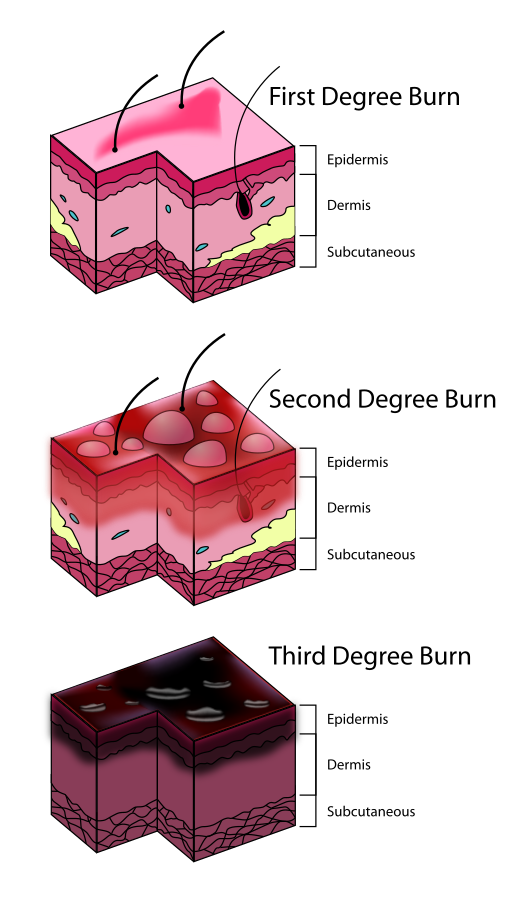
The presentation of burns is determined by the classification. See below:

Superficial (first-degree burn)

  • Layers affected: Epidermis only
  • Appearance: Red, dry, no blisters
  • Pain level: Mild to moderate
  • Healing time: 3-7 days without scarring
  • Example: Sunburn

Partial-thickness (second-degree burn)

a. Superficial partial-thickness

  • Layers affected: Epidermis + upper dermis
  • Appearance: Red, moist, blisters present
  • Pain level: Very painful (nerve endings intact)
  • Healing time: 10-21 days, minimal to no scarring

b. Deep partial-thickness

  • Layers affected: Epidermis + deeper dermis
  • Appearance: Mottled red/white, sluggish blanching
  • Pain level: Less painful (nerve endings damaged)
  • Healing time: 3-6 weeks, scarring likely

Full-thickness (third-degree burn)

  • Layers affected: Epidermis + entire dermis (may extend to subcutaneous tissue)
  • Appearance: White, leathery, charred, dry
  • Pain level: Painless (nerve endings destroyed)
  • Healing time: Requires skin grafting, significant scarring

Subdermal (fourth-degree burn)

  • Layers affected: Epidermis, dermis, fat, muscle, and bone
  • Appearance: Black, charred, necrotic tissue
  • Pain level: Painless (complete nerve destruction)
  • Healing time: Requires surgical intervention; amputation is possible

Use of grafts for burns

Grafts are required for deep partial thickness and full thickness burns to aid in re-epithelialization. Grafts can be taken from different areas of the body, another individual’s body, or another species. Below are different types of grafts:

  • Autografts: taken from the individual
  • Allografts: taken from cadavers
  • Xenografts: taken from another species (i.e., pig, cow)

Interventions for burns

The specific interventions for burns are determined by the depth of the wound and/or the level of tissue damage. See below for specifics regarding interventions:

Classification of burns
By - The original uploader was K. Aainsqatsi at English Wikipedia., Transferred from en.wikipedia to Commons., CC BY-SA 3.0
/
Wikimedia Commons
/
CC BY-SA 3.0

First-degree burns

  • Cool the burn with cool water.
  • Apply over-the-counter pain relievers, such as NSAIDs or acetaminophen.
  • Use aloe vera gel or petroleum jelly to soothe the burn.

Second-degree burns

  • In addition to the above, apply a topical antibiotic cream, such as Bacitracin or Neomycin.
  • Cover the burn with a non-stick dressing.
  • Use silver sulfadiazine cream to prevent infection.
Definitions
Bacitracin
Topical antibiotic that stops bacterial growth in a wound.
Neomycin
Topical antibiotic that fights infection, primarily against gram-negative bacteria.
Silver sulfadiazine
Antimicrobial cream used to prevent and treat infection in second- and third-degree burns.
Mafenide acetate
Antimicrobial cream that reduces bacterial load in burn tissue to promote healing of deep burns.

Third-degree burns

  • Intravenous fluids and pain relievers
  • Antibiotic therapy
  • Topical dressings, such as mafenide acetate or silver sulfadiazine
  • Surgery may be necessary to remove the burned skin and graft new skin.

Other medications

  • Corticosteroids may be used to reduce inflammation.
  • Antihistamines may be used to relieve itching.
  • Sedatives may be used to manage pain and anxiety.

Because corticosteroids can slow wound healing and topical antimicrobials such as silver can be cytotoxic with prolonged use, the PTA should report use of these medications to the supervising PT if wound progress stalls or worsens.

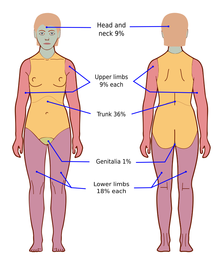
Rule of nines for burn assessment

The rule of nines is a quick method to estimate the total body surface area (TBSA) affected by burns. It differs for adults and children due to variations in body proportions.

Rule of nines for adults

In adults, the body is divided into sections, each representing 9% or a multiple of 9% of the total body surface area:

Body part Percentage (%) of TBSA
Head & neck 9% (4.5% front, 4.5% back)
Each arm 9% (4.5% front, 4.5% back)
Each leg 18% (9% front, 9% back)
Anterior torso 18%
Posterior torso 18%
Perineum (genital area) 1%

Example: A burn covering the entire front of one leg and half the anterior torso would be 9% + 9% = 18% TBSA.

Rule of nines for adults
By - Jmarchn, Own work,CC BY-SA 3.0
/
Wikimedia Commons
/
CC BY-SA 3.0

Rule of nines for children

Children have a larger head-to-body ratio, so the Rule of Nines is adjusted:

Body part Percentage (%) of TBSA
Head & neck 18% (9% front, 9% back)
Each arm 9% (4.5% front, 4.5% back)
Each leg 14% (7% front, 7% back)
Anterior torso 18%
Posterior torso 18%

Example: A child with burns covering the entire head and one arm would have 18% + 9% = 27% TBSA.

Other common skin conditions

Impetigo

Impetigo is a bacterial skin infection caused by Staphylococcus aureus or Streptococcus pyogenes. It is highly contagious and commonly affects children. The hallmark feature is honey-colored, crusted lesions that typically appear around the mouth, nose, and hands. Mild itching may be present, but systemic symptoms like fever are rare. Since impetigo is superficial, it does not cause deep tissue damage and is easily treatable with antibiotics.

Cellulitis

Cellulitis is a bacterial skin infection that occurs in the deeper layers of the skin and subcutaneous tissue. It is most commonly caused by Staphylococcus aureus or Streptococcus pyogenes. Unlike impetigo, cellulitis presents with diffuse redness, warmth, swelling, and pain, and it may be accompanied by systemic symptoms such as fever, chills, and malaise. It commonly affects the lower extremities, but can occur anywhere. Cellulitis can spread quickly and requires antibiotics, and in severe cases, hospitalization may be necessary.

Herpes zoster (shingles)

Herpes zoster, or shingles, is a viral infection caused by the reactivation of the varicella-zoster virus (the same virus that causes chickenpox). It presents as a painful, blistering rash that follows a dermatomal pattern - meaning it is limited to one side of the body along a nerve path. Before the rash appears, individuals may experience burning, tingling, or flu-like symptoms. Unlike impetigo and cellulitis, herpes zoster is not a bacterial infection and requires antiviral treatment. A significant complication is post-herpetic neuralgia, which can cause chronic nerve pain even after the rash has resolved.

Dermatitis (eczema)

Dermatitis is a non-infectious inflammatory skin condition caused by an immune response to allergens, irritants, or chronic skin sensitivity. It presents as red, dry, scaly, and itchy skin and is often associated with pruritus (intense itching). Unlike the other conditions, dermatitis is not contagious and does not involve bacterial or viral pathogens. Common triggers include allergens, soaps, stress, and climate changes. Treatment involves moisturizers, corticosteroids, and avoiding triggers.

Psoriasis

Psoriasis is a chronic autoimmune skin condition that causes rapid skin cell turnover, leading to the buildup of thick, scaly plaques on the skin. It is characterized by red, inflamed patches with silvery-white scales, often appearing on the scalp, elbows, knees, and lower back. The condition is not contagious and can be triggered by stress, infections, medications, and environmental factors. Psoriasis is associated with immune system dysfunction, specifically involving T-cells and inflammatory cytokines. It can also be linked to psoriatic arthritis, which affects the joints. Treatment includes topical steroids, phototherapy, and systemic immunosuppressants.

Scleroderma

Scleroderma is a chronic autoimmune connective tissue disease characterized by abnormal collagen deposition, leading to skin thickening, fibrosis, and vascular dysfunction. It can affect the skin, blood vessels, and internal organs, including the lungs, heart, kidneys, and gastrointestinal tract.

There are two main types:
1. Localized scleroderma - Primarily affects the skin and underlying tissues, often in morphea or linear forms.
2. Systemic sclerosis (systemic scleroderma) - Involves widespread fibrosis affecting the skin and internal organs.

Common symptoms include tight, hard skin, Raynaud’s phenomenon, joint pain, and digestive issues. The exact cause is unknown, but it is believed to involve immune system dysfunction and genetic/environmental factors. There is no cure, but treatment focuses on managing symptoms and slowing disease progression with immunosuppressants, vasodilators, and physical therapy.

Burns overview

  • Causes: thermal, friction, electrical, chemical, radiation
  • Classification based on skin layers affected and presentation

Superficial (first-degree) burns

  • Epidermis only
  • Red, dry, no blisters; mild-moderate pain
  • Heals 3-7 days, no scarring (e.g., sunburn)

Superficial partial-thickness (second-degree)

  • Epidermis + upper dermis
  • Red, moist, blisters; very painful (nerves intact)
  • Heals 10-21 days, minimal/no scarring

Deep partial-thickness (second-degree)

  • Epidermis + deeper dermis
  • Mottled red/white, sluggish blanching; less painful (nerve damage)
  • Heals 3-6 weeks, scarring likely

Full-thickness (third-degree) burns

  • Epidermis + entire dermis, possibly subcutaneous tissue
  • White, leathery, charred, dry; painless (nerves destroyed)
  • Requires skin grafting, significant scarring

Subdermal (fourth-degree) burns

  • Involves epidermis, dermis, fat, muscle, bone
  • Black, charred, necrotic; painless (complete nerve destruction)
  • Requires surgery; amputation possible

Grafts for burns

  • Needed for deep partial-thickness and full-thickness burns
  • Autograft: from patient’s own body
  • Allograft: from cadaver
  • Xenograft: from another species (pig, cow)

Burn interventions by severity

  • First-degree: cool water, NSAIDs/acetaminophen, aloe vera/petroleum jelly
  • Second-degree: add topical antibiotics (Bacitracin, Neomycin), non-stick dressing, silver sulfadiazine
  • Third-degree: IV fluids, pain relievers, antibiotics, mafenide acetate/silver sulfadiazine, surgery/grafting

Key burn medications

  • Bacitracin: topical antibiotic, stops bacterial growth
  • Neomycin: fights gram-negative infection
  • Silver sulfadiazine: antimicrobial for 2nd/3rd-degree burns
  • Mafenide acetate: reduces bacterial load in deep burns
  • Corticosteroids: reduce inflammation (can slow healing)
  • Antihistamines: relieve itching
  • Sedatives: manage pain/anxiety
  • PTA must report corticosteroid/antimicrobial use if healing stalls

Rule of nines (adults)

  • Estimates total body surface area (TBSA) burned
  • Head/neck: 9%; each arm: 9%; each leg: 18%
  • Anterior torso: 18%; posterior torso: 18%; perineum: 1%

Rule of nines (children)

  • Adjusted for larger head-to-body ratio
  • Head/neck: 18%; each arm: 9%; each leg: 14%
  • Anterior/posterior torso: 18% each

Impetigo

  • Bacterial (Staph aureus/Strep pyogenes), highly contagious, common in children
  • Honey-colored crusted lesions near mouth/nose/hands
  • Superficial, treatable with antibiotics; fever rare

Cellulitis

  • Bacterial infection of deeper skin/subcutaneous tissue
  • Diffuse redness, warmth, swelling, pain; may have systemic symptoms (fever, chills)
  • Commonly lower extremities; requires antibiotics, possible hospitalization

Herpes zoster (shingles)

  • Viral reactivation of varicella-zoster virus
  • Painful blistering rash in dermatomal pattern (one side of body)
  • Prodrome: burning/tingling before rash
  • Requires antivirals; risk of post-herpetic neuralgia (chronic nerve pain)

Dermatitis (eczema)

  • Non-infectious inflammatory response to allergens/irritants
  • Red, dry, scaly, itchy skin (pruritus)
  • Not contagious; treated with moisturizers, corticosteroids, trigger avoidance

Psoriasis

  • Chronic autoimmune condition, rapid skin cell turnover
  • Red plaques with silvery-white scales (scalp, elbows, knees, lower back)
  • Linked to T-cell/cytokine dysfunction; may cause psoriatic arthritis
  • Treated with topical steroids, phototherapy, immunosuppressants

Scleroderma

  • Chronic autoimmune connective tissue disease; abnormal collagen deposition
  • Two types: localized (skin/tissue only) vs. systemic sclerosis (organs involved)
  • Symptoms: tight/hard skin, Raynaud’s phenomenon, joint pain, GI issues
  • No cure; managed with immunosuppressants, vasodilators, PT

Sign up for free to take 8 quiz questions on this topic

Previous
Next  | 6.2 Lymphedema
All rights reserved ©2016 - 2026 Achievable, Inc.

Burns and other common skin conditions

Burns

Causes: Thermal, friction, electrical, chemical, radiation

The presentation of burns is determined by the classification. See below:

Superficial (first-degree burn)

  • Layers affected: Epidermis only
  • Appearance: Red, dry, no blisters
  • Pain level: Mild to moderate
  • Healing time: 3-7 days without scarring
  • Example: Sunburn

Partial-thickness (second-degree burn)

a. Superficial partial-thickness

  • Layers affected: Epidermis + upper dermis
  • Appearance: Red, moist, blisters present
  • Pain level: Very painful (nerve endings intact)
  • Healing time: 10-21 days, minimal to no scarring

b. Deep partial-thickness

  • Layers affected: Epidermis + deeper dermis
  • Appearance: Mottled red/white, sluggish blanching
  • Pain level: Less painful (nerve endings damaged)
  • Healing time: 3-6 weeks, scarring likely

Full-thickness (third-degree burn)

  • Layers affected: Epidermis + entire dermis (may extend to subcutaneous tissue)
  • Appearance: White, leathery, charred, dry
  • Pain level: Painless (nerve endings destroyed)
  • Healing time: Requires skin grafting, significant scarring

Subdermal (fourth-degree burn)

  • Layers affected: Epidermis, dermis, fat, muscle, and bone
  • Appearance: Black, charred, necrotic tissue
  • Pain level: Painless (complete nerve destruction)
  • Healing time: Requires surgical intervention; amputation is possible

Use of grafts for burns

Grafts are required for deep partial thickness and full thickness burns to aid in re-epithelialization. Grafts can be taken from different areas of the body, another individual’s body, or another species. Below are different types of grafts:

  • Autografts: taken from the individual
  • Allografts: taken from cadavers
  • Xenografts: taken from another species (i.e., pig, cow)

Interventions for burns

The specific interventions for burns are determined by the depth of the wound and/or the level of tissue damage. See below for specifics regarding interventions:

First-degree burns

  • Cool the burn with cool water.
  • Apply over-the-counter pain relievers, such as NSAIDs or acetaminophen.
  • Use aloe vera gel or petroleum jelly to soothe the burn.

Second-degree burns

  • In addition to the above, apply a topical antibiotic cream, such as Bacitracin or Neomycin.
  • Cover the burn with a non-stick dressing.
  • Use silver sulfadiazine cream to prevent infection.
Definitions
Bacitracin
Topical antibiotic that stops bacterial growth in a wound.
Neomycin
Topical antibiotic that fights infection, primarily against gram-negative bacteria.
Silver sulfadiazine
Antimicrobial cream used to prevent and treat infection in second- and third-degree burns.
Mafenide acetate
Antimicrobial cream that reduces bacterial load in burn tissue to promote healing of deep burns.

Third-degree burns

  • Intravenous fluids and pain relievers
  • Antibiotic therapy
  • Topical dressings, such as mafenide acetate or silver sulfadiazine
  • Surgery may be necessary to remove the burned skin and graft new skin.

Other medications

  • Corticosteroids may be used to reduce inflammation.
  • Antihistamines may be used to relieve itching.
  • Sedatives may be used to manage pain and anxiety.

Because corticosteroids can slow wound healing and topical antimicrobials such as silver can be cytotoxic with prolonged use, the PTA should report use of these medications to the supervising PT if wound progress stalls or worsens.

Rule of nines for burn assessment

The rule of nines is a quick method to estimate the total body surface area (TBSA) affected by burns. It differs for adults and children due to variations in body proportions.

Rule of nines for adults

In adults, the body is divided into sections, each representing 9% or a multiple of 9% of the total body surface area:

Body part Percentage (%) of TBSA
Head & neck 9% (4.5% front, 4.5% back)
Each arm 9% (4.5% front, 4.5% back)
Each leg 18% (9% front, 9% back)
Anterior torso 18%
Posterior torso 18%
Perineum (genital area) 1%

Example: A burn covering the entire front of one leg and half the anterior torso would be 9% + 9% = 18% TBSA.

Rule of nines for children

Children have a larger head-to-body ratio, so the Rule of Nines is adjusted:

Body part Percentage (%) of TBSA
Head & neck 18% (9% front, 9% back)
Each arm 9% (4.5% front, 4.5% back)
Each leg 14% (7% front, 7% back)
Anterior torso 18%
Posterior torso 18%

Example: A child with burns covering the entire head and one arm would have 18% + 9% = 27% TBSA.

Other common skin conditions

Impetigo

Impetigo is a bacterial skin infection caused by Staphylococcus aureus or Streptococcus pyogenes. It is highly contagious and commonly affects children. The hallmark feature is honey-colored, crusted lesions that typically appear around the mouth, nose, and hands. Mild itching may be present, but systemic symptoms like fever are rare. Since impetigo is superficial, it does not cause deep tissue damage and is easily treatable with antibiotics.

Cellulitis

Cellulitis is a bacterial skin infection that occurs in the deeper layers of the skin and subcutaneous tissue. It is most commonly caused by Staphylococcus aureus or Streptococcus pyogenes. Unlike impetigo, cellulitis presents with diffuse redness, warmth, swelling, and pain, and it may be accompanied by systemic symptoms such as fever, chills, and malaise. It commonly affects the lower extremities, but can occur anywhere. Cellulitis can spread quickly and requires antibiotics, and in severe cases, hospitalization may be necessary.

Herpes zoster (shingles)

Herpes zoster, or shingles, is a viral infection caused by the reactivation of the varicella-zoster virus (the same virus that causes chickenpox). It presents as a painful, blistering rash that follows a dermatomal pattern - meaning it is limited to one side of the body along a nerve path. Before the rash appears, individuals may experience burning, tingling, or flu-like symptoms. Unlike impetigo and cellulitis, herpes zoster is not a bacterial infection and requires antiviral treatment. A significant complication is post-herpetic neuralgia, which can cause chronic nerve pain even after the rash has resolved.

Dermatitis (eczema)

Dermatitis is a non-infectious inflammatory skin condition caused by an immune response to allergens, irritants, or chronic skin sensitivity. It presents as red, dry, scaly, and itchy skin and is often associated with pruritus (intense itching). Unlike the other conditions, dermatitis is not contagious and does not involve bacterial or viral pathogens. Common triggers include allergens, soaps, stress, and climate changes. Treatment involves moisturizers, corticosteroids, and avoiding triggers.

Psoriasis

Psoriasis is a chronic autoimmune skin condition that causes rapid skin cell turnover, leading to the buildup of thick, scaly plaques on the skin. It is characterized by red, inflamed patches with silvery-white scales, often appearing on the scalp, elbows, knees, and lower back. The condition is not contagious and can be triggered by stress, infections, medications, and environmental factors. Psoriasis is associated with immune system dysfunction, specifically involving T-cells and inflammatory cytokines. It can also be linked to psoriatic arthritis, which affects the joints. Treatment includes topical steroids, phototherapy, and systemic immunosuppressants.

Scleroderma

Scleroderma is a chronic autoimmune connective tissue disease characterized by abnormal collagen deposition, leading to skin thickening, fibrosis, and vascular dysfunction. It can affect the skin, blood vessels, and internal organs, including the lungs, heart, kidneys, and gastrointestinal tract.

There are two main types:
1. Localized scleroderma - Primarily affects the skin and underlying tissues, often in morphea or linear forms.
2. Systemic sclerosis (systemic scleroderma) - Involves widespread fibrosis affecting the skin and internal organs.

Common symptoms include tight, hard skin, Raynaud’s phenomenon, joint pain, and digestive issues. The exact cause is unknown, but it is believed to involve immune system dysfunction and genetic/environmental factors. There is no cure, but treatment focuses on managing symptoms and slowing disease progression with immunosuppressants, vasodilators, and physical therapy.

Key points

Burns overview

  • Causes: thermal, friction, electrical, chemical, radiation
  • Classification based on skin layers affected and presentation

Superficial (first-degree) burns

  • Epidermis only
  • Red, dry, no blisters; mild-moderate pain
  • Heals 3-7 days, no scarring (e.g., sunburn)

Superficial partial-thickness (second-degree)

  • Epidermis + upper dermis
  • Red, moist, blisters; very painful (nerves intact)
  • Heals 10-21 days, minimal/no scarring

Deep partial-thickness (second-degree)

  • Epidermis + deeper dermis
  • Mottled red/white, sluggish blanching; less painful (nerve damage)
  • Heals 3-6 weeks, scarring likely

Full-thickness (third-degree) burns

  • Epidermis + entire dermis, possibly subcutaneous tissue
  • White, leathery, charred, dry; painless (nerves destroyed)
  • Requires skin grafting, significant scarring

Subdermal (fourth-degree) burns

  • Involves epidermis, dermis, fat, muscle, bone
  • Black, charred, necrotic; painless (complete nerve destruction)
  • Requires surgery; amputation possible

Grafts for burns

  • Needed for deep partial-thickness and full-thickness burns
  • Autograft: from patient’s own body
  • Allograft: from cadaver
  • Xenograft: from another species (pig, cow)

Burn interventions by severity

  • First-degree: cool water, NSAIDs/acetaminophen, aloe vera/petroleum jelly
  • Second-degree: add topical antibiotics (Bacitracin, Neomycin), non-stick dressing, silver sulfadiazine
  • Third-degree: IV fluids, pain relievers, antibiotics, mafenide acetate/silver sulfadiazine, surgery/grafting

Key burn medications

  • Bacitracin: topical antibiotic, stops bacterial growth
  • Neomycin: fights gram-negative infection
  • Silver sulfadiazine: antimicrobial for 2nd/3rd-degree burns
  • Mafenide acetate: reduces bacterial load in deep burns
  • Corticosteroids: reduce inflammation (can slow healing)
  • Antihistamines: relieve itching
  • Sedatives: manage pain/anxiety
  • PTA must report corticosteroid/antimicrobial use if healing stalls

Rule of nines (adults)

  • Estimates total body surface area (TBSA) burned
  • Head/neck: 9%; each arm: 9%; each leg: 18%
  • Anterior torso: 18%; posterior torso: 18%; perineum: 1%

Rule of nines (children)

  • Adjusted for larger head-to-body ratio
  • Head/neck: 18%; each arm: 9%; each leg: 14%
  • Anterior/posterior torso: 18% each

Impetigo

  • Bacterial (Staph aureus/Strep pyogenes), highly contagious, common in children
  • Honey-colored crusted lesions near mouth/nose/hands
  • Superficial, treatable with antibiotics; fever rare

Cellulitis

  • Bacterial infection of deeper skin/subcutaneous tissue
  • Diffuse redness, warmth, swelling, pain; may have systemic symptoms (fever, chills)
  • Commonly lower extremities; requires antibiotics, possible hospitalization

Herpes zoster (shingles)

  • Viral reactivation of varicella-zoster virus
  • Painful blistering rash in dermatomal pattern (one side of body)
  • Prodrome: burning/tingling before rash
  • Requires antivirals; risk of post-herpetic neuralgia (chronic nerve pain)

Dermatitis (eczema)

  • Non-infectious inflammatory response to allergens/irritants
  • Red, dry, scaly, itchy skin (pruritus)
  • Not contagious; treated with moisturizers, corticosteroids, trigger avoidance

Psoriasis

  • Chronic autoimmune condition, rapid skin cell turnover
  • Red plaques with silvery-white scales (scalp, elbows, knees, lower back)
  • Linked to T-cell/cytokine dysfunction; may cause psoriatic arthritis
  • Treated with topical steroids, phototherapy, immunosuppressants

Scleroderma

  • Chronic autoimmune connective tissue disease; abnormal collagen deposition
  • Two types: localized (skin/tissue only) vs. systemic sclerosis (organs involved)
  • Symptoms: tight/hard skin, Raynaud’s phenomenon, joint pain, GI issues
  • No cure; managed with immunosuppressants, vasodilators, PT

More from Integumentary system

  • Skin structure, wound healing, and dressings
  • Debridement and wound classification by etiology