Achievable logoAchievable logo
NPTE-PTA
Sign in
Sign up
Purchase
Textbook
Practice exams
Support
How it works
Exam catalog
Mountain with a flag at the peak
Textbook
Introduction
1. Cardiopulmonary system
2. Pulmonary system
3. Neuromuscular system
3.1 Central nervous system
3.2 Anatomy and function of spinal cord
3.3 Peripheral nervous system
3.4 Compare and contrast central nervous systems pathologies
3.5 Peripheral nervous system conditions
3.6 Other neurological conditions
3.7 Interventions for neurological conditions
3.8 Vestibular system
4. Pediatrics
5. Musculoskeletal system
6. Other system
7. Non systems
Wrapping up
Achievable logoAchievable logo
3.8 Vestibular system
Achievable NPTE-PTA
3. Neuromuscular system
Our NPTE-PTA course is now in "early access" - get 50% off for a limited time.

Vestibular system

12 min read
Font
Discuss
Share
Feedback

Vestibular disorders

The vestibular system is a system composed of the inner ear and nerves that assist with orienting the head on the body when changes to the position of the eye or head occur. Stereocilia are mechanoreceptors within the ear that sense head position, head movement, and when the body is in motion. Head position is sensed by the utricle and macule. Head movement is sensed via the semicircular canals. The movement of fluid within the semicircular canals generates nerve impulses that send impulses to cranial nerve VIII (vestibulocochlear nerve) and then brainstem and cerebellum.

Inner ear anatomy
Inner ear anatomy
Definitions
Macule
The ear’s sensory organ that detects horizontal movement

The ear is the sensory organ that detects horizontal movement.

Saccule
The ear sensory organ that detects vertical movement

Semicircular canal

Three bony fluid-filled channels within the inner ear provide information to the brain regarding equilibrium.

Otoliths
Structures with the saccule, macule, and utricle that sense changes in gravity and movement
Otolith membrane
Gelatinous membrane within the inner ear that, when activated, provides the brain with information on the magnitude and direction of force applied to head movement
Endolymph
Sensory fluid within the inner ear that provides information regarding hearing and balance

Vestibular dysfunction can develop at any age. Factors precipitating vestibular disorders can be external or internal. External factors could be whiplash, a fall, an airplane ride, or pollutants. Internal factors may be infections, use of medications, stroke, brain injury, or migraine. Vestibular dysfunctions can further be divided into central and peripheral disorders- management of symptoms will be based on the classification of vestibular disorder.

Generalized symptoms of vestibular disorders are as follows:

  • Dizziness
  • Balance deficits
  • Vertigo
  • Blurred vision
  • Recurrent falls
  • Nausea and vomiting

The examination for vestibular dysfunction is critical in determining the type and the treatment options.

The following are ways to determine the type of disorder:

  • Inquire about symptoms and duration of symptoms
  • Conduct vestibular ocular- reflex testing by assessing nystagmus, clarity of vision with head movements , and body movements
  • Vertebral artery testing
  • Sensory testing
    • Clinical test of sensory interaction on balance (CTSIB)
  • Functional testing- transfers, gait, dynamic sitting, and dynamic balance
  • Vestibulospinal reflex testing: examining the posture and balance in all positions
  • Positional changes and their impact on symptoms
  • Cervical range of motion
  • Outcome measures as appropriate, such as the dynamic gait index
Definitions
Vestibular ocular reflex
Gaze stabilization during head movements; intact reflex means that the individual can move their head while keeping their eyes gazing at a fixed point
Vertebral artery testing
Tests the vertebral artery blood flow; done to ensure cervical motion needed to perform vestibular testing does not impede blood flow
Vestibulospinal reflex
Reflex that provides stabilization of the head over the body when movement of the body occurs; provides postural stability of the head over the body
Clinical test of sensory interaction on balance (CTSIB)
Used to assess which system an individual relies on, such as vestibular, somatosensory, or visual. The test is conducted as follows:
  • Patients stand with their hands at their sides, feet together, and perform the following 6 sensory conditions:
    • Stand on a firm surface, eyes open- testing the vestibular system
    • Stand on a firm surface, eyes closed- testing the vestibular system
    • Stand on a firm surface, visual conflict dome- testing the visual system
    • Stand on a foam surface, visual conflict dome - testing the visual system
    • Stand on a foam surface, eyes open - testing the somatosensory system
    • Stand on a foam surface, eyes closed - testing the somatosensory system

Central vestibular disorders

A central vestibular disorder exists when the brain is unable to process information regarding balance from the inner ear due to central nervous system dysfunction. Deficits are likely located in the brainstem or cerebellum. Causes of central vestibular disorder can be multiple sclerosis, CVA, neuroma, vestibular schwannoma, or neurofibromatosis.

Definitions

Neuroma

Benign tumors that grow on nerves

Vestibular schwannoma

Benign tumors that affect nerves in the inner ear

Neurofibromatosis
A genetic disorder in which tumors grow within the body

Central vestibular dysfunction symptoms include:

  • Nystagmus in vertical, horizontal, or rotational planes
  • Non-fatigable nystagmus
  • No hearing loss or tinnitus
  • Vertigo is sometimes present
  • Disequilibrium is severe
  • Can be associated with other cranial nerve or cerebellum signs
  • Visual fixation and head position have no impact

Treatment for central vestibular dysfunction:

  • Identify triggers, if any, that are noted to be external
  • Gaze stabilization exercises to assist with improving the vestibular ocular reflex
  • Coordination activities to address cerebellar symptoms
  • Adaptation exercises to decrease dizziness to allow for retraining of the brain
  • Substitution of visual and somatosensory systems in the presence of vestibular dysfunction

Peripheral vestibular dysfunction

Peripheral vestibular dysfunction is a condition in which the inner ear or vestibular nerve is impaired. Due to this impairment, the information sent to the brain is false and causes generalized symptoms of dizziness, vertigo, disorientation, blurred vision, and balance impairments. Causes of peripheral vestibular dysfunction are labyrinthitis, Meniere’s disease, vestibular neuritis, endolymph dysfunction, toxicity from medication, or aging. Most peripheral vestibular dysfunctions will be unilateral.

Definitions
Labyrinthitis
Inflammation of the inner ear
Meniere’s disease
Progressive hearing disorder that causes vertigo, hearing loss, and tinnitus
Vestibular neuritis
Inflammation of the vestibular nerve
Endolymph dysfunction
Fluid imbalance in the ear that can cause vertigo and balance disorders such as benign paroxysmal positional vertigo (BPPV)
Benign paroxysmal positional vertigo (BPPV)
Displacement of otoconia within the endolymph fluid of the semicircular canals that leads to a disruption of the signals sent to the brain when the head changes position

Symptoms of peripheral vestibular dysfunction

  • Nystagmus is horizontal and torsional
  • Nystagmus decreases with visual fixation
  • Nystagmus is fatiguable and lasts less than one minute
  • Vertigo is always present
  • Loss of hearing
  • Ringing in the ears
  • Disequilibrium
  • Feeling of fullness in the ear

Differential diagnosis of peripheral vestibular dysfunction with intervention strategies

Benign paroxysmal positional vertigo (BPPV)

Peripheral vestibular disorders
  • Benign paroxysmal positional vertigo (BPPV)
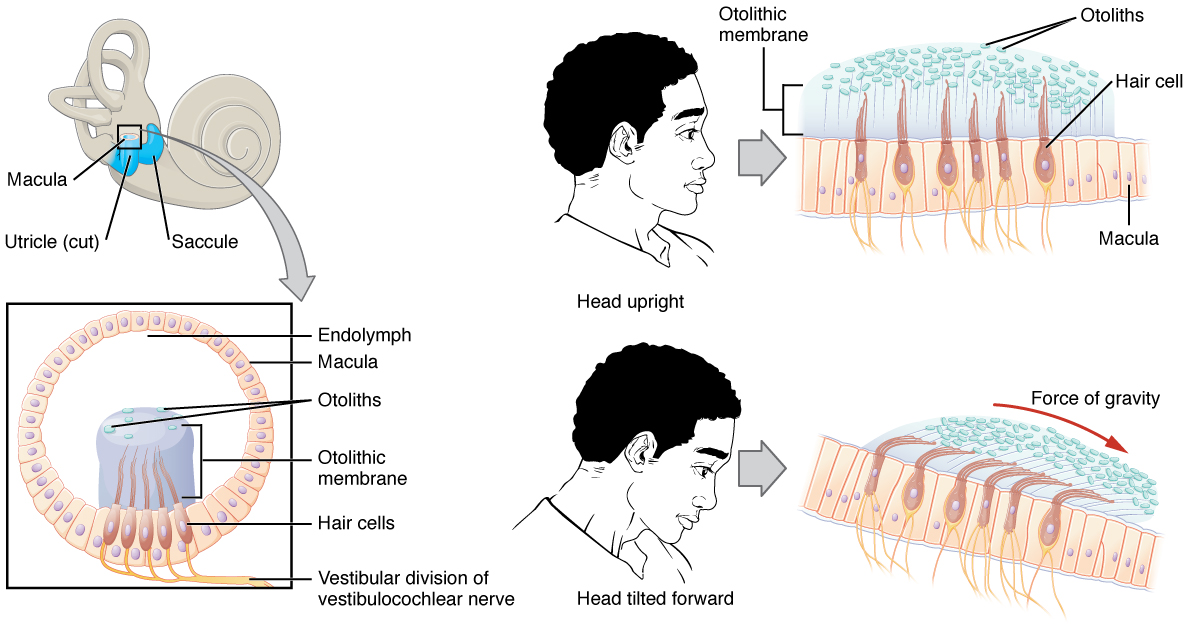
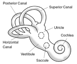
    • An endolymph disorder causing brief, intense vertigo due to head position change- the otoconia within the semicircular canals become dislodged
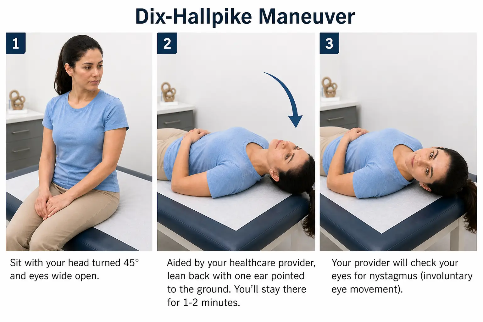
    • Confirmation of BPPV is done by performing the Dix-Hallpike - before completing, the therapist must perform a vertebral artery test and ensure appropriate cervical range of motion is present
      • Patient positioned in a long sitting with head turned to 45 degrees towards the affected side
      • Patient is quickly lowered to supine with head remaining in 45 degrees and hanging over the edge of the mat- therapist is observing for nystagmus
        • Nystagmus will resolve within 30-60 seconds
        • A positive test occurs when the patient experiences nystagmus and vertigo with a head position change
      • Repeat test on contralateral side
Dix hallpike
Dix hallpike

Understanding what nystagmus means in relation to BPPV

  • Ageotrophic
    • Fasting beating phase of the nystagmus is moving away from the ground (towards the upper ear)- this indicates lateral canal BPPV
  • Geotropic
    • Fast beating phase of nystagmus is moving towards the ground (towards the lower ear) - this indicates posterior canal BPPV

Differentiation of BPPV and canals impacted

  • Lateral canal BPPV
    • Can manifest as ageotrophic or geotrophic horizontal nystagmus
    • Treatment is with the ith log roll maneuver or barbeque roll to move otoconia into position (treatments are performed as home exercise programs)
      • Log roll maneuver
        • Lie on your back with your head centered and neck flexed about 30 degrees
        • Turn your head 90 degrees toward the affected ear
        • Hold for 30 seconds, or until vertigo and nystagmus stop
        • Turn your head back to center
        • Turn your head 90 degrees away from the affected ear
        • Lie on your stomach with your elbows bent
        • Roll back onto your back toward the affected ear
      • Barbeque roll maneuver (only for left lateral canal BPPV)
        • Lie on your left-hand side and wait for 30 seconds.
        • Roll onto your back and wait for 30 seconds.
          • Roll onto your right-hand side and wait for 30 seconds.
          • Tuck your chin down slightly. Roll onto your stomach and prop yourself up on your elbows. Wait for 30 seconds.
          • Roll onto your left-hand side and wait for 30 seconds.
          • Slowly return to a sitting position. Keep your chin level.
          • Sit for five minutes, and then repeat the previous step
          • Sit for 15 minutes to finish.
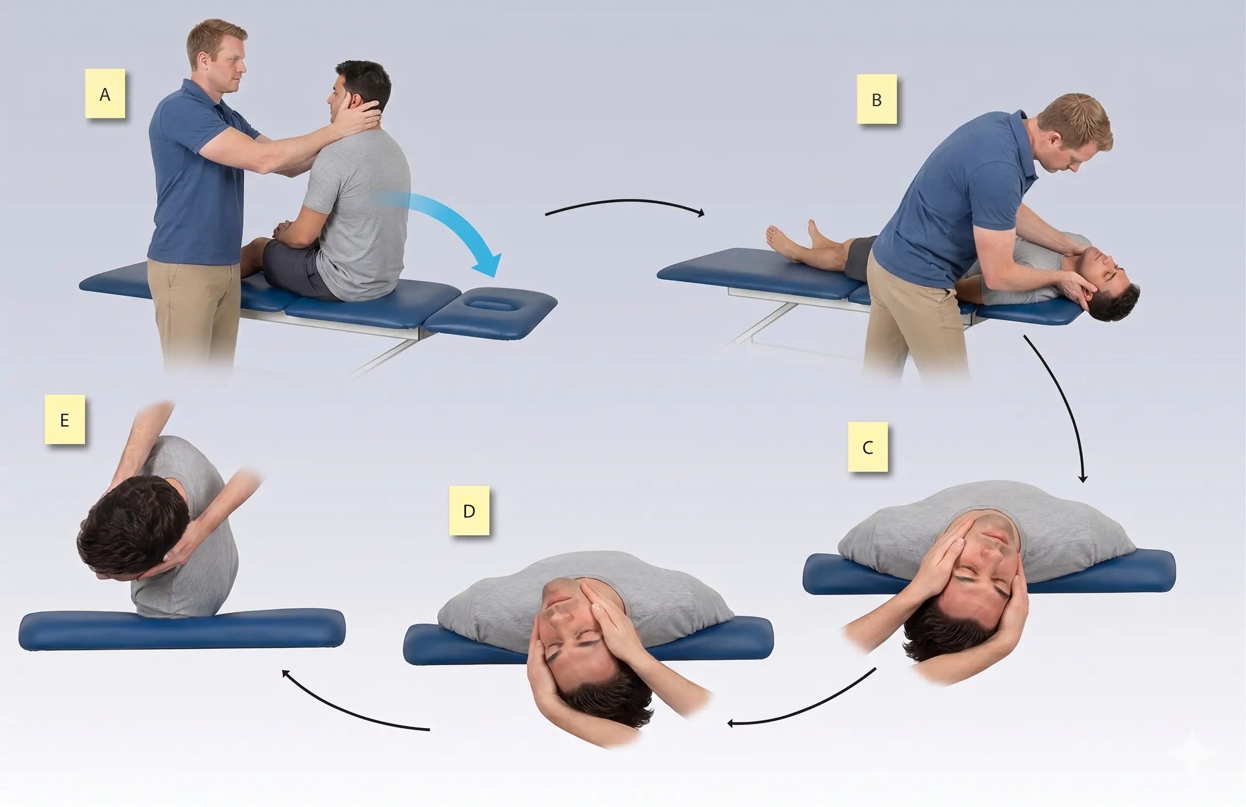
      • Posterior canal BPPV (most common BPPV)
        • Typically presents with rotary, geotrophic nystagmus
        • The epley maneuver is performed to reposition the otoconia
          • Sit in a long sitting position
          • Turn your head toward the side that causes vertigo.
          • Quickly lie you down on your back with your head in the same position just off the edge of the table. You will likely feel more intense vertigo symptoms at this point.
          • Slowly move your head to the opposite side.
          • Turn your body so that it is in line with your head. You will be lying on your side with your head and body facing to the side.
          • Sit upright.
      • Anterior canal BPPV (least common BPPV)
        • Typically presents with vertical downbeat nystagmus
        • The Yacovino maneuver is performed to reposition otoconia
          • Sit on the edge of a bed with your head in a neutral position.
          • Quickly lie back onto your back, allowing your head to hang slightly below horizontal, ensuring your neck is supported by the bed.
          • Stay in this head-hanging position until dizziness subsides, usually for 30 seconds or more.
          • While still lying down, slowly move your head into a chin-to-chest position, keeping your body relaxed.
          • Slowly return to a sitting position while maintaining the chin-to-chest position
Interventions for BPPV

Meniere’s disease

Meniere’s disease is a chronic inner ear disorder with common symptoms of fullness in the ear, ringing in the ear, loss of hearing, and vertigo due to abnormal fluid buildup in the ear. Episodes of vertigo can last for minutes to days. Due to the chronicity of the disease, management is to reduce the frequency of attacks and manage symptoms.

The management of symptoms includes

  • Low sodium diet- to reduce fluid levels
  • Regular exercise- improve balance and reduce stress
  • Management of stress- can exacerbate an episode of vertigo
  • Diuretics- reduce the fluid within the ear
  • Surgery- drain excess fluid or remove the damaged portion of the inner ear

Physical therapy management of individuals with Meniere’s disease is symptom-based, and episodes of care are short. The focus will primarily be to assist with any balance deficits, muscle weakness, provide an individualized home exercise program, and reinforce patient education.

Vestibular neuritis

Vestibular neuritis is inflammation of the vestibular nerve, which can be caused by viral infection, autoimmune disorders, or bacterial infections. The symptoms are sudden onset of severe vertigo (spinning sensation), nausea and vomiting, dizziness and lightheadedness, difficulty walking, and nystagmus. Symptoms are typically sudden and improve with medical intervention over a few days.

Treatment options will include:

  • Antibiotics for the management of infection
  • Corticosteroid medication for the management of inflammation
  • Anti-nausea medication for persistent nausea and vomiting

Physical therapy interventions to treat vestibular neuritis are rare, as the symptoms resolve quickly. If any balance or coordination deficits continue, then physical therapy interventions will be prescribed at that time.

Labyrinthitis

Labyrinthitis is an infection of the inner ear caused by head trauma, viral infections, bacterial infections, or autoimmune disorders. The symptoms are vertigo, nausea and vomiting, balance problems, hearing loss, tinnitus (ringing in the ears), and ear pain. Most symptoms resolve within a few weeks, but some permanent hearing loss may be present.

Treatment interventions include:

  • Antihistamines reduce the vertigo symptoms
  • Anti-nausea drugs for treatment of nausea and vomiting
  • Corticosteroids for treatment of inflammation
  • Antibiotics for treatment of infection

Sign up for free to take 12 quiz questions on this topic

All rights reserved ©2016 - 2026 Achievable, Inc.