Peripheral nervous system
The peripheral nervous system is a collection of nerves that connect the brain and spinal cord to other body parts (peripheral system).
Components of the peripheral nervous system
There are four (4) major components of the peripheral nervous system: cranial nerves, spinal nerves, peripheral nerves, and the nerves that comprise the autonomic nervous system.
- Cranial nerves
- 12 pairs of nerves arising from the brainstem responsible for sensory and motor functions of the head and neck
- List of cranial nerves
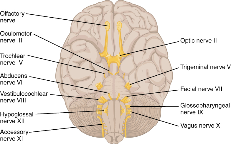
The peripheral nervous system includes twelve cranial nerves that control sensory and motor functions of the head and neck. Refer to the textbook to view the full list of each nerve’s function.
{`    * **Olfactory nerve I:**
        * Sensory only
            * Function: smell
    * **Optic nerve - II:**
        * Sensory only
            * Function: visual acuity
    * **Oculomotor nerve- III:**
        * Motor only
            * Function: moves the eye up, down, and in (medially); elevation of the eyelid, and constriction of the pupil.
    * **Trochlear nerve -IV:**
        * Motor only
            * Function: move the adducted eye down (torsional movement)
    * **Trigeminal nerve - V:**
        * Sensory and motor
            * Sensory -touch, pain, and temperature to the face; ophthalmic, maxillary, and mandibular regions
           * Motor - muscles of mastication

    * **Abducens nerve - VI:**
        * Motor only
            * Function: moves the eye out (laterally)
    * **Facial nerve - VII:**
        * Sensory and motor
            * Function: sensory- taste to the anterior two-thirds of the tongue; motor- facial expressions, closing eyes tightly

    * **Vestibulocochlear nerve- VIII:**
        * Sensory only
            * Function: balance and hearing acuity
    * **Glossopharyngeal nerve - IX:**
        * Sensory and motor
            * Function: sensory- taste to the posterior one-third of the tongue; motor- gag reflex, pharynx, and larynx control
    * **Vagus nerve- X:**
        * Sensory and motor
            * Sensory: small area of taste on the back of the tongue. Primary parasympathetic autonomic nervous system supply to internal organs assists with digestion, aids in slowing heart rate, and bronchial constriction
        * Motor- pharynx and larynx control (swallowing and speech)

    * **Accessory nerve- XI:**
        * Motor only
            * Function: controls the trapezius and sternocleidomastoid muscle
    * **Hypoglossal nerve - XII:**
        * Motor only
            * Function: controls tongue movements
`}
Spinal nerves
- Thirty-one (31) pairs of nerves exiting the varying vertebral areas - divided into dorsal (sensory) and ventral (motor) roots
- Each spinal nerve corresponds with a segment of the spinal cord (i.e., 8 cervical, 12 thoracic, 5 lumbar, 5 sacral, and 1 coccygeal)
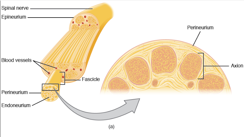
Structure of peripheral nerves
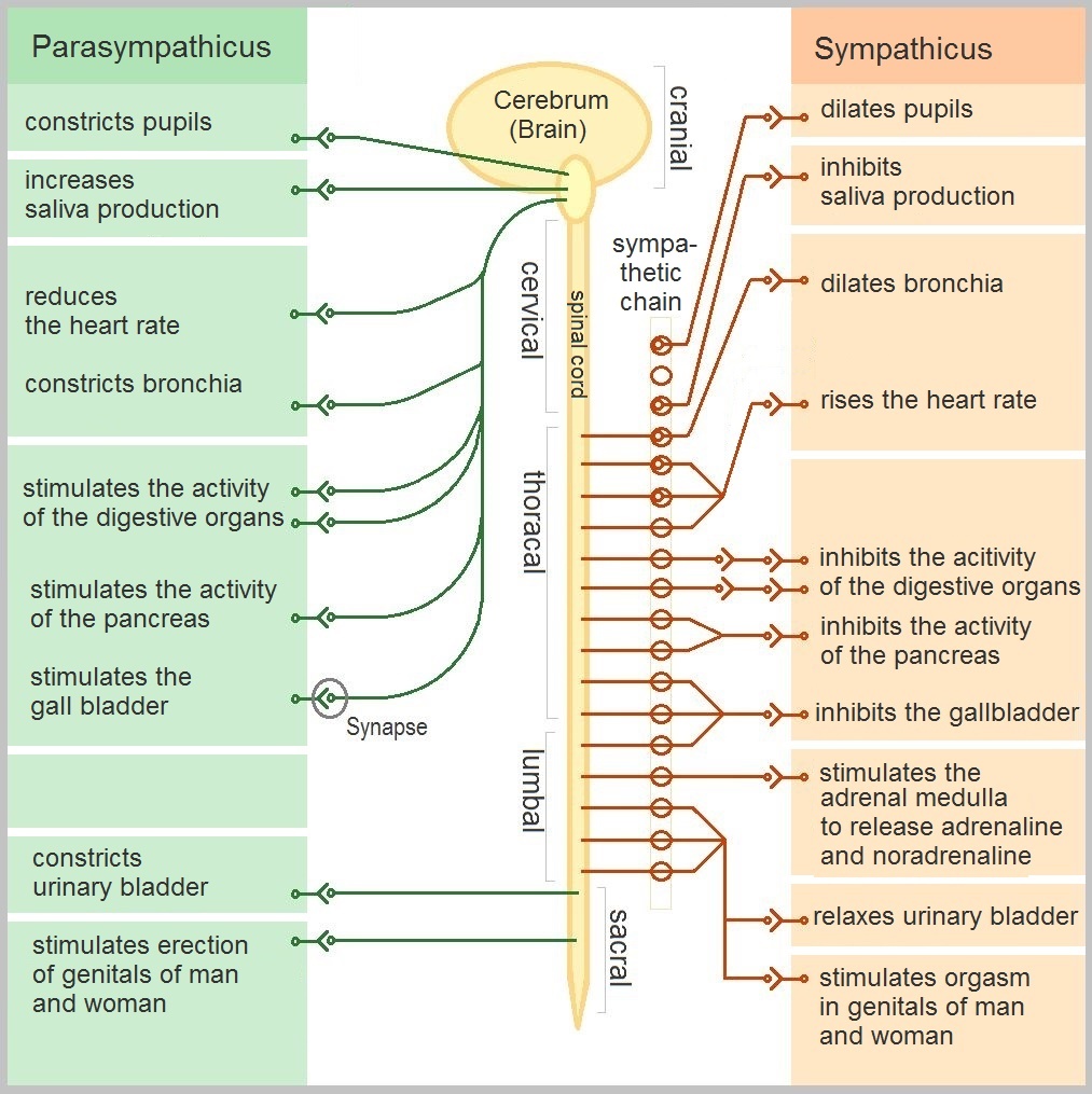
Autonomic nervous system
Dermatomes and myotomes
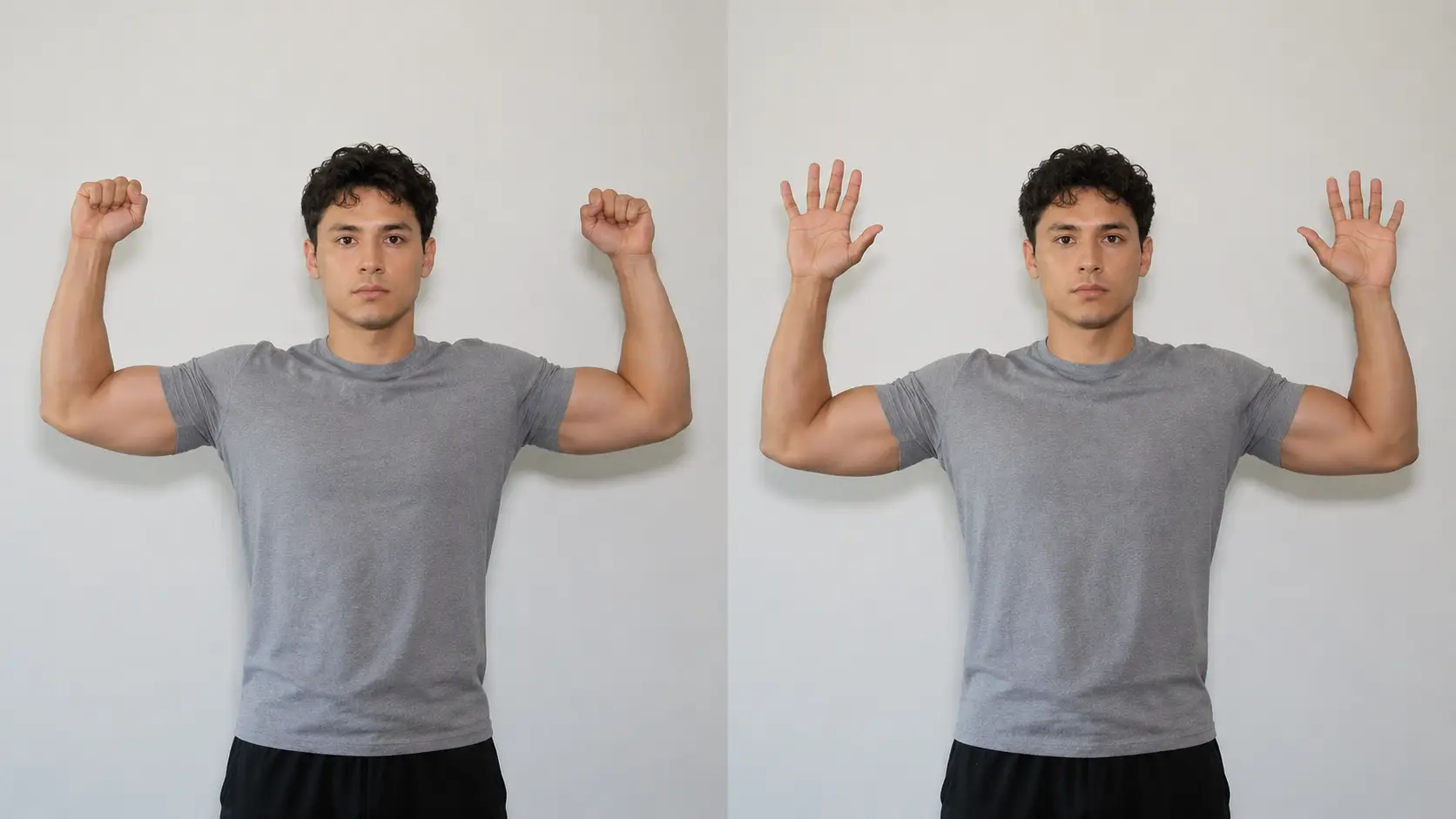
Myotomes
-
Upper extremity
- C1- cervical rotation
- C2-C4- shoulder elevation
- C5- shoulder abduction/elbow flexion
- C6- wrist extension
- C7- elbow extension/wrist flexion
- C8-digit flexion/thumb extension
- T1- finger adduction
-
Lower extremity
- L1-L2: hip flexion
- L3: knee extension
- L4: knee extension/ankle dorsiflexion
- L5: ankle dorsiflexion/great toe extension
- S1: ankle eversion/ankle plantar flexion
- S2: ankle plantar flexion
Spinal reflexes
- Spinal reflexes
- C5: biceps reflex
- C6: brachioradialis reflex
- C7: triceps reflex
- L3-L4: patellar reflex
- L5: semitendinosus reflex
- S1-S2: achilles reflex
Example of reflexes working in the spinal cord below:
Brachial, lumbar, and sacral plexuses
The brachial, lumbar, and sacral plexuses are a network of nerves that form a connection between the peripheral nerves and the central nervous system. The peripheral nerves innervating all muscles are derived from the brachial, lumbar, and sacral plexuses.
Brachial plexus
Muscles Innervated by the Brachial Plexus
Roots (C5-T1)
- Dorsal scapular nerve (C5):
- Rhomboid major
- Rhomboid minor
- Levator scapulae
- Long thoracic nerve (C5-C7):
- Serratus anterior
Trunks
- Suprascapular nerve (Upper trunk: C5-C6):
- Supraspinatus
- Infraspinatus
- Nerve to subclavius (Upper trunk: C5-C6):
- Subclavius
Divisions
- No direct muscle innervations from divisions.
Lateral Cord
- Lateral pectoral nerve (C5-C7):
- Pectoralis major (clavicular head)
- Musculocutaneous nerve (C5-C7):
- Biceps brachii
- Brachialis
- Coracobrachialis
Medial Cord
- Medial pectoral nerve (C8-T1):
- Pectoralis major (sternal head)
- Pectoralis minor
- Medial cutaneous nerves (arm & forearm):
- No motor function (sensory only)
- Ulnar nerve (C8-T1):
- Flexor carpi ulnaris
- Medial half of flexor digitorum profundus
- Most intrinsic hand muscles:
- Hypothenar muscles (abductor digiti minimi, flexor digiti minimi brevis, opponens digiti minimi)
- Adductor pollicis
- 3rd and 4th lumbricals
- Palmar and dorsal interossei
Posterior Cord
- Upper subscapular nerve (C5-C6):
- Subscapularis
- Thoracodorsal nerve (C6-C8):
- Latissimus dorsi
- Lower subscapular nerve (C5-C6):
- Subscapularis
- Teres major
- Axillary nerve (C5-C6):
- Deltoid
- Teres minor
- Radial nerve (C5-T1):
- All muscles in the posterior arm and forearm:
- Triceps brachii
- Anconeus
- Brachioradialis
- Extensor muscles of wrist and fingers (e.g., extensor carpi radialis longus/brevis, extensor digitorum, extensor carpi ulnaris, extensor pollicis muscles)
- Supinator
- All muscles in the posterior arm and forearm:
- Posterior interosseous nerve (C7-C8):
- Majority of muscles in the posterior (extensor) compartment of the forearm:
- Extensor digitorum communis, extensor digiti minimi, extensor carpi ulnaris, extensor pollicis longus, extensor pollicis brevis, and abductor pollicis longus.
- Majority of muscles in the posterior (extensor) compartment of the forearm:
Median Nerve (from both Lateral and Medial Cords)
- Most anterior forearm muscles:
- Pronator teres
- Flexor carpi radialis
- Palmaris longus
- Flexor digitorum superficialis
- Lateral half of flexor digitorum profundus
- Flexor pollicis longus
- Pronator quadratus
- Thenar muscles:
- Abductor pollicis brevis
- Flexor pollicis brevis (superficial head)
- Opponens pollicis
- 1st and 2nd lumbricals
- Anterior Interosseous Nerve (AIN) (C7-T1):
- Motor nerve branching from the median nerve:
- Flexor pollicis longus, flexor digitorum profundus, and pronator quadratus.
- Motor nerve branching from the median nerve:
Lumbar plexus
Muscles Innervated by the Lumbar Plexus (L1-L4)
The lumbar plexus gives rise to several nerves that supply the abdomen, groin, and thigh. Refer to the textbook to view the full list of muscles each nerve innervates.
Iliohypogastric Nerve (L1)
- Internal oblique
- Transversus abdominis
Genitofemoral Nerve (L1-L2)
- Cremaster muscle (in males; part of spermatic cord)
Lateral Femoral Cutaneous Nerve (L2-L3)
- No motor innervation - sensory only
Femoral Nerve (L2-L4)
- Iliacus
- Pectineus (also sometimes by the obturator nerve)
- Sartorius
- Quadriceps femoris group:
- Rectus femoris
- Vastus lateralis
- Vastus medialis
- Vastus intermedius
Obturator Nerve (L2-L4)
- Obturator externus
- Adductor longus
- Adductor brevis
- Adductor magnus (adductor portion)
- Gracilis
- Pectineus (occasionally - (occasionally shared with femoral nerve)
Sensory nerves of lumbar plexus
Lateral Femoral Cutaneous Nerve (L2-L3)
No motor innervation - sensory only, lateral aspect of the thigh
Saphenous Nerve (L3-L4)
No motor innervation - sensory only, medial aspect of the lower leg
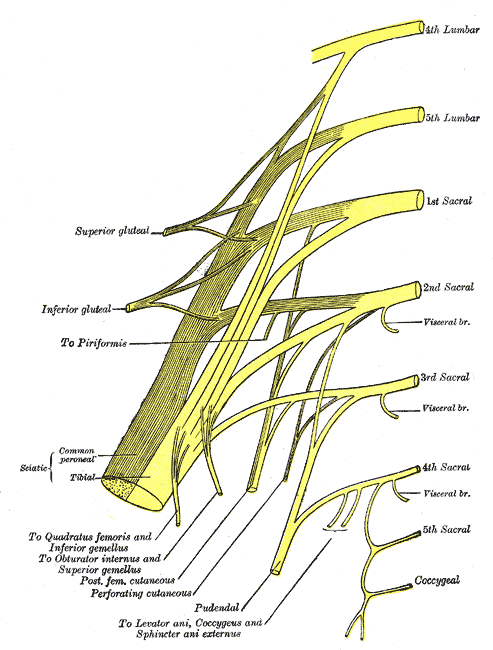
Sacral plexus
Muscles Innervated by the Sacral Plexus (L4-S3)
Superior gluteal nerve (L4-S1)
- Gluteus medius and minimus
- Tensor fasciae latae
Inferior gluteal nerve (L5-S2)
- Gluteus maximus
The sciatic nerve splits into tibial and common peroneal divisions that supply the muscles of the lower leg and foot. Refer to the textbook to view the full list of muscles each division innervates.
Sciatic Nerve (L4-S3)
- Tibial and Common Peroneal (Fibular) divisions
- Tibial Division
- Major Proximal Muscle
- Biceps femoris long head
- Semitendinosus
- Semimembranosus
- Gastrocnemius
- Soleus
- Plantaris
- Foot and Ankle Muscles
- Tibialis posterior
- Flexor hallucis longus (FHL)
- Flexor digitorum longus (FDL)
- Intrinsic foot muscles (medial & lateral plantar branches):
- Abductor hallucis
- Flexor digitorum brevis
- Flexor hallucis brevis
- Quadratus plantae
- Adductor hallucis
- Lumbricals 1-4
- Interossei (dorsal & plantar)
- Abductor digiti minimi
- Flexor digiti minimi brevis
- Opponens digiti minimi
- Major Proximal Muscle
- Common Peroneal (Fibular) Division
- Major Proximal Muscle
- Biceps femoris short head
- Foot and Ankle Muscles
- Deep fibular (anterior compartment):
- Tibialis anterior (TA)
- Extensor hallucis longus (EHL)
- Extensor digitorum longus (EDL)
- Peroneus (fibularis) tertius
- Extensor digitorum brevis (EDB)
- Extensor hallucis brevis (EHB)
- Superficial fibular (lateral compartment):
- Peroneus (fibularis) longus
- Peroneus (fibularis) brevis
- Major Proximal Muscle
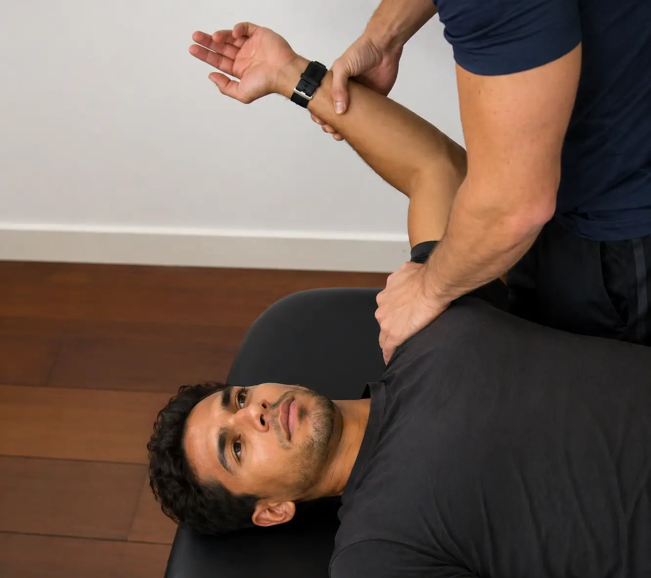
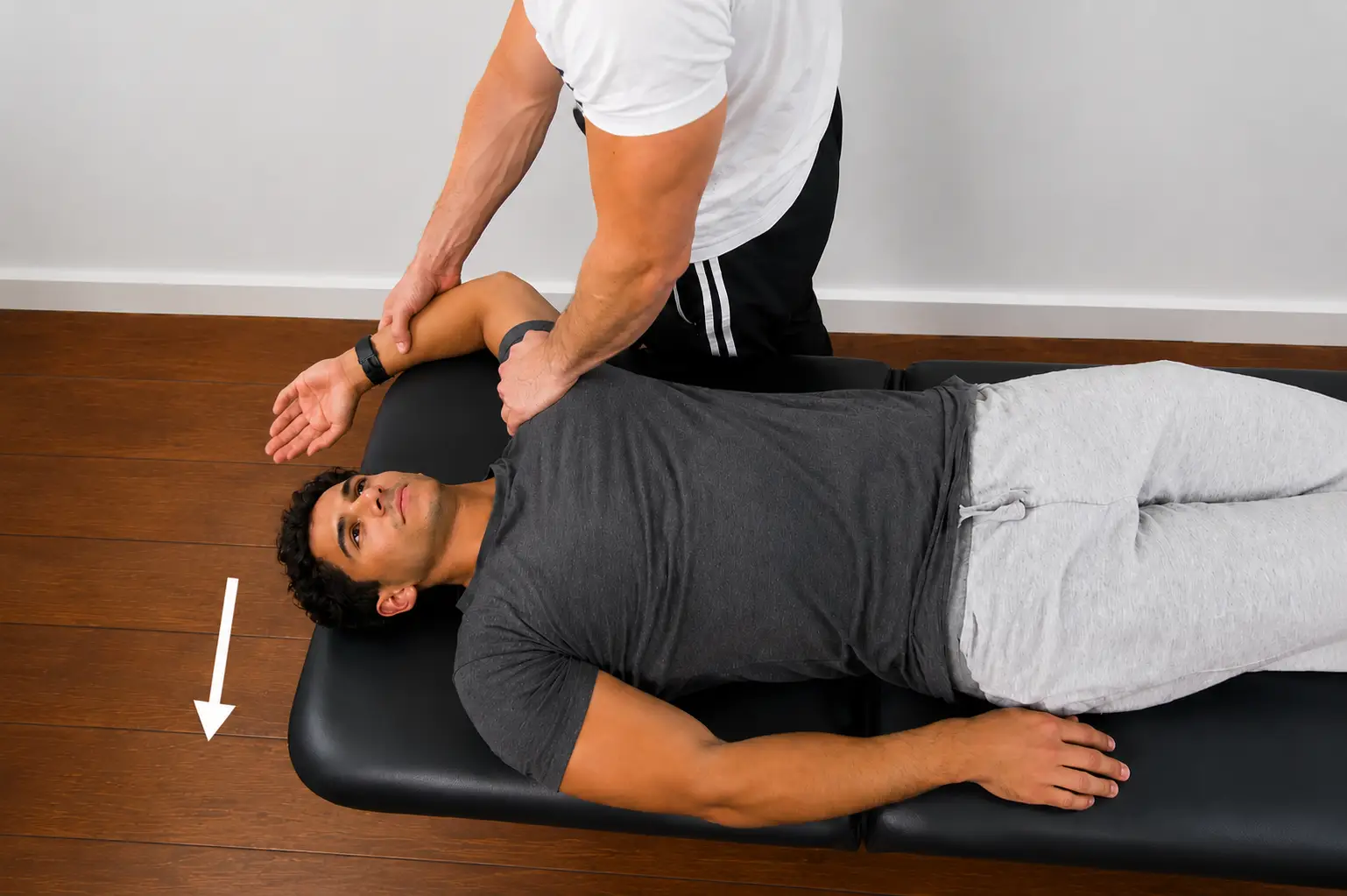
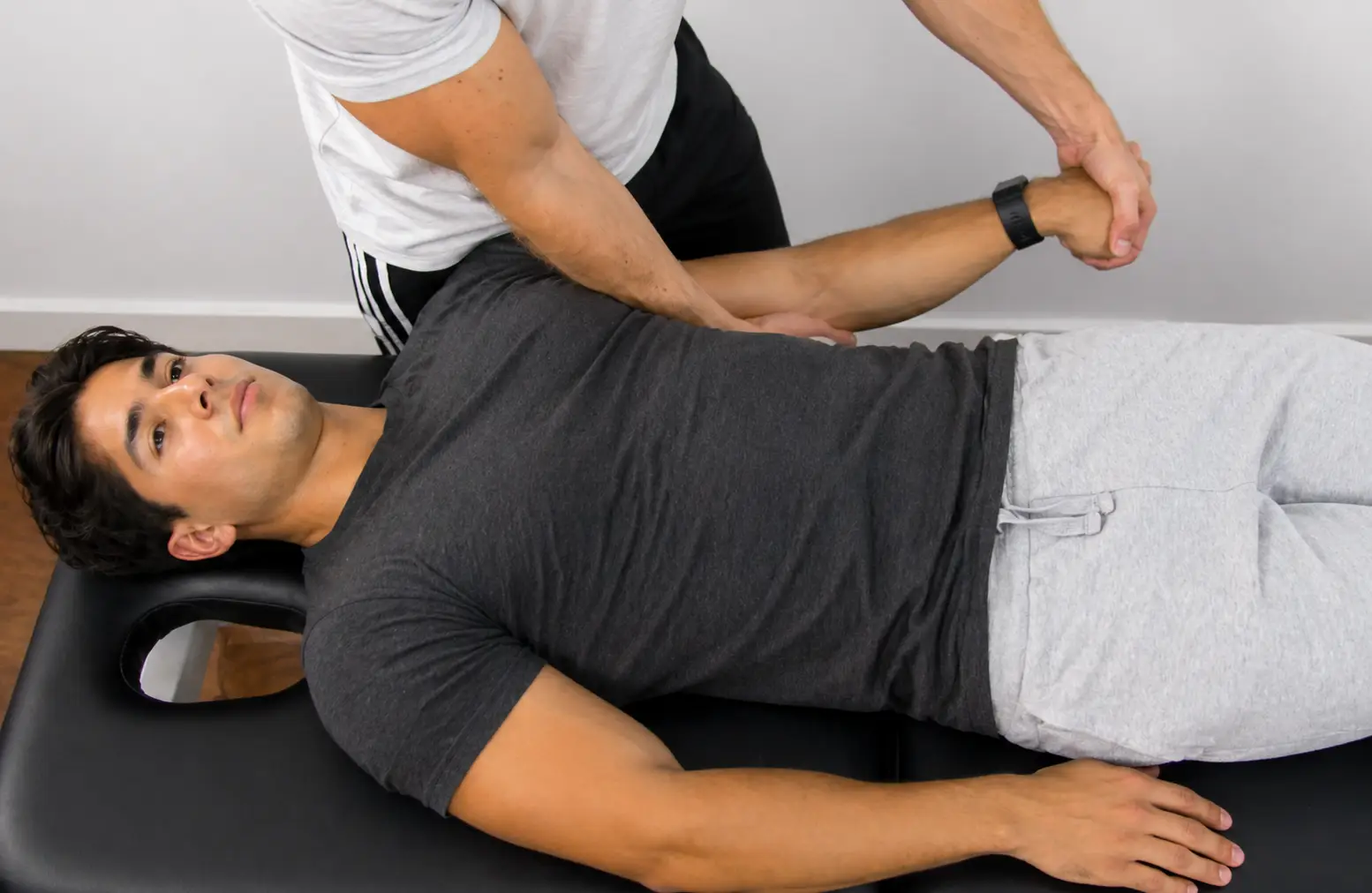
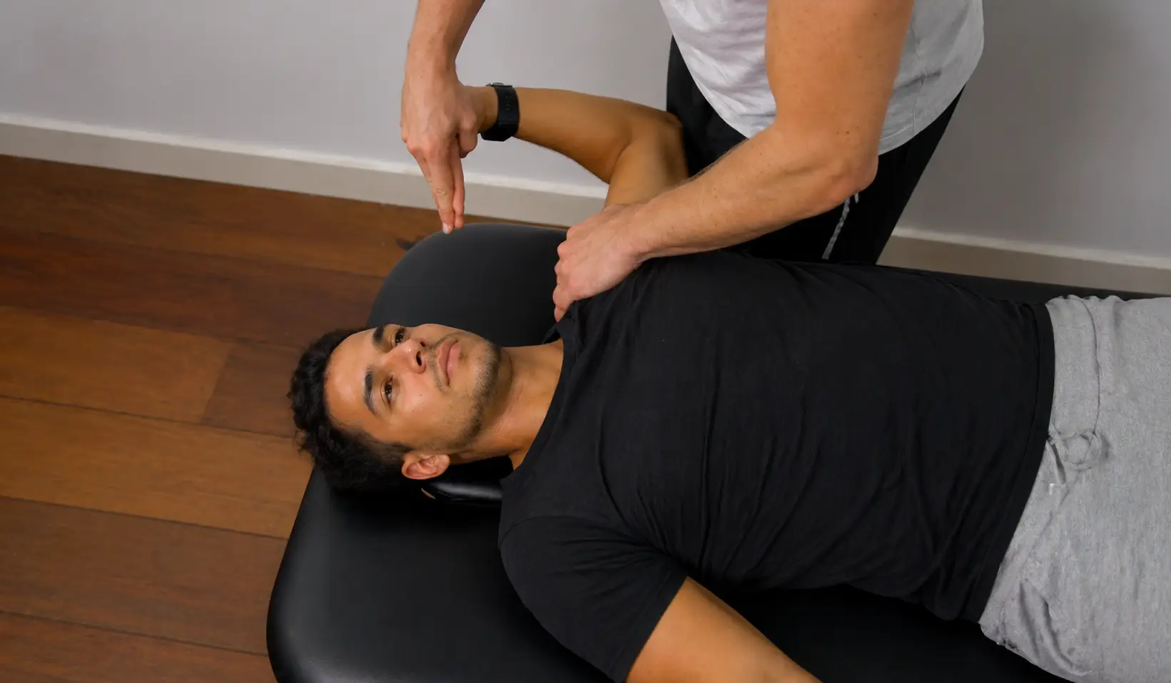
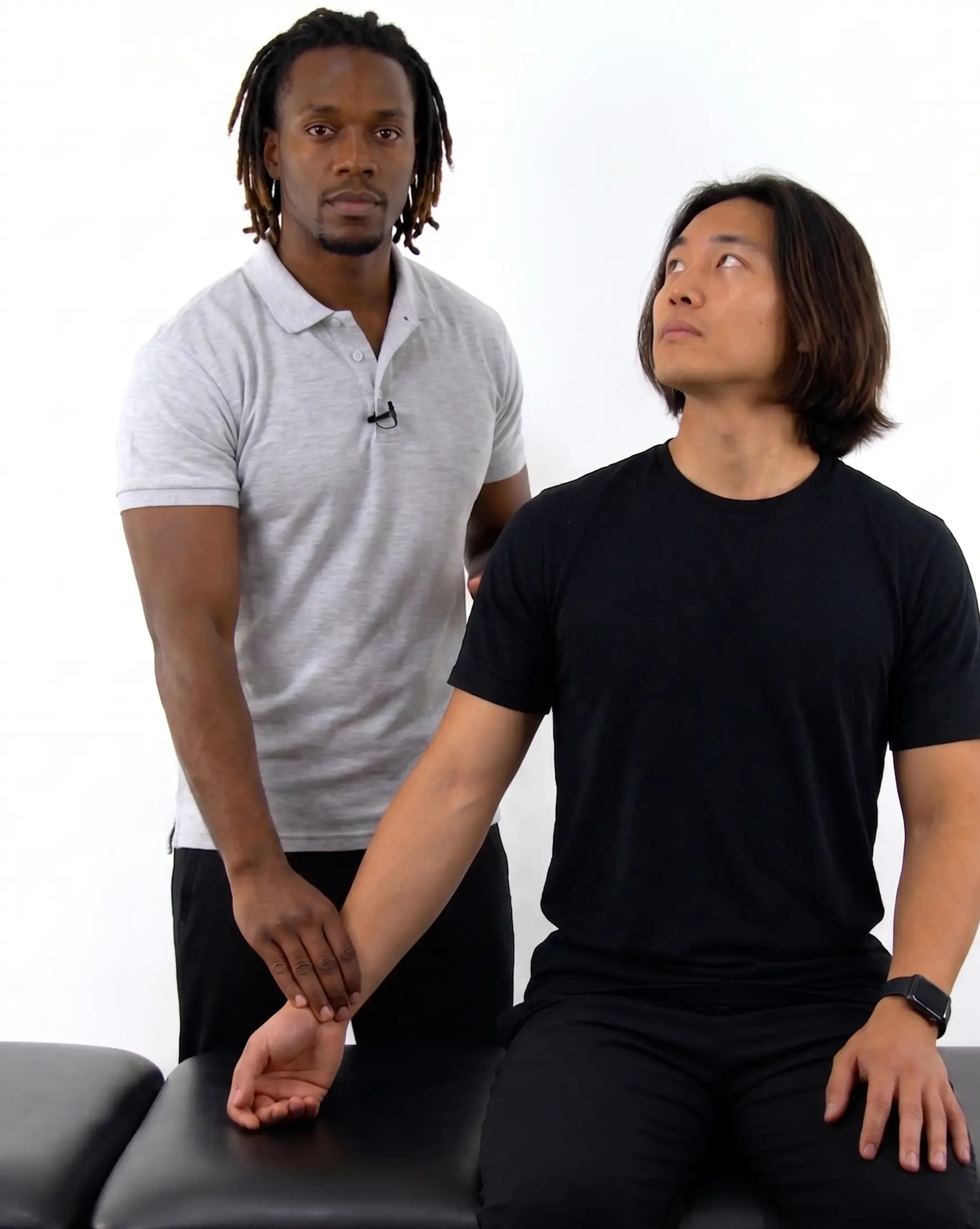
Upper limb tension tests
- Upper limb tension test 1 (ULTT1)- median and anterior interosseous nerve
- Cervical spine: contralateral lateral flexion
- Shoulder: depression and abduction to 110 degrees
- Elbow: extension
- Forearm: supination
- Wrist: extension
- Fingers and thumb: extension
- Upper limb tension test 2 (ULTT 2)- median, axillary, and musculocutaneous nerve
- Cervical spine: contralateral lateral flexion
- Shoulder: depression and abduction to 10 degrees, lateral rotation
- Elbow: extension
- Forearm: supination
- Wrist: extension
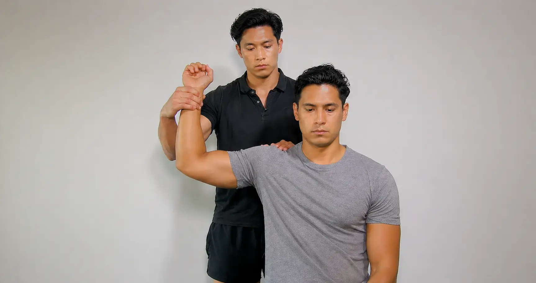
- Upper limb tension test 3 (ULTT 3)- radial nerve
- Cervical spine: contralateral lateral flexion
- Shoulder: depression and abduction to 10 degrees; internal rotation
- Elbow: extension
- Forearm: pronation
- Wrist: flexion with ulnar deviation
- Fingers and thumb: flexion
- Upper limb tension test 4 (ULTT 4)- ulnar nerve
- Cervical spine: contralateral lateral flexion
- Shoulder: depression and abduction (10 - 90 degrees) with hand to ear
- Elbow: flexion
- Forearm: pronation
- Wrist: extension and radial deviation
- Fingers and thumb: extension
Thoracic outlet syndrome
- Adson’s test (compression of the subclavian artery and/or nerves as it passes through the interscalene space)
- Patient sitting with radial nerve palpated; head rotated toward the extremity being tested with the shoulder extended and externally rotated; extend the head
- Positive: reproduction of neurological (pain, weakness, numbness, and loss of hand coordination) and vascular symptoms (loss of radial pulse)
- Patient sitting with radial nerve palpated; head rotated toward the extremity being tested with the shoulder extended and externally rotated; extend the head
- Roos elevated arm test (nerves and/or blood vessels in the space between the collarbone and the first rib are compressed)
- Patient standing with shoulders fully externally rotated, 90 degrees abducted, elbows flexed to 90 degrees- patient then rapidly opens and closes hand for 3 minutes
- Positive: reproduction of neurological (pain, weakness, numbness, and loss of hand coordination) and vascular symptoms (loss of radial pulse)
- Patient standing with shoulders fully externally rotated, 90 degrees abducted, elbows flexed to 90 degrees- patient then rapidly opens and closes hand for 3 minutes
- Wright test (compression at the space behind the pectoralis minor muscle)
- Patient seated with passive movement of the arm into abduction and external rotation
- Positive: reproduction of neurological (pain, weakness, numbness, and loss of hand coordination) and vascular symptoms (loss of radial pulse)
- Patient seated with passive movement of the arm into abduction and external rotation
- Costoclavicular test (compression of the neurovascular bundle between the clavicle and first rib)
- To perform the test, the patient sits, the therapist assists the patient in performing the following 4 movements: scapula retraction, scapula depression, elevation, and protraction - the patient holds each position for up to 30 seconds, while the patient rests his or her forearms on his thighs
- Positive: reproduction of neurological (pain, weakness, numbness, and loss of hand coordination) and vascular symptoms (loss of radial pulse)
- To perform the test, the patient sits, the therapist assists the patient in performing the following 4 movements: scapula retraction, scapula depression, elevation, and protraction - the patient holds each position for up to 30 seconds, while the patient rests his or her forearms on his thighs