Central nervous system
Upper motor neuron units
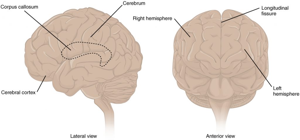
Anatomy of the brain
- The brain is divided into two (2) hemispheres, divided externally by the longitudinal fissure and internally by the corpus callosum
- Right hemisphere:
- Spatial awareness
- Emotional processing
- Facial recognition
- Creativity
- Abstract thought
- Controls the left side of the body
- Left hemisphere
- Language
- Logic and reasoning
- Analytical thinking
- Controls the right side of the body
- Right hemisphere:
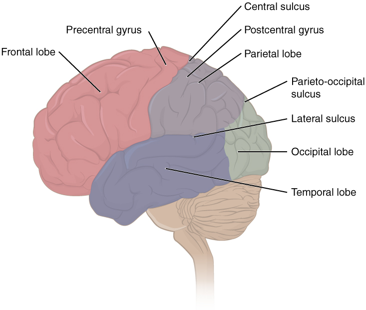
- The brain is divided into six (6) lobes with distinct characteristics that work collectively to assist function:
- Frontal lobe:
- Executive function
- Voluntary movement
- Problem solving
- Learning
- Behavior
- Impulse control
- Personality
- Social behavior
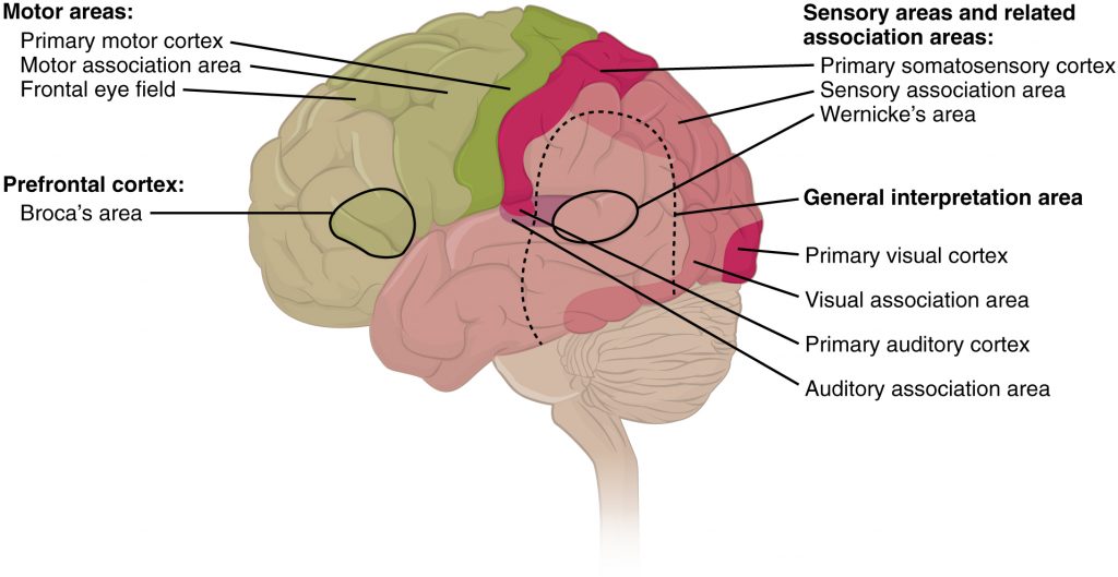
- Expressive language (Broca’s area)
- Parietal lobe:
- Awareness of somatic sense — touch, pain, temperature, pressure, vibration
- Processing somatic sensation- analyzing, recognizing, and developing of memory of somatic sense
- Spatial and body awareness
- Coordination of visual, auditory, and somatosensory stimuli
- Temporal lobe:
- Hearing
- Receptive language (Wernicke’s area)
- Memory
- Declarative: memory regarding names of people, places, or things
- Procedural: memory of how to perform activities such as brushing teeth, putting on makeup, etc.
- Occipital lobe:
- Awareness of visual stimuli
- Processing of visual stimuli
- Cerebellum:
- Motor learning
- Coordinate movement
- Balance and equilibrium
- Proprioception sense
- Maintain posture
- Brainstem:
- Ascending and descending tracts are located in the brainstem
- Heart rate and respiration rate
- Sleep and wake cycles
- Digestion
- Body temperature
- Vomiting
- Swallowing
- Frontal lobe:
Other important brain structures
-
Thalamus
- Receives sensory information from the body
- Sends these signals to the appropriate areas of the cerebral cortex for further processing.
- Helps coordinate movements by sending signals to the motor cortex.
- Assists with memory processing
-
Hypothalamus
- Maintains homeostasis within the body by regulation of hormones
-
Basal ganglia
- Initiation of movements
- Assists with maintaining posture and muscle tone
- Assists with controlling voluntary movements
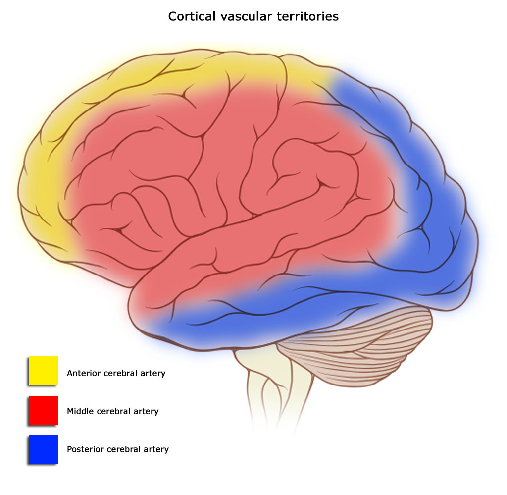
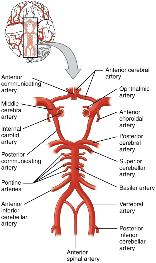
Arterial circulation of the brain
Circle of Willis
- A network of arteries that provides blood supply to the brain
- Major arteries
- Anterior cerebral artery (ACA)
- Supplies frontal, prefrontal, and supplementary motor cortex, as well as circulation to primary motor and sensory cortex
- Injury to the ACA can cause hemiparesis with weakness of the lower extremity > upper extremity, with sparing of face, apraxia, abulia, akinetic mutism, and urinary incontinence
- Anterior communicating artery
- The blood supply that connects the left and right ACA
- Injury to the anterior communicating artery can cause visual disturbances, memory deficits, cognitive impairment, severe headache, altered mental status, and impaired executive function
- Middle cerebral artery
- Blood supply to the frontal, temporal, parietal, and deeper structures
- Injury to the middle cerebral artery can cause hemiparesis with weakness of the upper extremity > lower extremity; it innervates both Broca’s area and Wernicke’s area, and neglect
- Internal carotid artery
- Provides oxygenation to the brain
- Injury can lead to blurred vision, confusion, memory loss, hemiparesis, or sudden death
- Posterior cerebral artery
- Supplies blood to the occipital and temporal lobes
- Injury can cause visual field loss, visual impairment, headache, confusion, and memory impairment
- Posterior communicating artery
- Connects the internal carotid artery to the posterior cerebral artery
- Injury can cause visual field loss, ptosis, diplopia, headache, confusion, memory impairment, hemiparesis
- Anterior cerebral artery (ACA)
Other Important arteries of the brain
- Basilar artery
- Supplies: Brainstem, occipital lobe, cerebellum, thalamus, medial temporal lobes
- Functions: Supports autonomic regulation
- Injury Signs: Hemiparesis or quadriparesis, facial paralysis, dizziness, headache, dysarthria
- Involvement: Combines with vertebral artery → vertebrobasilar system → damage to both can cause locked-in syndrome
- Vertebral artery
- Supplies: Brain and spinal cord
- Injury Signs: Headache, neck pain, dysarthria, dysphagia, seizures, impaired coordination, sensory/motor deficits to the face and body
- Involvement: Joins basilar artery → vertebrobasilar system → dual injury may result in locked-in syndrome
- Posterior inferior cerebellar communicating artery (PICA, Wallenburg syndrome, or Lateral medullary syndrome)
- Supplies blood flow to the medulla, fourth ventricle, and cerebellum
- Injury can cause diplopia, ptosis, facial pain, vertigo, slurred speech, hoarseness, balance deficits, and sensory deficits on the same-side face and the contralateral body