A CVA is a result of ischemia (blood clot) or hemorrhage (rupture in blood vessels) in the brain causing sudden, focal neurological deficits. Ischemic strokes are more likely to occur in contrast to hemorrhagic strokes.
The risk factors contributing to the likelihood of developing a stroke are due to hypertension, arteriosclerosis, diabetes, cardiac disease, hyperlipidemia, smoking, sedentary lifestyle, and previous history of TIA.
Ischemic strokes can be medically managed with tissue plasminogen activator (tPa) if given within the first 12 hours after onset of symptoms. Post 12 hour management is management via anticoagulant and antihypertensive medications.
Hemorrhagic strokes are medically managed via craniotomy or arterial clipping of bleeding arteries. Management also involves antihypertensive and epileptic medications.
Cardinal symptoms of stroke
Sudden weakness and/or numbness
Difficulty speak
Difficulty walking
Confusion
Visual changes
Facial drooping
Neurological deficits associated with stroke
Left hemisphere injury
Right side hemiplegia
Right side hemisensory
Speech-language deficits
Trouble planning/sequencing movement
Difficulty processing
Right hemisphere injury
Left side hemiplegia
Left side hemisensory
Visual-perceptual deficits
Poor judgement
Impulsive
Abstract concepts difficulty to comprehend
Difficulty perceiving emotions
Middle cerebral artery stroke
Contralateral hemiplegia with upper extremity weakness greater than lower extremity weakness
Contralateral paresthesia with upper extremity weakness greater than lower extremity weakness
Homonymous hemianopsia
Motor speech deficits
Broca’s aphasia
Receptive speech deficits
Wernicke’s aphasia
Loss of gaze to opposite side
Anterior cerebral artery stroke
Contralateral hemiplegia with lower extremity weakness greater than upper extremity weakness
Contralateral paresthesia with lower extremity weakness greater than upper extremity weakness
Urinary incontinence
Apraxia
Mutism (less verbal)
Akinetic (less mobile)
Posterior cerebral artery stroke
Contralateral sensory loss
Involuntary movements
Intention tremors- unintentional movement of extremity when performing a task
Choreoathetosis- irregular, involuntary movements that can be chorea or writhing
Thalamic pain: chronic, burning, or constrictive pain all over the body
Oculomotor nerve palsy
Generalized brainstem deficits
Vertebral-basilar artery injured- occlusion to large portion of vertebral- basilar artery
Locked in syndrome develops
Paralysis of all muscles with except eye movement
Cognition remains intact
Ventral pons area deficits
Millard-Gilbert syndrome
Basilar artery injured- small branch of occlusion
Impacts the facial and abducens cranial nerves, as well as corticospinal tract
Facial muscles and lateral rectus impacted on ipsilateral side
Inability to abduct eye on ipsilateral side
Weakness of upper and lower extremities contralateral side (hemiplegia)
Lateral medulla deficits
Lateral medullary syndrome (Wallenburg syndrome or PICA syndrome)
Impacts the posterior inferior cerebellar artery
Deficits in cranial nerves- trigeminal and vagus on ipsilateral
Deficits in pain and temperature
Decreased gag reflex
Nystagmus on ipsilateral side
Horner’s syndrome on ipsilateral side
Diplopia (double vision), anhidrosis (inability to sweat), ptosis (drooping of eyelid)
Pain and temperature of contralateral
Hemiparesis contralateral
Brunnstrom stages for recovery
The Brunnstrom stages of recovery are a guide to describe the motor recovery status post stroke. Individuals post stroke may progress through all stages or remain at a certain level for an extended period of time. There is no timetable for recovery.
Stage 1: Flaccidity, with little or no voluntary movement
Stage 2: Spasticity appears, and voluntary movement is possible
Stage 3: Spasticity increases, and patients can voluntarily perform limb synergies
Stage 4: Spasticity decreases, and patients can perform movement combinations that are not synergies
Stage 5: Patients can perform complex movement combinations
Stage 6: Spasticity disappears
Stage 7: Patients return to normal function
Synergy patterns
Synergy patterns are abnormal muscle patterns developing status-post stroke. The two synergy patterns that exist are flexion and extension synergy patterns. Below are descriptions of muscle activation during the synergy patterns.
Flexion synergy
Upper limb
Scapula: retraction and/or elevation
Shoulder: abduction and external rotation
Elbow: flexion
Forearm: supination
Lower limb
Hip: flexion, abduction, and external rotation
Knee: flexion
Foot and ankle: dorsiflexion
Extension synergy
Upper limb
Scapula: protraction and/or depression
Shoulder: adduction and internal rotation
Elbow; extension
Forearm: pronation
Lower limb
Hip: extension, adduction, and internal rotation
Knee: extension
Foot and ankle: plantarflexion
Homonymous hemianopia
Key features
Cause: Most commonly due to lesions in the optic tract, optic radiation, or occipital lobe on the opposite side of the vision loss (e.g., a left-sided brain lesion causes right homonymous hemianopsia).
Common causes: Stroke (especially affecting the posterior cerebral artery), traumatic brain injury, brain tumors.
Symptoms:
Bumping into objects on the affected side
Reading difficulties (especially when vision loss is on the right)
Difficulty with driving or navigating environments
Diagnosis: Confirmed with visual field testing (perimetry).
Rehabilitation focus:
Visual scanning training
Compensatory strategies (e.g., turning the head to scan the blind side)
Environmental modifications
Apraxia
Ideomotor apraxia
Clinical presentation:
A patient can describe a movement but cannot perform it when asked.
May improve with automatic or habitual actions.
Common errors include awkward or incorrect limb positioning during tasks like waving or brushing teeth.
Common causes:
Left parietal lobe lesions
Stroke, particularly in the dominant hemisphere
Ideational apraxia
Clinical presentation
Misuse of objects (e.g., attempting to write with a fork)
Skipping essential steps in a task (e.g., putting on shoes before socks)
Incoherent task sequencing (e.g., pouring juice after trying to drink from the empty cup)
Common causes:
Extensive damage to the left hemisphere
Dementia or widespread cortical disease
Traumatic brain injury (TBI)
Types of injury:
Open head injury: skull fracture that results in brain exposure
Increased risk for development of infection
Closed head injury: no skull fracture or exposed brain
Increased risk for intracranial pressure
Pathophysiology:
Local brain injury
Damage to a specific region of the brain due to bruising, bleeding, laceration, or swelling
Coup-countercoup injury
Injury in which the brain is damaged due to point of impact and opposite point of contact due to acceleration of brain in skull
Diffuse axonal injury
Tearing of axons and small vessels due to acceleration of brain in skull leading to neuronal death
Edema
Increased swelling due to increased intracranial pressure typically from increased volume of cerebrospinal fluid in ventricles
Hypoxic-ischemic injury
Loss of cerebral circulation due to compromise typically from deficits in cardiovascular and respiratory systems
Stratification of brain injury
Brain injuries can be stratified into three (3) categories: mild, moderate, and severe based on symptomatology present. Stratification can be done with use of Glasgow Coma Scale (GCS):
Mild TBI (i.e. concussion)
GCS score: 13-15
Loss of consciousness: 0-30 minutes
Alteration of consciousness: brief; >24 hours
Post-traumatic amnesia: <1 day
Imaging: normal
Recovery: full recovery of all physical and cognitive with patient seen in outpatient setting
Moderate TBI
GCS score: 9-12
Loss of consciousness: >30 minutes; less than 24 hours
Alteration of consciousness: >24 hours
Post-traumatic amnesia: 1-7 days
Imaging: normal or abnormal
Recovery: potential to have full recovery of physical and cognitive function with intense rehabilitation of inpatient rehabilitation or skilled nursing facility
Severe TBI
GCS score: 8 or less
Loss of consciousness: >24 hours
Alteration of consciousness: >24 hours
Post-traumatic amnesia: >7 days
Imaging: normal or abnormal
Recovery: permanent physical and cognitive impairment; typically in nursing home setting or home setting with total care
Behavioral stratification of traumatic brain injury
Ranchos los amigos cognitive scale utilized to classify behaviors through a predictable sequence. Individuals progress through levels in sequence but can plateau at any point. Level of behavior:
Level I- No response
Cognitive assistance needed: total assistance with all cognitive functions
Response to stimuli: inconsistent responses to stimuli
Single step commands: unable to follow single step commands
Characteristics of level: individuals typically in comatose state
Level II- Generalized response
Cognitive assistance needed: total assistance with all cognitive functions
Response to stimuli: consistent, generalized response to stimuli
Single step commands: unable to follow single step commands
Characteristics of level: individuals in vegetative state
Level III- Localized response
Cognitive assistance needed: total assistance with all cognitive functions
Response to stimuli: localized response to stimuli
Single step commands: unable to follow single step commands
Characteristics of level: individuals within minimally conscious state; able to inconsistently track objects and respond to their name
Level IV- Confused, agitated
Cognitive assistance needed: maximal assistance for cognitive function
Response to stimuli: localized response
Single step commands: unable to follow single step command
Characteristics of level: confused, combative, easily agitated, hypersexual behaviors
Level V- Confused, inappropriate
Cognitive assistance needed: maximal assistance for cognitive function
Response to stimuli: localized response
Single step commands: unable to follow single step command
Characteristics of level: confused, inappropriate behaviors (combative and hypersexual behaviors have subsided), severely impaired memory
Level VI- Confused, appropriate
Cognitive assistance needed: moderate assistance for cognitive function
Response to stimuli: localized response
Single step commands: able to follow single step commands
Characteristics of level: remote memory more consistent than recent memory, requires assistance with problem solving, minimal carry-over with new tasks
Level VII- Automatic, appropriate
Cognitive assistance needed: minimal assistance for cognitive function
Response to stimuli: localized response
Single step commands: able to follow single step commands
Characteristics of level: improve carry over with activity, minimal assistance for learning new tasks, increased awareness of deficits
Level VIII- Purposeful and appropriate
Cognitive assistance needed: stand by assistance for cognitive function
Response to stimuli: localized response
Single step commands: able to follow single step commands
Characteristics of level: able to integrate new and old memory into making decisions, able to make adjustments to behavior in social interaction with minimal assistance
Spinal cord injury
Partial or complete disruption of the spinal cord resulting in paralysis, sensory loss, altered spinal reflexes, and altered autonomic function.
The causes of spinal cord injury can be falls, motor vehicle accidents, penetrating wounds, disc prolapse, or vascular compromise.
Pathophysiology:
Primary injury: direct injury to the spinal cord or disruption of vascular supply
Secondary injury: edema, demyelination, or necrosis of axons
Severity of injury:
Complete: no sensory or motor below level of injury; no sacral sparing
Incomplete: inconsistent sensory or motor below the level of injury; sacral sparing present
American spinal cord injury (ASIA) levels of injury
A- Complete
No motor or sensory preserved below the level of injury
No motor or sensory in sacral segment S4-S5 (sacral sparing)
B- Incomplete
Sensory but no motor below the level of injury
Sensory to sacral segments S4-S5 but no motor to these segments
C- Incomplete
Motor function is present below the level of injury with major muscle have a manual muscle test grade less than 3
D- Incomplete
Motor function is present below the level of injury with major muscles have a manual muscle grade greater than 3
E- Normal
Motor and sensory are normal
Classifications of incomplete spinal cord injuries
Central cord syndrome
Etiology; Cervical hyperextension
Deficits:
Loss of bilateral pain and temperature contralateral (spinothalamic tract)
Loss of bilateral motor function - primarily upper extremities (corticospinal tract)
Intact:
Preservation of proprioceptions, vibratory sense, and kinesthesia (dorsal column lateral meniscus)
Anterior cord syndrome
Etiology Cervical flexion, vascular compromise
Deficits:
Loss of motor function bilaterally below the level of injury (corticospinal tract)
Loss of bilateral pain and temperature (spinothalamic)
Intact: : Preservation of proprioceptions, vibratory sense, and kinesthesia (dorsal column lateral meniscus)
Brown-sequard syndrome
Etiology: Penetrating wound from gunshot or stab wound
Deficits:
Ipsilateral loss of two-point discrimination, pressure, vibration, and proprioception (dorsal column lateral meniscus)
Ipsilateral loss of motor function (corticospinal tract)
Contralateral loss of pain and temperature (spinothalamic)
Intact: None
Posterior cord syndrome
Etiology: Cervical hyperextension
Deficits:
Bilateral loss of proprioception, vibration, pressure (dorsal column lateral meniscus)
Intact:
Preservation of motor function (corticospinal)
Pain and temperature (spinothalamic)
Cauda equina injury (classified as lower motor neuron lesion)
Intact: Variable dependent upon severity of injury
Associated disorders with spinal cord injury
Spinal shock:
Immediately after injury a time of absent reflexes and flaccidity due to the body working to protect itself after injury
Timeframe can vary from 24 hours to 24 weeks
Difficult to get accurate ASIA assessment due to spinal shock and changes in level of injury
Autonomic dysreflexia
Medical emergency for individuals with T6 or above injury
Caused by noxious stimuli in which spinal cord injured individual is unable to physically
Noxious stimulus causes activation of sympathetic nervous system and due to spinal cord injury the parasympathetic nervous system is unable to be enacted due to injury to spinal cord (activation of parasympathetic system will cancel out the effects of sympathetic nervous system
Noxious stimulus can be constipation, catheter malfunction, pressure injuries, tight clothes, sitting on sharp object
Symptoms: headache, increased blood pressure, bradycardia, diaphoresis above the level of injury, flushing below the level of injury, seizures. If left untreated, it can lead to death.
Medical management: sit patient up and assess for noxious stimulus
Spasticity
Increased tone to extremities or trunk due to consistent contraction; leads to range of motion deficits and functional impairment
Will only be seen in upper motor neuron lesions
Occurs most often in incomplete lesions
Utilize Modified Ashworth Scale to assess
Heterotrophic ossification
Abnormal bone growth in muscle
Symptoms: firmness at muscle site, pain with palpation, pain with muscle stretch or palpation
Typically involves the quadriceps and brachialis muscles
Deep vein thrombosis:
Medical emergency
Development due to immobility of extremities which places individuals at increased risk for blood clot development
C6: independent with sliding board transfers on level surfaces
C7 and beyond: independent with transfers without sliding board on unlevel surfaces
Wheelchair use
C1-C5: power wheelchair use with use of head control, mouth control, or joystick
C6: manual wheelchair on level surfaces with use of wheel projections and large knobby wheels- independent status
C7 and beyond:: manual wheelchair usage without adaptations in all surfaces- independent status
Gait
T12:
Exercise only ambulation due to high energy needs
Hip-knee-ankle orthosis (HKAFO) with hip locked in extension or reciprocating gait orthosis (RGO) necessary for gait
Manual wheelchair used primarily
L1-L2:
Exercise only ambulation due to high energy needs
Hip-knee-ankle orthosis with hip unlocked to allow for flexion to occur necessary for ambulation
Manual wheelchair used primarily
L3:
Household ambulation
Ankle foot orthosis (AFO) used to assist with knee control
Manual wheelchair used in community
L4 and below:
Community ambulation
Ankle foot orthosis (AFO) used to assist with ankle control
Manual wheelchair used as needed
Parkinson’s disease
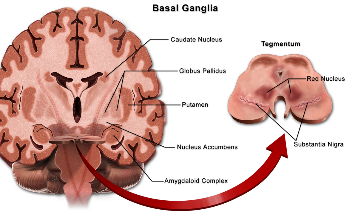
Basal ganglia
Common impairments of Parkinson’s disease
Resting tremor
Impaired postural reflexes
Cogwheel rigidity
Bradykinesia
Slowed reaction time
Masked face
Dysarthria
Hypophonia (decreased speech volume)
Contractures in flexor and adductor muscles
Postural deficits- rounded shoulders, kyphosis
Restrictive lung disease
Visual impairment
Dementia in later stages
Standardized test for Parkinson’s disease
Hoehn & Yahr classification of disability
Stage 1
Disability: Minimal or absent
Extent of disability: Unilateral
Stage 2
Disability: Minimal
Extent of disability: Midline involvement and bilateral
Stage 3
Disability: Impaired postural reflexes, unsteadiness when rising from chair; continues to be independent and work
Extent of disability: Midline involvement and bilateral
Stage 4
Disability: All symptoms present; ambulation only with assistive device
Extent of disability: Midline involvement and bilateral
Stage 5
Disability: All symptoms present; confined to wheelchair or bed
Extent of disability: Midline involvement and bilateral
Retropulsion pull test
Testing Procedures:
The subject stands in a comfortable stance with eyes open (have feet shoulder width apart if they assume an unusually wide or narrow stance).
The examiner stands behind the subject.
The subject is instructed to do whatever it takes to not fall and are told that the examiner will catch them if they do fall.
The examiner gives a sudden, brief backward pull to the shoulders with sufficient force to cause the subject to have to regain their balance.
The subject should not know exactly when the pull is coming.
Interpretation of results: scoring is from 0 to 4:
0 = recovers independently may take 1 or 2 steps or an ankle reaction
1 = three steps or more backward but recovers independently
2 = retropulsion, needs to be assisted to prevent fall
3 = very unstable, tends to lose balance spontaneously
4 = unable to stand without assistance (UPDRS method)
Medication for Parkinson’s disease
Levodopa
Mechanism of action: relieve symptoms of Parkinson’s disease by turning levodopa into dopamine (due to lack of dopamine in basal ganglia)
Side effects: mental confusion, hallucinations, postural hypotension, restlessness, abnormal movements
On and off time: Levodopa should be given 1 hour prior to initiation of activity for optimal effects; medication will wear off and symptoms will occur
Anticholinergic medications
Mechanism of action: decrease tremors by blocking cholinergic production in basal ganglia
Side effects: dry mouth, urine retention, constipation
Huntington’s disease
Symptoms
Involuntary writhing movements (choreic movements) at rest
Muscle rigidity
Poor balance
Difficulty with swallowing/speaking
Impaired executive function
Decline in overall mental health
Functional decline
Physical therapy considerations with Huntington’s disease
Balance/gait exercise- using weighted walker
Postural stability
Family education/training as needed
No modalities indicated
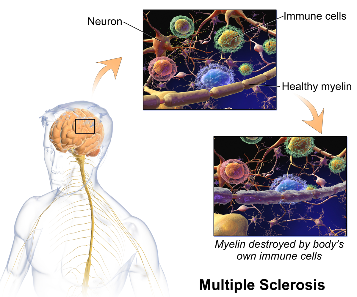
Multiple sclerosis (MS)
Types of Multiple sclerosis
Relapsing-remitting
Characterized by cycles of disease exacerbation followed by periods of remission; varied levels of disability
Primary progressive
Characterized by disease consistently being present without remission; permanent disability results
Secondary-progressive
Characterized by disease beginning as cyclic relapse/remitting initially followed by continuous progression of disease without remission; permanent disability results
Progressive-relapsing
Characterized by progressive disease with unpredictable times of remission; permanent disability results
Anatomy of multiple sclerosis
Symptoms of multiple sclerosis- symptoms will vary depending on location of plaques
Weakness
Spasticity
Hyperreflexia
Impaired coordination
Visual deficits
Ataxia
Vestibular dysfunction
Dysarthria
Paresthesia
Lhermitte’s sign: electric shock like symptoms resulting from neck flexion
Special considerations with multiple sclerosis
Avoidance of precipitating factors:
Stress
Trauma
Pregnancy
Trauma
Heat
Hyperventilation
Dehydration
Increase exertion
Amyotrophic lateral sclerosis (ALS)
Symptoms
Asymmetrical weakness
Facial weakness
Difficulty with swallowing
Hyperreflexia
Spasticity
Compromised cranial nerve integrity
Stages of ALS
Stage 1- early stage
Mild weakness or stiffness in the hands, feet, or limbs
May have difficulty with fine motor tasks (e.g., writing, buttoning)
Stage 2- middle severe
Weakness spreads to other parts of the body
Difficulty walking, speaking, or swallowing
May require assistance with daily activities
Stage 3- late state severe:
Late Stage Severe weakness and paralysis, Difficulty breathing, and Need for a wheelchair and ventilator.
Stage 4- end stage
Total paralysis
Loss of cognitive function in some cases may occur but rare
Medical management is treatment of symptoms as there is no cure for ALS. Motor function progressively diminishes until the individual becomes totally dependent for activities of daily living and mobility while on ventilator support. Physical therapy assists with prescription of assistive devices and family education/training as appropriate.
Epilepsy
Symptoms:
Altered consciousness
Convulsions
Sensory phenomena: heightened somatosensory, visual, auditory, or olfactory senses