


This browser is too old to run Achievable.

Update it, or open this page in a current version of Chrome, Safari, Firefox or Edge. On an iPhone or iPad, update iOS: every browser there runs on the version of Safari that comes with it.

Achievable logoAchievable logo
NPTE-PTA
Sign in
Sign up
Purchase
Textbook
Practice exams
Support
How it works
Resources
Exam catalog
Mountain with a flag at the peak
Textbook
Introduction
1. Cardiovascular system
2. Pulmonary system
3. Neuromuscular system
3.1 Central nervous system
3.2 Anatomy and function of spinal cord
3.3 Peripheral nervous system
3.4 Stroke deficits and recovery
3.5 Traumatic brain injury
3.6 Spinal cord injury
3.7 Neurodegenerative disorders and epilepsy
3.8 Peripheral nervous system conditions
3.9 Other neurological conditions
3.10 Interventions for neurological conditions
3.11 Vestibular system
4. Pediatrics
5. Musculoskeletal system
6. Other systems
7. Non-systems
Wrapping up
Achievable logoAchievable logo
3.2 Anatomy and function of spinal cord
Achievable NPTE-PTA
3. Neuromuscular system
Our NPTE-PTA course is now in "early access" - get 50% off for a limited time.

Anatomy and function of spinal cord

8 min read
Font
Discuss
Share
Feedback

Primary function of the spinal cord

  • Acts as a relay pathway between the peripheral system and the brain
    • Provides sensory information regarding the environment to the brain
    • Manages motor commands to the peripheral system in reaction to the environment
    • Coordinates simple reflexes that do not need to go to the brain, such as withdrawal from pain, patellar reflex, etc.

PTA scope of practice: The PTA collects data and recognizes and communicates changes in a patient’s status to the supervising PT; evaluating the patient, interpreting findings, and changing the plan of care remain the PT’s responsibility. Dermatomal sensory testing and deep tendon reflex testing rest on this anatomy; when a PTA collects these findings, a new or changed deficit is reported to the supervising PT, who interprets it. Supervision and delegation rules are covered in PTA responsibilities, research, and ADA.

Anatomy of the spinal cord

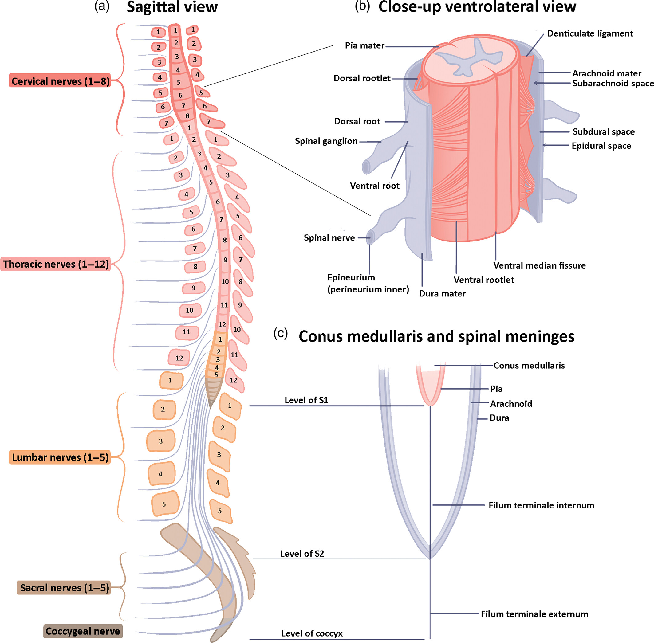
  • Cervical spine
    • Eight nerves that provide innervation to the neck and upper extremity
  • Thoracic spine
    • Twelve nerves that provide innervation to the upper chest, chest, abdomen, and upper back
  • Lumbar spine
    • Five nerves that provide innervation to the lower abdominal wall, groin, and front and inner thigh, and (with the sacral nerves) the leg and foot
  • Sacral spine
    • Five nerves that provide innervation to the buttocks, back of the thigh, much of the leg and foot (including the sole and outer foot), and the perineum, and that carry bowel, bladder, and sexual function (S2-S4)
  • Coccygeal spine
    • One nerve that provides innervation to the perineal area
Spinal cord sections
Sheryl Tan, Faull L, Curtis A, The Anatomical record
/
Wikimedia Commons
/
CC BY 4.0

Protective layers of the spinal cord

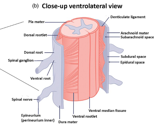
  • Dura mater
    • Outermost protective layer
    • Space between dura and the bony walls of the vertebral canal referred to as epidural space, which contains adipose tissue and blood vessels
  • Arachnoid mater
    • Middle layer with a web-like appearance
    • Space between arachnoid and pia referred to as subarachnoid space and contains cerebrospinal fluid
  • Pia mater
    • Innermost layer that is in direct contact with the spinal cord
Spinal cord anatomy
Spinal cord anatomy
Sheryl Tan, Faull L, Curtis A, The Anatomical record
/
Wikimedia Commons
/
CC BY 4.0
Definitions
Dorsal root
Sensory (afferent) nerve that carries information from the periphery into the spinal cord.
Ventral root
Motor (efferent) nerve that carries commands from the spinal cord out to the periphery.
Spinal nerve
Pathway for sensory and motor information to move between the peripheral system and the spinal cord. The dorsal (sensory) and ventral (motor) roots join together to form this mixed spinal nerve.
Denticulate ligament
Ligament that provides stabilization of the spinal cord.
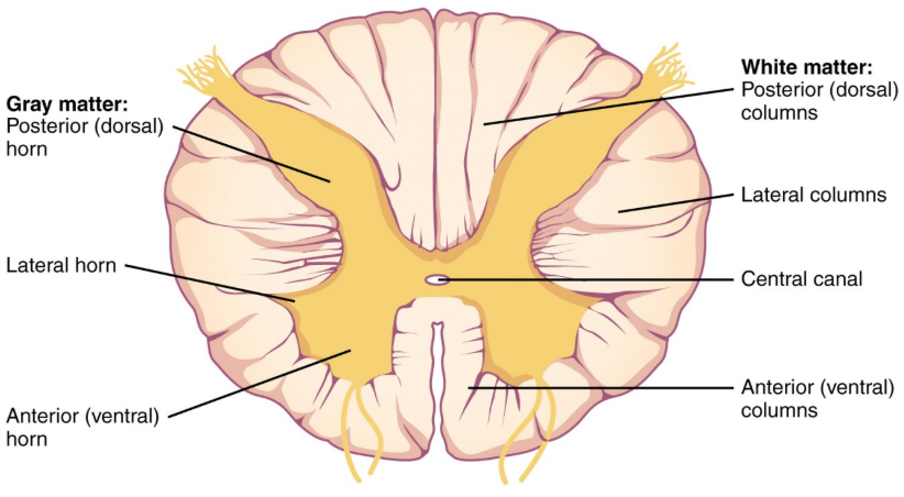
Posterior (dorsal) gray horn
Cell bodies of second-order sensory neurons and interneurons that receive and process input arriving through the dorsal root. The cell bodies of the first-order (primary) sensory neurons sit outside the cord, in the dorsal root ganglion.
Lateral gray horn
Cell bodies of preganglionic sympathetic neurons; present only in the thoracic and upper lumbar (T1-L2) segments of the spinal cord.
Anterior (ventral) gray horn
Cell bodies of the lower motor neurons that control skeletal muscles.
Posterior (dorsal) white column
Nerve fibers containing sensory information from the body, such as two-point discrimination, fine touch, vibration, and proprioception.
Lateral white column
Contains both ascending sensory fibers (the lateral spinothalamic tract, carrying pain and temperature) and descending motor fibers (the lateral corticospinal tract, controlling voluntary movement of the extremities).
Anterior (ventral) white column
Contains both descending motor fibers (the anterior corticospinal tract, which carries commands to axial muscles and decussates near its target, remaining largely ipsilateral within the cord) and ascending sensory fibers (the anterior spinothalamic tract, which carries crude touch and pressure and crosses in the spinal cord to ascend contralaterally to the brain).
Sympathetic nervous system
A division of the autonomic nervous system that prepares the body to respond to physical activity, psychological stress, or perceived danger by initiating the “fight-or-flight” response.
Parasympathetic nervous system
A division of the autonomic nervous system that promotes recovery, conservation of energy, and homeostasis after stress or exertion - often called the “rest-and-digest” system.

The sympathetic nervous system increases heart rate, blood pressure, and respiratory rate while redirecting blood away from digestion and toward working muscles. The parasympathetic nervous system reverses these effects, slowing heart rate and blood pressure while promoting digestion and waste elimination.

Cross section of spinal column
Anatomy & Physiology by Lindsay M. Biga, Sierra Dawson, Amy Harwell, Robin Hopkins, Joel Kaufmann, Mike LeMaster, Philip Matern, Katie Morrison-Graham, Devon Quick & Jon Runyeon
/
Oregon State University
/
CC BY 4.0

Spinal tracts

Pathways of spinal cord
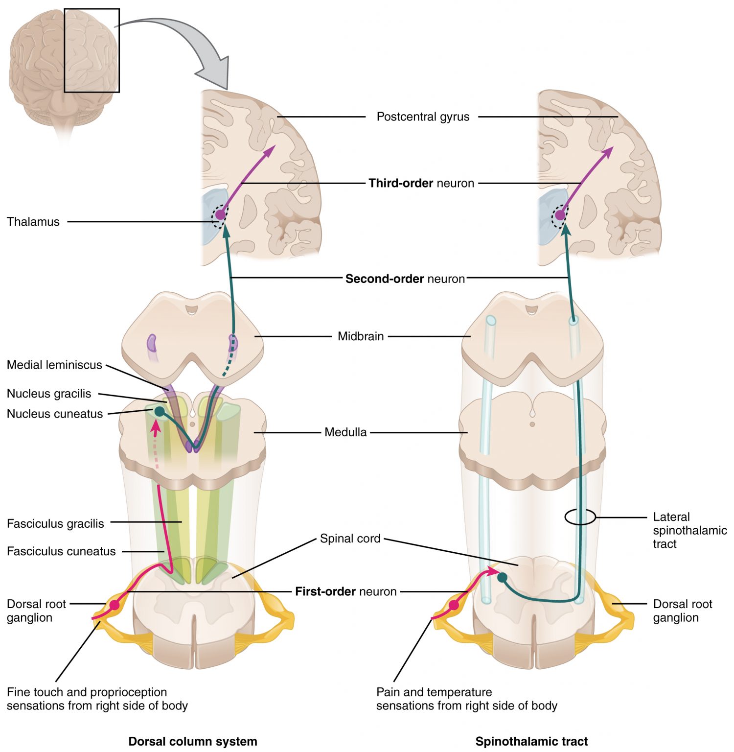
Anatomy & Physiology by Lindsay M. Biga, Sierra Dawson, Amy Harwell, Robin Hopkins, Joel Kaufmann, Mike LeMaster, Philip Matern, Katie Morrison-Graham, Devon Quick & Jon Runyeon
/
Oregon State University
/
CC BY 4.0
  • Ascending tracts
    • Carry sensory information such as touch, pain, and temperature from the peripheral system to the brain
      • Dorsal column-medial lemniscal: transmits light touch, deep pressure, two-point discrimination, vibratory, and conscious proprioceptive information to the cerebrum via the thalamus; ascends on the same (ipsilateral) side of the spinal cord and crosses in the medulla to the contralateral side
      • Anterior and lateral spinothalamic: transmit pain, crude touch, and temperature information to the cerebrum via the thalamus; crosses in the spinal cord (anterior white commissure), typically within one to two segments of entry, to the contralateral side
      • Spinocerebellar: transmits proprioceptive information to the cerebellum; does not cross, stays on the same side
  • Descending tracts
    • Carry motor information from the brain to the peripheral system to give voluntary commands of movement of axial and skeletal muscles
    • Anterior and lateral corticospinal: transmit commands to move axial and/or skeletal muscles
      • Anterior corticospinal: motor system that descends uncrossed in the front of the spinal cord, controlling trunk/axial muscles for posture
      • Lateral corticospinal: motor system where fibers cross over (decussate) in the lower medulla, forming the lateral tract, which controls movement of the extremities - particularly fine, coordinated movements - on the opposite (contralateral) side of the body
    • Reticulospinal: a descending pathway from the pontine and medullary reticular formation that helps regulate muscle tone and posture, unlike the corticospinal tract, which carries voluntary motor commands

Arterial circulation of the spinal cord

  • Three longitudinal arteries of the spinal cord: one anterior and two posterior spinal arteries
    • All three run along the surface of the cord within the subarachnoid space (between the arachnoid and pia mater), closely covered by the pia; the anterior spinal artery lies in the anterior median fissure
    • Anterior spinal artery
      • Supplies blood to the anterior two-thirds of the spinal cord, except the dorsal column
    • Posterior spinal artery
      • Paired (left and right) posterior spinal arteries supply the posterior one-third of the spinal cord, including the dorsal columns and dorsal gray horns

Spinal cord function & PTA scope

  • Relays sensory info to brain and motor commands to periphery
  • Coordinates simple reflexes (withdrawal, patellar) without brain involvement
  • PTA collects data (e.g., dermatomal/DTR testing) and reports changes; PT interprets findings and modifies plan of care

Spinal cord regions

  • Cervical: 8 nerves — neck, upper extremity
  • Thoracic: 12 nerves — upper chest, chest, abdomen, upper back
  • Lumbar: 5 nerves — lower abdominal wall, groin, front and inner thigh, and (with sacral) leg and foot
  • Sacral: 5 nerves — buttocks, back of thigh, much of leg and foot, perineum; bowel, bladder, sexual function (S2-S4)
  • Coccygeal: 1 nerve — perineal area

Protective layers

  • Dura mater (outermost) → epidural space (fat, blood vessels)
  • Arachnoid mater (web-like, middle) → subarachnoid space (CSF)
  • Pia mater (innermost, directly on cord)

Spinal cord cross-sectional anatomy

  • Dorsal root = sensory (afferent); ventral root = motor (efferent); together form mixed spinal nerve
  • Denticulate ligament stabilizes cord
  • Gray matter horns:
    • Posterior (dorsal) horn: second-order sensory neuron and interneuron cell bodies (first-order are in the dorsal root ganglion)
    • Lateral horn: preganglionic sympathetic cell bodies (only T1–L2)
    • Anterior (ventral) horn: lower motor neuron cell bodies
  • White matter columns:
    • Posterior (dorsal): fine touch, vibration, proprioception, two-point discrimination
    • Lateral: ascending pain/temp (spinothalamic) + descending voluntary motor (corticospinal) to extremities
    • Anterior (ventral): descending motor to axial muscles (largely ipsilateral) + ascending crude touch/pressure (crosses in cord)
  • Autonomic divisions: sympathetic (“fight-or-flight,” ↑HR/BP/RR, ↓digestion) vs. parasympathetic (“rest-and-digest,” opposite effects)

Spinal tracts

  • Ascending (sensory, to brain):
    • Dorsal column-medial lemniscal: touch, pressure, vibration, proprioception; ipsilateral until crossing in medulla
    • Spinothalamic (anterior/lateral): pain, crude touch, temperature; crosses in spinal cord near entry level
    • Spinocerebellar: proprioception to cerebellum; stays ipsilateral
  • Descending (motor, from brain):
    • Anterior corticospinal: axial/trunk muscles, uncrossed
    • Lateral corticospinal: extremity movement, fine motor control, crosses (decussates) in lower medulla → contralateral control

Arterial supply

  • Anterior spinal artery: supplies anterior two-thirds of cord (excludes dorsal columns)
  • Posterior spinal arteries (paired): supply posterior one-third, including dorsal columns and dorsal gray horns

Sign up for free to take 15 quiz questions on this topic

Previous
Next  | 3.3.1 Components and nerve structure
All rights reserved ©2016 - 2026 Achievable, Inc.

Anatomy and function of spinal cord

Primary function of the spinal cord

  • Acts as a relay pathway between the peripheral system and the brain
    • Provides sensory information regarding the environment to the brain
    • Manages motor commands to the peripheral system in reaction to the environment
    • Coordinates simple reflexes that do not need to go to the brain, such as withdrawal from pain, patellar reflex, etc.

PTA scope of practice: The PTA collects data and recognizes and communicates changes in a patient’s status to the supervising PT; evaluating the patient, interpreting findings, and changing the plan of care remain the PT’s responsibility. Dermatomal sensory testing and deep tendon reflex testing rest on this anatomy; when a PTA collects these findings, a new or changed deficit is reported to the supervising PT, who interprets it. Supervision and delegation rules are covered in PTA responsibilities, research, and ADA.

Anatomy of the spinal cord

  • Cervical spine
    • Eight nerves that provide innervation to the neck and upper extremity
  • Thoracic spine
    • Twelve nerves that provide innervation to the upper chest, chest, abdomen, and upper back
  • Lumbar spine
    • Five nerves that provide innervation to the lower abdominal wall, groin, and front and inner thigh, and (with the sacral nerves) the leg and foot
  • Sacral spine
    • Five nerves that provide innervation to the buttocks, back of the thigh, much of the leg and foot (including the sole and outer foot), and the perineum, and that carry bowel, bladder, and sexual function (S2-S4)
  • Coccygeal spine
    • One nerve that provides innervation to the perineal area

Protective layers of the spinal cord

  • Dura mater
    • Outermost protective layer
    • Space between dura and the bony walls of the vertebral canal referred to as epidural space, which contains adipose tissue and blood vessels
  • Arachnoid mater
    • Middle layer with a web-like appearance
    • Space between arachnoid and pia referred to as subarachnoid space and contains cerebrospinal fluid
  • Pia mater
    • Innermost layer that is in direct contact with the spinal cord
Definitions
Dorsal root
Sensory (afferent) nerve that carries information from the periphery into the spinal cord.
Ventral root
Motor (efferent) nerve that carries commands from the spinal cord out to the periphery.
Spinal nerve
Pathway for sensory and motor information to move between the peripheral system and the spinal cord. The dorsal (sensory) and ventral (motor) roots join together to form this mixed spinal nerve.
Denticulate ligament
Ligament that provides stabilization of the spinal cord.
Posterior (dorsal) gray horn
Cell bodies of second-order sensory neurons and interneurons that receive and process input arriving through the dorsal root. The cell bodies of the first-order (primary) sensory neurons sit outside the cord, in the dorsal root ganglion.
Lateral gray horn
Cell bodies of preganglionic sympathetic neurons; present only in the thoracic and upper lumbar (T1-L2) segments of the spinal cord.
Anterior (ventral) gray horn
Cell bodies of the lower motor neurons that control skeletal muscles.
Posterior (dorsal) white column
Nerve fibers containing sensory information from the body, such as two-point discrimination, fine touch, vibration, and proprioception.
Lateral white column
Contains both ascending sensory fibers (the lateral spinothalamic tract, carrying pain and temperature) and descending motor fibers (the lateral corticospinal tract, controlling voluntary movement of the extremities).
Anterior (ventral) white column
Contains both descending motor fibers (the anterior corticospinal tract, which carries commands to axial muscles and decussates near its target, remaining largely ipsilateral within the cord) and ascending sensory fibers (the anterior spinothalamic tract, which carries crude touch and pressure and crosses in the spinal cord to ascend contralaterally to the brain).
Sympathetic nervous system
A division of the autonomic nervous system that prepares the body to respond to physical activity, psychological stress, or perceived danger by initiating the “fight-or-flight” response.
Parasympathetic nervous system
A division of the autonomic nervous system that promotes recovery, conservation of energy, and homeostasis after stress or exertion - often called the “rest-and-digest” system.

The sympathetic nervous system increases heart rate, blood pressure, and respiratory rate while redirecting blood away from digestion and toward working muscles. The parasympathetic nervous system reverses these effects, slowing heart rate and blood pressure while promoting digestion and waste elimination.

Spinal tracts

  • Ascending tracts
    • Carry sensory information such as touch, pain, and temperature from the peripheral system to the brain
      • Dorsal column-medial lemniscal: transmits light touch, deep pressure, two-point discrimination, vibratory, and conscious proprioceptive information to the cerebrum via the thalamus; ascends on the same (ipsilateral) side of the spinal cord and crosses in the medulla to the contralateral side
      • Anterior and lateral spinothalamic: transmit pain, crude touch, and temperature information to the cerebrum via the thalamus; crosses in the spinal cord (anterior white commissure), typically within one to two segments of entry, to the contralateral side
      • Spinocerebellar: transmits proprioceptive information to the cerebellum; does not cross, stays on the same side
  • Descending tracts
    • Carry motor information from the brain to the peripheral system to give voluntary commands of movement of axial and skeletal muscles
    • Anterior and lateral corticospinal: transmit commands to move axial and/or skeletal muscles
      • Anterior corticospinal: motor system that descends uncrossed in the front of the spinal cord, controlling trunk/axial muscles for posture
      • Lateral corticospinal: motor system where fibers cross over (decussate) in the lower medulla, forming the lateral tract, which controls movement of the extremities - particularly fine, coordinated movements - on the opposite (contralateral) side of the body
    • Reticulospinal: a descending pathway from the pontine and medullary reticular formation that helps regulate muscle tone and posture, unlike the corticospinal tract, which carries voluntary motor commands

Arterial circulation of the spinal cord

  • Three longitudinal arteries of the spinal cord: one anterior and two posterior spinal arteries
    • All three run along the surface of the cord within the subarachnoid space (between the arachnoid and pia mater), closely covered by the pia; the anterior spinal artery lies in the anterior median fissure
    • Anterior spinal artery
      • Supplies blood to the anterior two-thirds of the spinal cord, except the dorsal column
    • Posterior spinal artery
      • Paired (left and right) posterior spinal arteries supply the posterior one-third of the spinal cord, including the dorsal columns and dorsal gray horns
Key points

Spinal cord function & PTA scope

  • Relays sensory info to brain and motor commands to periphery
  • Coordinates simple reflexes (withdrawal, patellar) without brain involvement
  • PTA collects data (e.g., dermatomal/DTR testing) and reports changes; PT interprets findings and modifies plan of care

Spinal cord regions

  • Cervical: 8 nerves — neck, upper extremity
  • Thoracic: 12 nerves — upper chest, chest, abdomen, upper back
  • Lumbar: 5 nerves — lower abdominal wall, groin, front and inner thigh, and (with sacral) leg and foot
  • Sacral: 5 nerves — buttocks, back of thigh, much of leg and foot, perineum; bowel, bladder, sexual function (S2-S4)
  • Coccygeal: 1 nerve — perineal area

Protective layers

  • Dura mater (outermost) → epidural space (fat, blood vessels)
  • Arachnoid mater (web-like, middle) → subarachnoid space (CSF)
  • Pia mater (innermost, directly on cord)

Spinal cord cross-sectional anatomy

  • Dorsal root = sensory (afferent); ventral root = motor (efferent); together form mixed spinal nerve
  • Denticulate ligament stabilizes cord
  • Gray matter horns:
    • Posterior (dorsal) horn: second-order sensory neuron and interneuron cell bodies (first-order are in the dorsal root ganglion)
    • Lateral horn: preganglionic sympathetic cell bodies (only T1–L2)
    • Anterior (ventral) horn: lower motor neuron cell bodies
  • White matter columns:
    • Posterior (dorsal): fine touch, vibration, proprioception, two-point discrimination
    • Lateral: ascending pain/temp (spinothalamic) + descending voluntary motor (corticospinal) to extremities
    • Anterior (ventral): descending motor to axial muscles (largely ipsilateral) + ascending crude touch/pressure (crosses in cord)
  • Autonomic divisions: sympathetic (“fight-or-flight,” ↑HR/BP/RR, ↓digestion) vs. parasympathetic (“rest-and-digest,” opposite effects)

Spinal tracts

  • Ascending (sensory, to brain):
    • Dorsal column-medial lemniscal: touch, pressure, vibration, proprioception; ipsilateral until crossing in medulla
    • Spinothalamic (anterior/lateral): pain, crude touch, temperature; crosses in spinal cord near entry level
    • Spinocerebellar: proprioception to cerebellum; stays ipsilateral
  • Descending (motor, from brain):
    • Anterior corticospinal: axial/trunk muscles, uncrossed
    • Lateral corticospinal: extremity movement, fine motor control, crosses (decussates) in lower medulla → contralateral control

Arterial supply

  • Anterior spinal artery: supplies anterior two-thirds of cord (excludes dorsal columns)
  • Posterior spinal arteries (paired): supply posterior one-third, including dorsal columns and dorsal gray horns

More from Neuromuscular system

  • Central nervous system
  • Stroke deficits and recovery
  • Traumatic brain injury
  • Spinal cord injury
  • Neurodegenerative disorders and epilepsy