


This browser is too old to run Achievable.

Update it, or open this page in a current version of Chrome, Safari, Firefox or Edge. On an iPhone or iPad, update iOS: every browser there runs on the version of Safari that comes with it.

Achievable logoAchievable logo
NPTE-PTA
Sign in
Sign up
Purchase
Textbook
Practice exams
Support
How it works
Resources
Exam catalog
Mountain with a flag at the peak
Textbook
Introduction
1. Cardiovascular system
2. Pulmonary system
3. Neuromuscular system
3.1 Central nervous system
3.2 Anatomy and function of spinal cord
3.3 Peripheral nervous system
3.3.1 Components and nerve structure
3.3.2 Dermatomes, myotomes, reflexes, and plexuses
3.3.3 Upper limb tension tests and thoracic outlet syndrome
3.4 Stroke deficits and recovery
3.5 Traumatic brain injury
3.6 Spinal cord injury
3.7 Neurodegenerative disorders and epilepsy
3.8 Peripheral nervous system conditions
3.9 Other neurological conditions
3.10 Interventions for neurological conditions
3.11 Vestibular system
4. Pediatrics
5. Musculoskeletal system
6. Other systems
7. Non-systems
Wrapping up
Achievable logoAchievable logo
3.3.3 Upper limb tension tests and thoracic outlet syndrome
Achievable NPTE-PTA
3. Neuromuscular system
3.3. Peripheral nervous system
Our NPTE-PTA course is now in "early access" - get 50% off for a limited time.

Upper limb tension tests and thoracic outlet syndrome

9 min read
Font
Discuss
Share
Feedback

Upper limb tension tests

Definitions
Upper limb tension test
Assists with identifying peripheral nerve injury by placing the upper limb in a position that will stress the nerve. A test is positive when it reproduces the patient’s symptoms, shows a side-to-side difference, or when symptoms change with a sensitizing movement such as cervical side bending.

PTA scope of practice: The PTA collects data and recognizes and communicates changes in a patient’s status to the supervising PT; evaluating the patient, interpreting findings, and changing the plan of care remain the PT’s responsibility. When a PTA performs the upper limb tension tests and thoracic outlet tests in this chapter as part of data collection, a positive finding such as reproduced symptoms or a diminished radial pulse is reported to the supervising PT, not interpreted as a diagnosis. Supervision and delegation rules are covered in PTA responsibilities, research, and ADA.

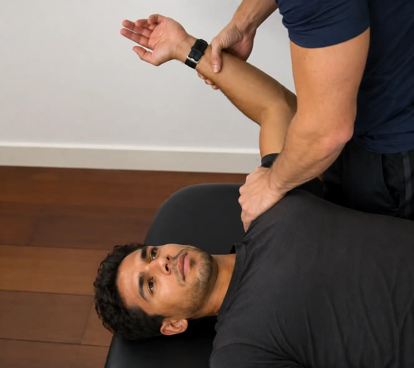
  • Upper limb tension test 1 (ULTT 1) - median and anterior interosseous nerve
    • Cervical spine: contralateral lateral flexion
    • Shoulder: depression and abduction to 110 degrees
    • Elbow: extension
    • Forearm: supination
    • Wrist: extension
    • Fingers and thumb: extension
Upper limb tension test 1 demonstrating median nerve bias with shoulder abduction, elbow extension, and wrist extension.
ULTT 1: Median nerve
Achievable
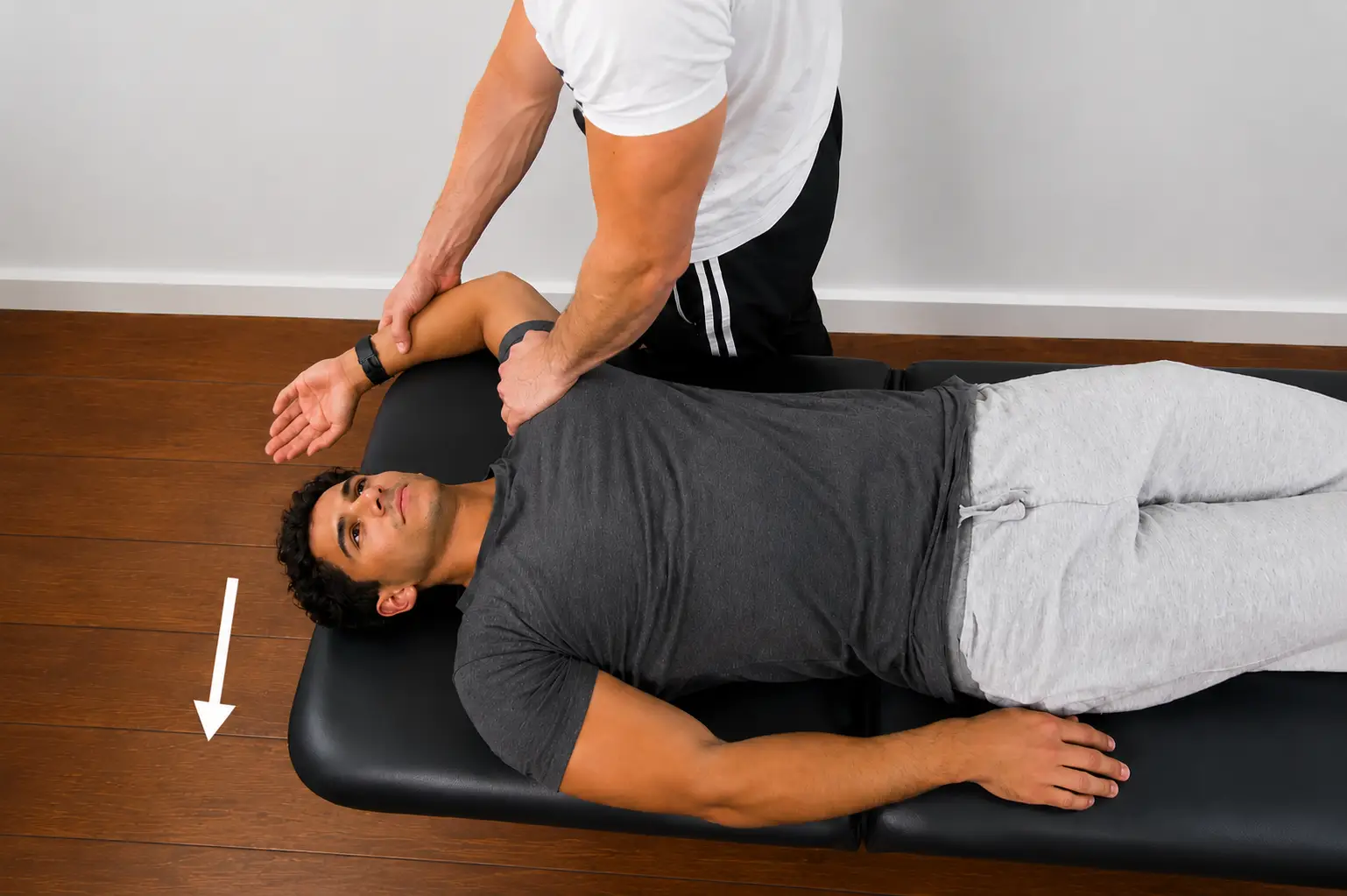
  • Upper limb tension test 2 (ULTT 2) - median, axillary, and musculocutaneous nerve
    • Cervical spine: contralateral lateral flexion
    • Shoulder: depression and abduction to 10 degrees, lateral rotation
    • Elbow: extension
    • Forearm: supination
    • Wrist: extension
Upper limb tension test 2 showing median nerve bias with shoulder depression and abduction, elbow extension, and forearm supination.
ULTT 2: Median, axillary, and musculocutaneous nerves
Achievable
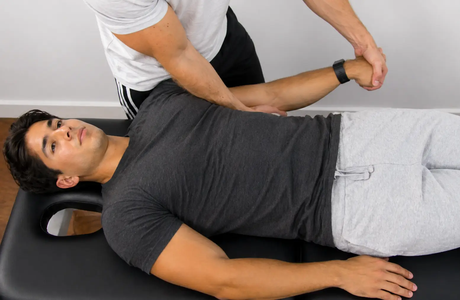
  • Upper limb tension test 3 (ULTT 3) - radial nerve
    • Cervical spine: contralateral lateral flexion
    • Shoulder: depression and abduction to 10 degrees; internal rotation
    • Elbow: extension
    • Forearm: pronation
    • Wrist: flexion with ulnar deviation
    • Fingers and thumb: flexion
Upper limb tension test 3 demonstrating radial nerve bias with shoulder depression, elbow extension, and forearm pronation.
ULTT 3: Radial nerve
Achievable
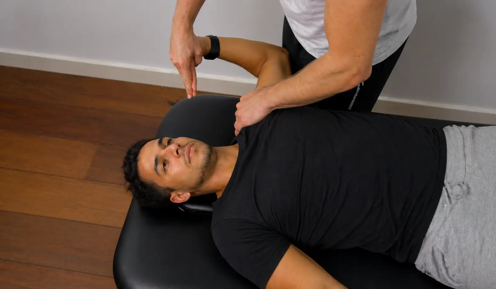
  • Upper limb tension test 4 (ULTT 4) - ulnar nerve
    • Cervical spine: contralateral lateral flexion
    • Shoulder: depression and abduction (10-90 degrees) with hand to ear; lateral rotation
    • Elbow: flexion
    • Forearm: pronation
    • Wrist: extension and radial deviation
    • Fingers and thumb: extension
Upper limb tension test 4 showing ulnar nerve bias with shoulder abduction, elbow flexion, and wrist extension.
ULTT 4: Ulnar nerve
Achievable

Comparing the four ULTTs

Test Shoulder Elbow Forearm Wrist
ULTT 1 (median/AIN) Depression, abduction 110° Extension Supination Extension
ULTT 2 (median/axillary/musculocutaneous) Depression, abduction 10°, lateral rotation Extension Supination Extension
ULTT 3 (radial) Depression, abduction 10°, internal rotation Extension Pronation Flexion with ulnar deviation
ULTT 4 (ulnar) Depression, abduction 10-90°, lateral rotation, hand to ear Flexion Pronation Extension and radial deviation
Sidenote
Alternate ULTT numbering

Some sources (e.g., Butler) label these tests ULTT 1, 2a, 2b, and 3 instead of ULTT 1-4; this chapter uses the more common Magee numbering (ULTT 1-4).

Thoracic outlet syndrome

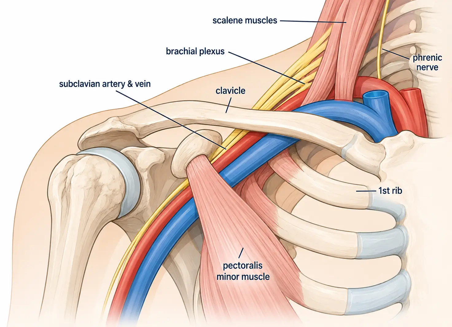
Labeled diagram of the thoracic outlet showing the clavicle, first rib, brachial plexus, subclavian vessels, and surrounding muscles.
Thoracic outlet anatomy
Achievable
Definitions
Thoracic outlet syndrome
A condition caused by compression of the brachial plexus, subclavian artery, and/or subclavian vein as they pass through the thoracic outlet - the space bounded by the clavicle, the first rib, and the scalene muscles.

Neurogenic TOS is the most common type and causes paresthesia and weakness, usually in the ulnar (C8-T1) distribution. Vascular TOS causes coolness, color change, swelling, or a diminished pulse. Contributing factors include a cervical rib, forward-head or drooped-shoulder posture, scalene tightness, and repetitive overhead work. Because sustained overhead positions and repetitive overhead work can provoke or worsen symptoms, interventions emphasize postural correction and scalene/pectoralis minor flexibility; progressive vascular signs noticed during treatment are reported to the supervising PT immediately.

These provocative tests position the patient to narrow a specific part of the thoracic outlet and check whether symptoms are reproduced. A positive finding is reproduction of neurological symptoms (pain, weakness, numbness, or loss of hand coordination) and/or vascular symptoms (a diminished or absent radial pulse); report positive findings to the supervising PT. Stop the test immediately if it produces new or worsening neurovascular symptoms, such as pallor, a lost pulse, or progressive numbness.

Comparing the four TOS tests

Test Compression site Key position
Adson’s test Interscalene triangle (between the scalene muscles) Head rotated toward the tested side, neck extended, shoulder extended and externally rotated, deep breath held
Costoclavicular test Costoclavicular space (between the clavicle and first rib) Military brace: shoulders retracted and depressed, chest lifted
Roos elevated arm test Costoclavicular space Shoulders abducted 90 degrees and externally rotated, elbows flexed 90 degrees, hands slowly open and close
Wright’s test Subpectoral space (behind pectoralis minor) Arm hyperabducted overhead with external rotation
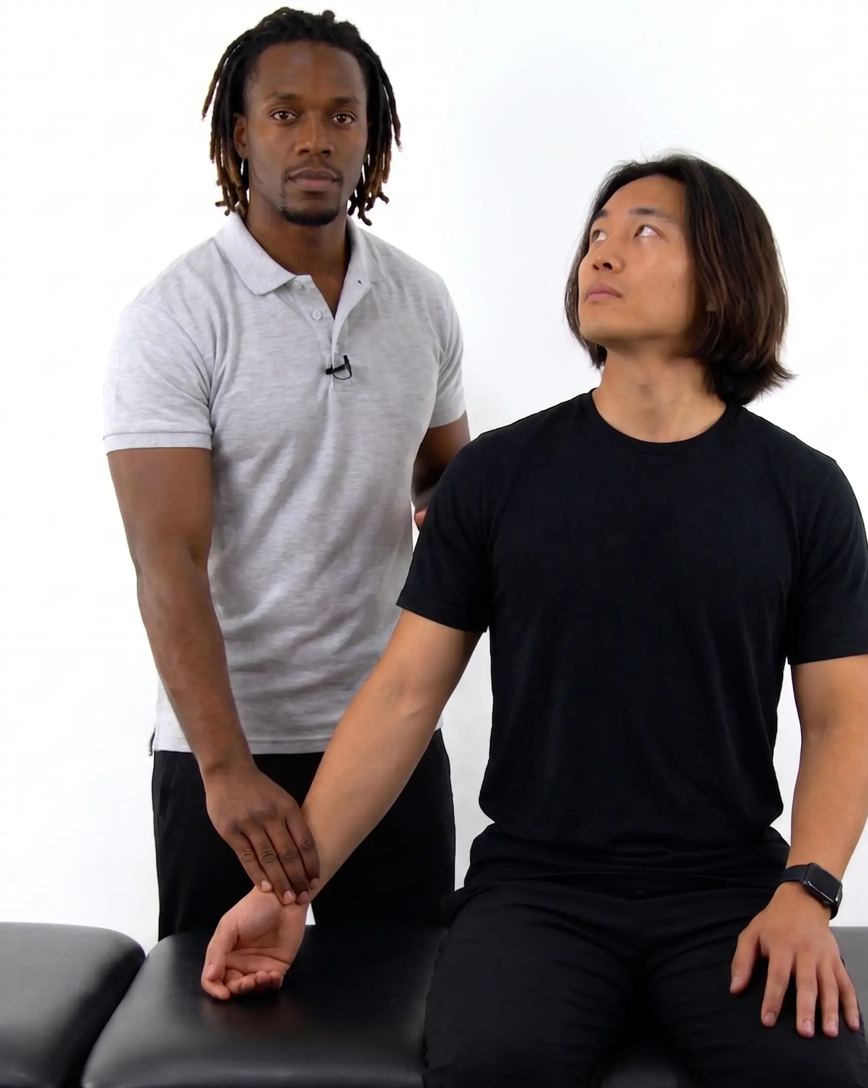
  • Adson’s test (compression of the subclavian artery and/or nerves as they pass through the interscalene space)
    • Patient seated with the radial pulse palpated; the patient takes and holds a deep breath while the head is rotated toward the extremity being tested, the shoulder is extended and externally rotated, and the head is extended
Adson's test for thoracic outlet syndrome with the arm extended and the head rotated toward the tested side.
Thoracic outlet syndrome - Adson's test
Achievable
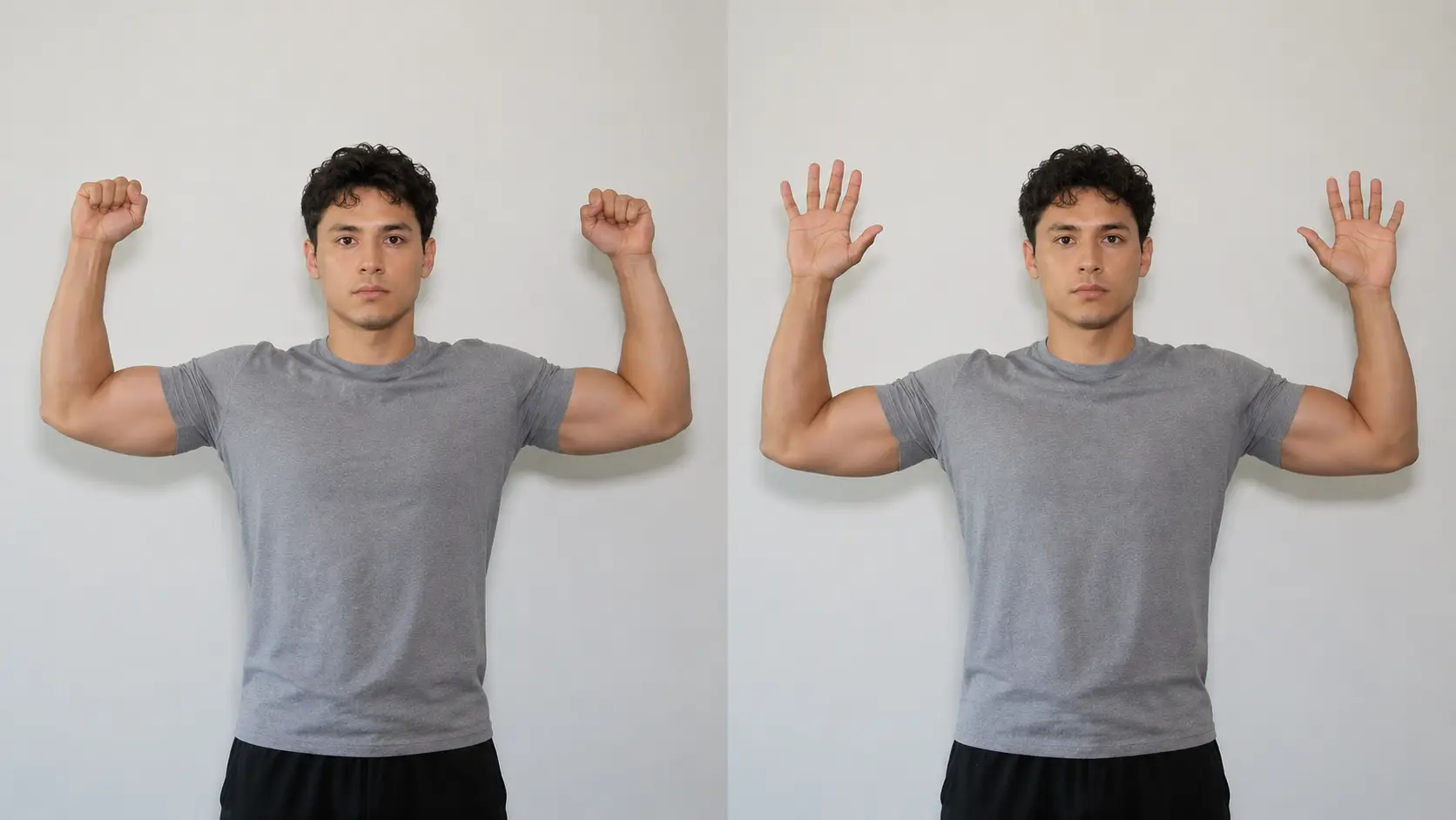
  • Roos elevated arm test (nerves and/or blood vessels in the space between the clavicle and the first rib are compressed)
    • Patient standing with shoulders fully externally rotated, 90 degrees abducted, elbows flexed to 90 degrees; the patient then slowly opens and closes their hands for 3 minutes
      • Positive: reproduction of symptoms, heaviness or fatigue in the arms, or inability to maintain the position for the full 3 minutes
Roos test for thoracic outlet syndrome with both arms held in 90 degrees of shoulder abduction and external rotation while repeatedly opening and closing the hands.
Thoracic outlet syndrome - Roos test
Achievable
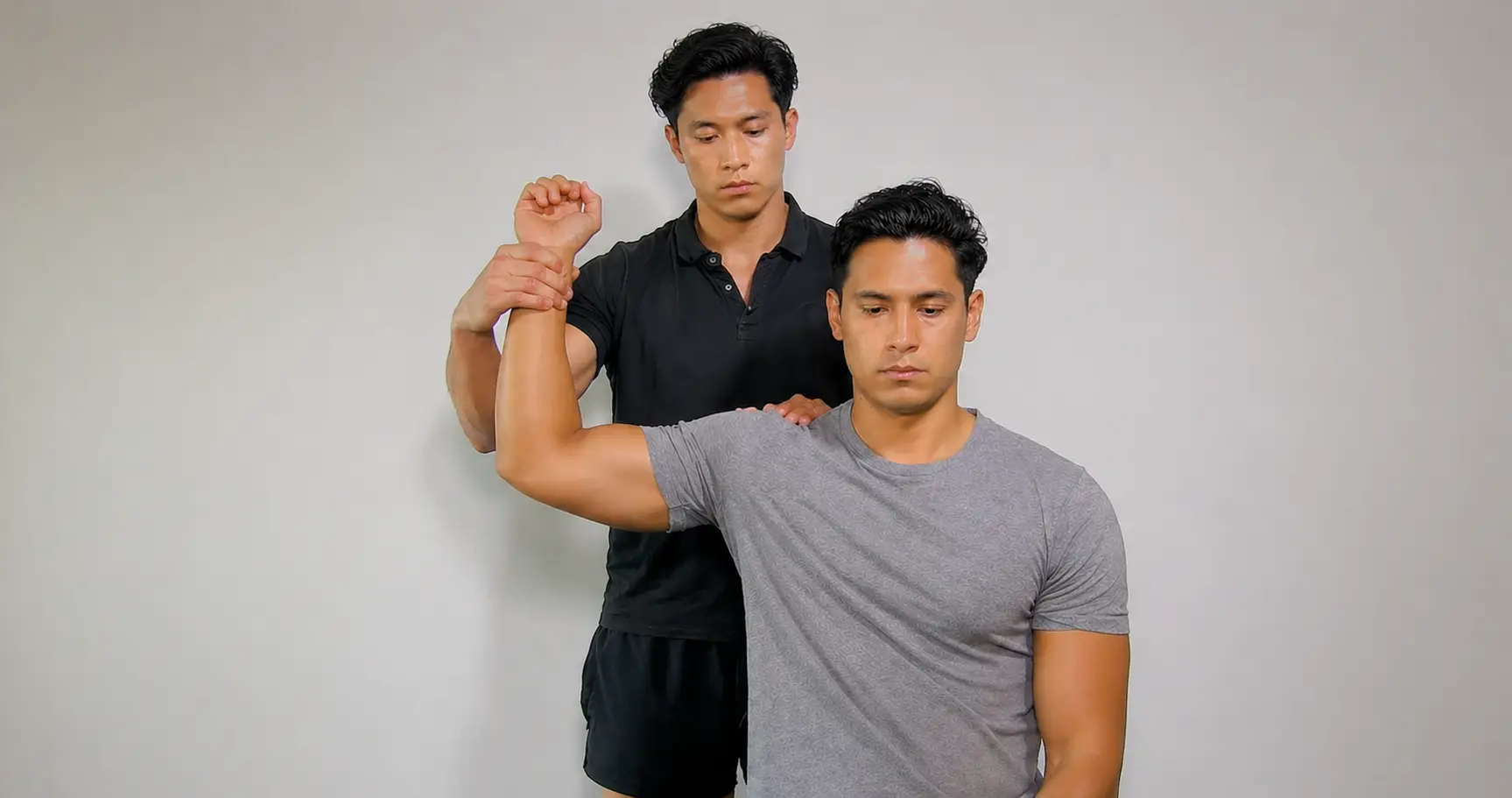
  • Wright’s test (compression at the space behind the pectoralis minor muscle)
    • Patient seated with the radial pulse palpated; the arm is passively hyperabducted overhead (about 180 degrees) with external rotation and held for 1-2 minutes
Wright's test for thoracic outlet syndrome with the arm hyperabducted and externally rotated overhead.
Thoracic outlet syndrome - Wright's test
Achievable
  • Costoclavicular test (compression of the neurovascular bundle between the clavicle and first rib)
    • Patient seated with the radial pulse palpated; the patient retracts and depresses the shoulders while lifting the chest into an exaggerated military brace posture and holds for up to 1 minute
Costoclavicular test for thoracic outlet syndrome with the shoulders retracted and depressed in an exaggerated military posture.
Thoracic outlet syndrome - Costoclavicular test
Achievable

Upper limb tension tests (ULTT) - purpose and PTA role

  • Position upper limb to stress a peripheral nerve; positive = reproduces symptoms, side-to-side difference, or change with sensitizing movement (e.g., cervical side bending)
  • PTA collects data and reports positive findings (reproduced symptoms, diminished pulse) to supervising PT; interpretation/diagnosis remains PT’s responsibility
  • Also called ULTT 1, 2a, 2b, 3 in Butler’s system (this chapter uses Magee’s ULTT 1-4)

ULTT patterns by nerve

  • ULTT 1 (median/AIN): shoulder abducted 110°, elbow extension, forearm supination, wrist/finger extension
  • ULTT 2 (median/axillary/musculocutaneous): shoulder abducted 10° with lateral rotation, elbow extension, forearm supination, wrist extension
  • ULTT 3 (radial): shoulder internal rotation, elbow extension, forearm pronation, wrist flexion with ulnar deviation, finger flexion
  • ULTT 4 (ulnar): hand-to-ear position, elbow flexion (only test with flexed elbow), forearm pronation, wrist extension with radial deviation
  • Key differentiator: elbow flexion = ulnar; wrist flexion = radial; all others use wrist extension

Thoracic outlet syndrome (TOS) - overview

  • Compression of brachial plexus and/or subclavian artery/vein within thoracic outlet (bounded by clavicle, first rib, scalenes)
  • Neurogenic TOS (most common): paresthesia/weakness, typically ulnar (C8-T1) distribution
  • Vascular TOS: coolness, color change, swelling, diminished pulse
  • Contributing factors: cervical rib, forward-head/drooped-shoulder posture, scalene tightness, repetitive overhead work
  • Treatment focus: postural correction, scalene/pec minor flexibility; report progressive vascular signs immediately

TOS provocative tests - compression sites and positions

  • Positive = reproduced neuro symptoms (pain, weakness, numbness, poor coordination) and/or vascular symptoms (diminished/absent radial pulse); stop test if new/worsening neurovascular signs appear (pallor, lost pulse, progressive numbness)
  • Adson’s test: interscalene triangle; head rotated/extended toward tested side, shoulder extended/externally rotated, deep breath held
  • Costoclavicular test: costoclavicular space; military brace posture (shoulders retracted/depressed, chest lifted)
  • Roos elevated arm test: costoclavicular space; shoulders abducted/externally rotated 90°, elbows flexed 90°, open/close hands for 3 minutes
  • Wright’s test: subpectoral space (behind pec minor); arm hyperabducted overhead (~180°) with external rotation, held 1-2 minutes

Sign up for free to take 7 quiz questions on this topic

Previous
Next  | 3.4 Stroke deficits and recovery
All rights reserved ©2016 - 2026 Achievable, Inc.

Upper limb tension tests and thoracic outlet syndrome

Upper limb tension tests

Definitions
Upper limb tension test
Assists with identifying peripheral nerve injury by placing the upper limb in a position that will stress the nerve. A test is positive when it reproduces the patient’s symptoms, shows a side-to-side difference, or when symptoms change with a sensitizing movement such as cervical side bending.

PTA scope of practice: The PTA collects data and recognizes and communicates changes in a patient’s status to the supervising PT; evaluating the patient, interpreting findings, and changing the plan of care remain the PT’s responsibility. When a PTA performs the upper limb tension tests and thoracic outlet tests in this chapter as part of data collection, a positive finding such as reproduced symptoms or a diminished radial pulse is reported to the supervising PT, not interpreted as a diagnosis. Supervision and delegation rules are covered in PTA responsibilities, research, and ADA.

  • Upper limb tension test 1 (ULTT 1) - median and anterior interosseous nerve
    • Cervical spine: contralateral lateral flexion
    • Shoulder: depression and abduction to 110 degrees
    • Elbow: extension
    • Forearm: supination
    • Wrist: extension
    • Fingers and thumb: extension
  • Upper limb tension test 2 (ULTT 2) - median, axillary, and musculocutaneous nerve
    • Cervical spine: contralateral lateral flexion
    • Shoulder: depression and abduction to 10 degrees, lateral rotation
    • Elbow: extension
    • Forearm: supination
    • Wrist: extension
  • Upper limb tension test 3 (ULTT 3) - radial nerve
    • Cervical spine: contralateral lateral flexion
    • Shoulder: depression and abduction to 10 degrees; internal rotation
    • Elbow: extension
    • Forearm: pronation
    • Wrist: flexion with ulnar deviation
    • Fingers and thumb: flexion
  • Upper limb tension test 4 (ULTT 4) - ulnar nerve
    • Cervical spine: contralateral lateral flexion
    • Shoulder: depression and abduction (10-90 degrees) with hand to ear; lateral rotation
    • Elbow: flexion
    • Forearm: pronation
    • Wrist: extension and radial deviation
    • Fingers and thumb: extension

Comparing the four ULTTs

Test Shoulder Elbow Forearm Wrist
ULTT 1 (median/AIN) Depression, abduction 110° Extension Supination Extension
ULTT 2 (median/axillary/musculocutaneous) Depression, abduction 10°, lateral rotation Extension Supination Extension
ULTT 3 (radial) Depression, abduction 10°, internal rotation Extension Pronation Flexion with ulnar deviation
ULTT 4 (ulnar) Depression, abduction 10-90°, lateral rotation, hand to ear Flexion Pronation Extension and radial deviation
Sidenote
Alternate ULTT numbering

Some sources (e.g., Butler) label these tests ULTT 1, 2a, 2b, and 3 instead of ULTT 1-4; this chapter uses the more common Magee numbering (ULTT 1-4).

Thoracic outlet syndrome

Definitions
Thoracic outlet syndrome
A condition caused by compression of the brachial plexus, subclavian artery, and/or subclavian vein as they pass through the thoracic outlet - the space bounded by the clavicle, the first rib, and the scalene muscles.

Neurogenic TOS is the most common type and causes paresthesia and weakness, usually in the ulnar (C8-T1) distribution. Vascular TOS causes coolness, color change, swelling, or a diminished pulse. Contributing factors include a cervical rib, forward-head or drooped-shoulder posture, scalene tightness, and repetitive overhead work. Because sustained overhead positions and repetitive overhead work can provoke or worsen symptoms, interventions emphasize postural correction and scalene/pectoralis minor flexibility; progressive vascular signs noticed during treatment are reported to the supervising PT immediately.

These provocative tests position the patient to narrow a specific part of the thoracic outlet and check whether symptoms are reproduced. A positive finding is reproduction of neurological symptoms (pain, weakness, numbness, or loss of hand coordination) and/or vascular symptoms (a diminished or absent radial pulse); report positive findings to the supervising PT. Stop the test immediately if it produces new or worsening neurovascular symptoms, such as pallor, a lost pulse, or progressive numbness.

Comparing the four TOS tests

Test Compression site Key position
Adson’s test Interscalene triangle (between the scalene muscles) Head rotated toward the tested side, neck extended, shoulder extended and externally rotated, deep breath held
Costoclavicular test Costoclavicular space (between the clavicle and first rib) Military brace: shoulders retracted and depressed, chest lifted
Roos elevated arm test Costoclavicular space Shoulders abducted 90 degrees and externally rotated, elbows flexed 90 degrees, hands slowly open and close
Wright’s test Subpectoral space (behind pectoralis minor) Arm hyperabducted overhead with external rotation
  • Adson’s test (compression of the subclavian artery and/or nerves as they pass through the interscalene space)
    • Patient seated with the radial pulse palpated; the patient takes and holds a deep breath while the head is rotated toward the extremity being tested, the shoulder is extended and externally rotated, and the head is extended
  • Roos elevated arm test (nerves and/or blood vessels in the space between the clavicle and the first rib are compressed)
    • Patient standing with shoulders fully externally rotated, 90 degrees abducted, elbows flexed to 90 degrees; the patient then slowly opens and closes their hands for 3 minutes
      • Positive: reproduction of symptoms, heaviness or fatigue in the arms, or inability to maintain the position for the full 3 minutes
  • Wright’s test (compression at the space behind the pectoralis minor muscle)
    • Patient seated with the radial pulse palpated; the arm is passively hyperabducted overhead (about 180 degrees) with external rotation and held for 1-2 minutes
  • Costoclavicular test (compression of the neurovascular bundle between the clavicle and first rib)
    • Patient seated with the radial pulse palpated; the patient retracts and depresses the shoulders while lifting the chest into an exaggerated military brace posture and holds for up to 1 minute
Key points

Upper limb tension tests (ULTT) - purpose and PTA role

  • Position upper limb to stress a peripheral nerve; positive = reproduces symptoms, side-to-side difference, or change with sensitizing movement (e.g., cervical side bending)
  • PTA collects data and reports positive findings (reproduced symptoms, diminished pulse) to supervising PT; interpretation/diagnosis remains PT’s responsibility
  • Also called ULTT 1, 2a, 2b, 3 in Butler’s system (this chapter uses Magee’s ULTT 1-4)

ULTT patterns by nerve

  • ULTT 1 (median/AIN): shoulder abducted 110°, elbow extension, forearm supination, wrist/finger extension
  • ULTT 2 (median/axillary/musculocutaneous): shoulder abducted 10° with lateral rotation, elbow extension, forearm supination, wrist extension
  • ULTT 3 (radial): shoulder internal rotation, elbow extension, forearm pronation, wrist flexion with ulnar deviation, finger flexion
  • ULTT 4 (ulnar): hand-to-ear position, elbow flexion (only test with flexed elbow), forearm pronation, wrist extension with radial deviation
  • Key differentiator: elbow flexion = ulnar; wrist flexion = radial; all others use wrist extension

Thoracic outlet syndrome (TOS) - overview

  • Compression of brachial plexus and/or subclavian artery/vein within thoracic outlet (bounded by clavicle, first rib, scalenes)
  • Neurogenic TOS (most common): paresthesia/weakness, typically ulnar (C8-T1) distribution
  • Vascular TOS: coolness, color change, swelling, diminished pulse
  • Contributing factors: cervical rib, forward-head/drooped-shoulder posture, scalene tightness, repetitive overhead work
  • Treatment focus: postural correction, scalene/pec minor flexibility; report progressive vascular signs immediately

TOS provocative tests - compression sites and positions

  • Positive = reproduced neuro symptoms (pain, weakness, numbness, poor coordination) and/or vascular symptoms (diminished/absent radial pulse); stop test if new/worsening neurovascular signs appear (pallor, lost pulse, progressive numbness)
  • Adson’s test: interscalene triangle; head rotated/extended toward tested side, shoulder extended/externally rotated, deep breath held
  • Costoclavicular test: costoclavicular space; military brace posture (shoulders retracted/depressed, chest lifted)
  • Roos elevated arm test: costoclavicular space; shoulders abducted/externally rotated 90°, elbows flexed 90°, open/close hands for 3 minutes
  • Wright’s test: subpectoral space (behind pec minor); arm hyperabducted overhead (~180°) with external rotation, held 1-2 minutes

More from Peripheral nervous system

  • Components and nerve structure
  • Dermatomes, myotomes, reflexes, and plexuses