



Components and nerve structure
The peripheral nervous system (PNS) is all neural tissue outside the brain and spinal cord; it carries sensory input to the central nervous system and motor output to muscles, organs, and glands.
The contrasting upper motor neuron (UMN) presentation - weakness with spasticity, hyperreflexia, and a positive Babinski sign (without significant atrophy or fasciculations) rather than hypotonia and hyporeflexia - is covered in the upcoming central nervous system pathology chapters.
Here’s a side-by-side comparison of the two presentations:
| Sign | LMN lesion | UMN lesion |
|---|---|---|
| Tone | Hypotonia | Spasticity |
| Reflexes | Hyporeflexia | Hyperreflexia |
| Atrophy | Marked | Minimal |
| Fasciculations | Present | Absent |
| Babinski sign | Absent | Present |
Components of the peripheral nervous system
There are four major components of the peripheral nervous system: cranial nerves, spinal nerves, peripheral nerves, and the nerves that comprise the autonomic nervous system. A PTA screens each of these systems directly during data collection, so knowing their structure explains what a given exam finding means.
Cranial nerves
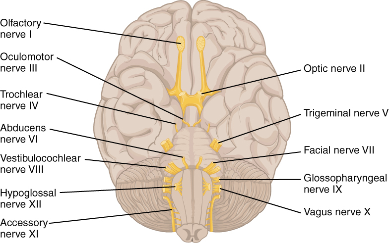
Twelve pairs of nerves arise from the brainstem, providing sensory and motor innervation to the head and neck.
Olfactory nerve - I
- Sensory only
- Function: smell
Optic nerve - II
- Sensory only
- Function: visual acuity
Oculomotor nerve - III
- Motor only
- Function: moves the eye up, down, and in (medially); elevation of the eyelid; constriction of the pupil
Trochlear nerve - IV
- Motor only
- Function: moves the eye down and in (innervates the superior oblique muscle)
Trigeminal nerve - V
- Sensory and motor
- Sensory: touch, pain, and temperature to the face; ophthalmic, maxillary, and mandibular regions
- The lingual nerve, a branch of the mandibular division (CN V3), carries general sensation (touch, pain, temperature) from the anterior two-thirds of the tongue, while taste from that area travels with the chorda tympani of the facial nerve
- Motor: muscles of mastication
Abducens nerve - VI
- Motor only
- Function: moves the eye out (laterally); the nerve begins at a nucleus in the dorsal pons and controls the lateral rectus muscle, which abducts the eye
- Palsy of the abducens nerve weakens the lateral rectus, so the affected eye turns inward and cannot abduct, causing horizontal double vision (diplopia)
- Palsy of the trochlear nerve (CN IV), which supplies the superior oblique, causes vertical and/or torsional double vision that is usually worse on downgaze
Facial nerve - VII
- Sensory and motor
- Sensory: taste to the anterior two-thirds of the tongue
- Motor: facial expressions, closing eyes tightly
Vestibulocochlear nerve - VIII
- Sensory only
- Function: balance and hearing acuity
Glossopharyngeal nerve - IX
- Sensory and motor
- Sensory: taste to the posterior one-third of the tongue
- Motor: gag reflex, pharynx, and larynx control
Vagus nerve - X
- Sensory and motor
- Sensory: small area of taste on the back of the tongue; sensation from the pharynx and thoracic and abdominal organs
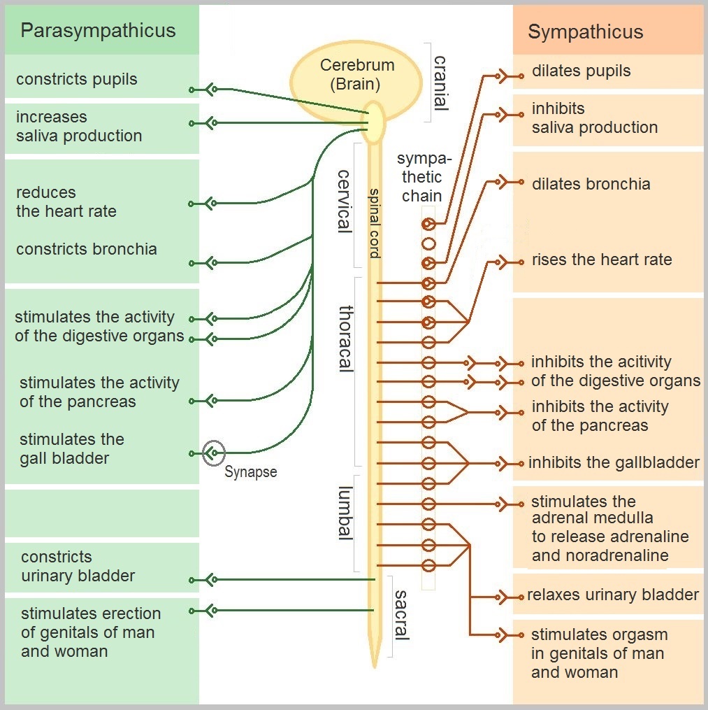
- Motor: pharynx and larynx control (swallowing and speech); primary parasympathetic supply to internal organs, slowing heart rate, constricting the bronchi, and assisting digestion
Accessory nerve - XI
- Motor only
- Function: controls the trapezius and sternocleidomastoid muscles
Hypoglossal nerve - XII
- Motor only
- Function: controls tongue movements
A PTA screens cranial nerve function as part of data collection:
| Cranial nerve | Quick screen |
|---|---|
| I - olfactory | Identify a familiar scent with the eyes closed |
| II - optic | Visual acuity and visual fields by confrontation |
| III, IV, VI - eye movement nerves | Follow a finger through an “H” pattern; pupillary light reflex |
| V - trigeminal | Light touch to the face; resisted jaw clench |
| VII - facial | Raise eyebrows, close eyes tightly, smile |
| VIII - vestibulocochlear | Finger-rub hearing test; observe balance |
| IX, X - glossopharyngeal, vagus | Gag reflex; say “ah” and watch for symmetric palate elevation |
| XI - accessory | Resisted shoulder shrug and head turn |
| XII - hypoglossal | Stick out the tongue and check for deviation |
A new or worsening finding is documented and reported to the supervising PT, who decides whether a formal re-evaluation is needed - the PTA’s role is to collect and report the finding, not to diagnose it.
Spinal nerves
Thirty-one pairs of spinal nerves (8 cervical, 12 thoracic, 5 lumbar, 5 sacral, 1 coccygeal) exit the spinal cord, one per cord segment. Each forms from a dorsal (sensory) root and a ventral (motor) root.
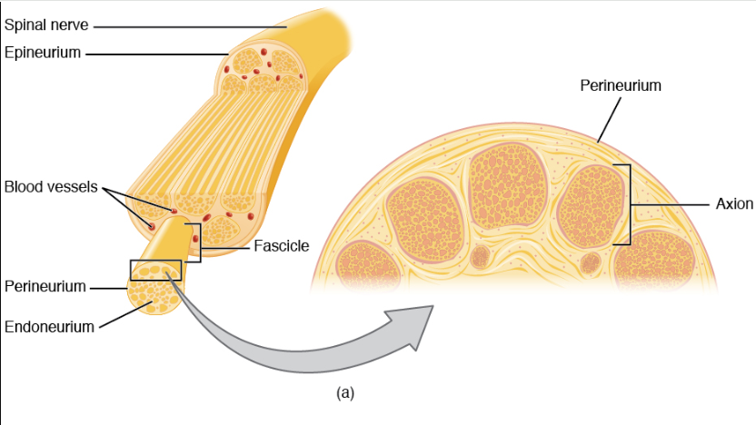
Structure of peripheral nerves
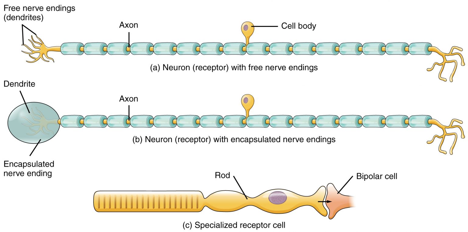
Peripheral nerves also carry sensory receptors - specialized endings such as mechanoreceptors, thermoreceptors, and nociceptors - that convert a specific stimulus (pressure, temperature, pain) into the electrical signal the nerve fiber carries to the CNS, as shown below.
Autonomic nervous system
The autonomic nervous system provides motor innervation to smooth muscle, cardiac muscle, and glands, giving unconscious control over these systems. It’s further divided into the sympathetic, parasympathetic, and enteric divisions, each of which works to maintain homeostasis in the body.