


This browser is too old to run Achievable.

Update it, or open this page in a current version of Chrome, Safari, Firefox or Edge. On an iPhone or iPad, update iOS: every browser there runs on the version of Safari that comes with it.

Achievable logoAchievable logo
NPTE-PTA
Sign in
Sign up
Purchase
Textbook
Practice exams
Support
How it works
Resources
Exam catalog
Mountain with a flag at the peak
Textbook
Introduction
1. Cardiovascular system
2. Pulmonary system
3. Neuromuscular system
3.1 Central nervous system
3.2 Anatomy and function of spinal cord
3.3 Peripheral nervous system
3.4 Stroke deficits and recovery
3.5 Traumatic brain injury
3.6 Spinal cord injury
3.7 Neurodegenerative disorders and epilepsy
3.8 Peripheral nervous system conditions
3.9 Other neurological conditions
3.10 Interventions for neurological conditions
3.11 Vestibular system
4. Pediatrics
5. Musculoskeletal system
6. Other systems
7. Non-systems
Wrapping up
Achievable logoAchievable logo
3.9 Other neurological conditions
Achievable NPTE-PTA
3. Neuromuscular system
Our NPTE-PTA course is now in "early access" - get 50% off for a limited time.

Other neurological conditions

10 min read
Font
Discuss
Share
Feedback

Cerebellar dysfunctions

Definitions
Cerebellar dysfunction
A condition in which the cerebellum has been damaged, leading to deficits in coordination, balance, and motor control. Common causes include stroke, tumor, infections, genetic disorders, or nutritional deficits.

Symptomatology is based on the part of the cerebellum affected.

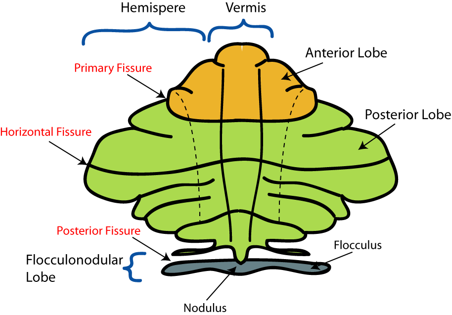
Parts of cerebellum
Parts of cerebellum
The original uploader was Nrets at English Wikipedia
/
Wikimedia Commons
/
CC BY-SA 3.0

Midline (vermis/flocculonodular) lesions mainly cause trunk instability, ataxic gait, and nystagmus; lateral hemisphere lesions mainly cause limb dysmetria, intention tremor, and dysdiadochokinesia. Cerebellar signs appear on the same side as the lesion, and the resulting gait and trunk instability increase fall risk, so guard closely during functional activities.

Common symptoms associated with cerebellar dysfunction

  • Ataxic gait
  • Trunk instability
  • Intention tremor
  • Dysdiadochokinesia
  • Dysmetria
  • Motor learning impairments
  • Cognitive deficits for processing and attention to tasks
  • Hypotonia
  • Weakness and fatigue
Definitions
Dysmetria
Errors of force, direction, and/or amplitude when performing a movement. Hypermetria is overshooting the target; hypometria is undershooting it.
Ataxia
Lack of muscle coordination, resulting in uncoordinated, clumsy movements.
Dysdiadochokinesia
Inability to perform rapid, alternating movements.
Dyssynergia
Lack of coordination between muscles.

Pitfall: cerebellar intention tremor worsens as the limb approaches its target, while a Parkinson’s resting tremor instead eases with voluntary movement. Likewise, cerebellar ataxia is unaffected by vision (Romberg negative), while sensory ataxia from a proprioceptive loss gets markedly worse with the eyes closed (Romberg positive).

Perceptual dysfunction

Definitions
Perceptual dysfunction
Impairments that cause mobility deficits and cannot be directly attributed to a loss of sensory, motor, or comprehension abilities. Individuals are unable to process the input of the environment, which leads to difficulty reacting appropriately to it.

Examples of perceptual dysfunctions are described below.

Spatial relation disorders

  • Causes: stroke, brain injury, Alzheimer’s disease, Parkinson’s disease
  • Definition: difficulty interpreting information about where an object is in relation to another object in the environment
  • Effects: dependent on the type of spatial disorder

Types of spatial relation disorders

  • Figure-ground discrimination disorder: inability to perceive an object against a background
    • Example: the hidden-object illustrations in “Where’s Waldo?” books
  • Spatial relation deficit disorder: inability to properly space objects in relation to one another
    • Example: an individual has difficulty understanding descriptive spatial terms such as on, over, below, between, or under - for example, understanding that “the book is on the table.”
  • Topographical disorientation disorder: inability to navigate a familiar route independently
    • Example: an individual who has difficulty recalling how many left turns to make when driving from home to the grocery store
  • Depth/distance perception disorder: unable to accurately judge depth or distance
    • Example: an individual who walks into a wall while ambulating due to an inability to judge distance accurately
  • Vertical disorientation disorder: unable to accurately determine what is upright
    • Example: an individual sits or stands leaning to one side while perceiving that they are upright

Agnosia

  • Causes: stroke, brain injury, brain abscess
  • Definition: inability to recognize familiar objects with one sensory modality
    • Example: unable to name a pencil in hand with eyes closed
  • Effects: needs to utilize more than one sensory modality when naming an object

Apraxia

  • Causes: stroke, brain injury, Alzheimer’s disease, Parkinson’s disease
  • Definition: difficulty performing purposeful movements when the normal range of motion, strength, and comprehension is present
  • Effects: breaking down complex movements to smaller movements, increasing verbal and visual cues, and increasing repetition

Types of apraxia

  • Ideomotor apraxia: difficulty translating the idea of a movement into motor commands. Individuals may struggle to perform tasks such as waving, blowing a kiss, or licking their lips.
  • Ideational apraxia: inability to plan and sequence complex movements, such as dressing or brushing teeth.
  • Constructional apraxia: a patient cannot draw, construct, or copy an object even though the task is understood and the physical ability to do it is intact.

Body scheme/image disorders

  • Causes: brain injury, brain abscess, developmental delay
  • Definition: difficulty with understanding body segments in relationship to the environment
  • Effects: dependent on the type of body scheme disorders

Types of body scheme/image disorders

  • Body scheme disorder (asomatognosia): inability to recognize a body part as your own
    • Example: an individual watches their hand typing but does not recognize it as part of their own body
  • Visual spatial neglect: ignoring one side of the body and any stimuli from that side
    • Example: a patient with stroke eats food from only one side of the plate or fails to groom or dress the neglected side of the body
    • Approach and cue the patient from the neglected side during mobility and ADL training to encourage attention to that side
  • Right/left discrimination disorder: unable to identify the left and right sides of the body
    • Example: an individual asked to step onto the step with the left foot but is consistently unable to identify their left lower extremity
  • Anosognosia: denial of neglect or lack of awareness of the severity of the dysfunction
    • Example: an individual attempts to transfer without assistance despite requiring maximum assistance, leading to injury; this is typically seen after brain injury, especially right-hemisphere stroke, and requires close supervision during transfers and mobility to prevent injury
    • Neglect and anosognosia often occur together after right-hemisphere stroke but are separate deficits: neglect is failing to attend to one side, while anosognosia is failing to recognize that a deficit exists, so a patient can have neglect and still be fully aware of it

Encephalitis

Definitions
Encephalitis
Inflammation of the brain, most often from infection (usually viral) but sometimes autoimmune.

Primary symptoms are headache, fever, body aches, fatigue, weakness, confusion, agitation, seizures, loss of sensation, and loss of consciousness.

Individuals with encephalitis require bed rest, fluids, and antiviral or anti-inflammatory medications. Physical therapy provides supportive treatment based on the functional deficits presented, but should stop the session and report to the supervising PT and nursing if the patient has a seizure, a declining level of consciousness, new confusion, or worsening headache or fever.

Meningitis is inflammation of the lining of the brain and spinal cord, and its common symptoms are fever, headache, and a stiff neck. Bacterial meningitis, including meningococcal meningitis, usually comes on quickly with fever and chills, severe headache, stiff neck and sensitivity to light.

Neurological conditions and post-COVID

COVID-19 infection increases the risk of secondary neurological conditions: stroke through clotting and reduced perfusion, and encephalitis, transverse myelitis, and Guillain-Barré syndrome through abnormal immune responses. Treatment addresses the secondary diagnosis using the approaches described throughout this chapter.

Bulbar palsy

Definitions
Bulbar palsy
A lower motor neuron lesion of the motor nuclei of cranial nerves IX, X, XI and XII in the medulla (the “bulb”).

A stroke in the medulla and motor neuron disease are common causes. A lateral medullary stroke (vertebral or posterior inferior cerebellar artery) affects nerves IX and X but spares the hypoglossal nucleus, so it causes dysphagia, hoarseness and dysarthria without tongue wasting. Dysarthria is difficulty saying words because of problems with the muscles used for speech; the muscles may be weak or completely paralyzed.

  • Bulbar palsy (lower motor neuron): severe dysphagia with a reduced or absent gag reflex, flaccid dysarthria, and tongue wasting and fasciculation
  • Pseudobulbar palsy (upper motor neuron): from lesions of the corticobulbar tracts on both sides (for example, bilateral strokes); spastic dysarthria, a spastic tongue without wasting or fasciculation, a brisk jaw jerk, and emotional lability
  • Bulbar-onset ALS: about 25-30% of ALS begins with dysarthria and dysphagia, with signs of both kinds, and it progresses faster than limb-onset ALS

Dysphagia is managed first with diet texture changes, swallowing exercises and compensatory strategies directed by the speech-language pathologist. A feeding tube, such as a PEG, is added when eating by mouth becomes unsafe or insufficient; in ALS it is placed before vital capacity falls below 50% of predicted. The main risks are aspiration pneumonia, malnutrition and dehydration.

PTA role: Follow the diet and positioning orders (upright for meals; nothing by mouth during therapy if the patient is NPO). Coughing or a wet voice with eating or drinking, a new fever, or a change in breathing is reported to the supervising PT.

Trigeminal neuralgia

Definitions
Trigeminal neuralgia
A chronic pain disorder in which the trigeminal nerve (cranial nerve V) is damaged - trigeminal is the main sensory nerve to the face.

The pain associated with trigeminal neuralgia is neuropathic and can last from a few seconds up to two minutes. It is caused by irritation of the trigeminal nerve, most often from compression by a blood vessel (see types below).

Symptoms

  • Sudden, intense pain to one side of the face
  • Typically pain-free between attacks; some patients (an atypical form) have continuous burning or aching background pain

The pain can be exacerbated by rubbing the cheek (when washing the face, shaving, or applying makeup), eating, drinking, talking, blowing the nose, brushing teeth, or exposure to wind. Patients can experience these attacks multiple times per day, with intervening periods of remission.

Types of trigeminal neuralgia

  • Primary: caused by compression of a blood vessel against the trigeminal nerve
  • Secondary: caused by stroke, multiple sclerosis, or tumor

Treatment by physical therapy may include the use of transcutaneous electrical stimulation (TENS) for pain relief, along with collecting data on and reporting other symptoms to the PT. Medical management may include nerve block, pain medications, or anticonvulsants.

Alzheimer’s disease

Definitions
Alzheimer’s disease
A progressive neurodegenerative disorder that slowly impairs executive function and memory, leading to complete debility.

Alzheimer’s disease affects multiple areas of the brain, especially the temporal (hippocampus) and parietal lobes. The typical age of onset is after 60 years old. Cholinergic neurons of the basal forebrain degenerate, and choline acetyltransferase, the enzyme that makes acetylcholine, is reduced.

Symptoms

  • Loss of declarative (episodic) memory, especially for recently learned information, is typically the earliest symptom; procedural memory is relatively preserved until later stages
  • Decreased spatial awareness
  • Word-finding difficulty
  • Changes in personality and behavior
  • Sundowning, a behavioral symptom that tends to emerge in later stages
    • Increased confusion/agitation at the time of sunset

Intervention strategies for individuals with Alzheimer’s disease

  • Sessions are to assist with maintaining the current level of function
  • Basic ADLs and mobility status
  • Providing assistive devices (AD) and durable medical equipment (DME) as appropriate
  • Management of behaviors
  • Be consistent in routine (blocked routine)
  • Closed environment to minimize distractions
  • De-escalate aggressive or combative behaviors by changing the current task
  • Re-orient as appropriate

Delirium begins suddenly, causes fluctuations in mental function, and is usually reversible, whereas dementia begins gradually, progresses slowly, and is usually irreversible. Vascular dementia may progress in steps: symptoms can worsen suddenly and then plateau or lessen somewhat, unlike other dementias, which tend to progress continuously.

PTA role: across all of the conditions in this chapter, the PTA carries out the plan of care the supervising PT has already established - collecting data on function, cognition, and behavior, and recognizing and reporting any changes to the PT. Evaluating, re-evaluating, and modifying the plan of care remain the PT’s responsibility.

Motor learning principles and detailed intervention progressions for these conditions are covered in the next chapter, Interventions for neurological conditions.

Cerebellar dysfunction

  • Midline/vermis lesions → trunk instability, ataxic gait, nystagmus; lateral hemisphere lesions → limb dysmetria, intention tremor, dysdiadochokinesia
  • Signs are ipsilateral to lesion; high fall risk, guard closely
  • Key terms: dysmetria (hyper/hypometria), ataxia, dysdiadochokinesia, dyssynergia
  • Differentiating tremors/ataxia: cerebellar intention tremor worsens approaching target (vs. Parkinson’s resting tremor eases with movement); cerebellar ataxia is Romberg negative (vision doesn’t matter) vs. sensory ataxia is Romberg positive (worsens eyes closed)

Perceptual dysfunction

  • Impairments in processing environmental input, not explained by sensory/motor/comprehension loss alone
  • Spatial relation disorders: figure-ground discrimination, spatial relation deficit, topographical disorientation, depth/distance perception, vertical disorientation
  • Agnosia: can’t recognize objects via one sensory modality; compensate with multiple senses
  • Apraxia: purposeful movement difficulty despite intact strength/ROM/comprehension
    • Ideomotor: can’t translate idea into motor command (e.g., waving)
    • Ideational: can’t plan/sequence complex tasks (e.g., dressing)
  • Body scheme/image disorders:
    • Asomatognosia: doesn’t recognize body part as own
    • Visual spatial neglect: ignores one side; cue/approach from neglected side
    • Right/left discrimination disorder
    • Anosognosia: unaware of deficit severity (distinct from neglect—can have neglect while aware of it); common post right-hemisphere stroke, requires close supervision to prevent injury

Encephalitis & post-COVID neuro conditions

  • Encephalitis = brain inflammation (usually viral infection, sometimes autoimmune); symptoms: headache, fever, confusion, seizures, LOC changes
  • Stop session and report to PT/nursing if seizure, declining consciousness, new confusion, or worsening fever/headache
  • COVID-19 increases risk of stroke (clotting), encephalitis, transverse myelitis, Guillain-Barré (abnormal immune response); treat per secondary diagnosis

Bulbar palsy

  • Bulbar palsy = LMN lesion of CN IX, X, XI, XII in medulla; causes: brainstem stroke, motor neuron disease
  • Bulbar (LMN): severe dysphagia, reduced/absent gag, flaccid dysarthria, tongue wasting/fasciculation
  • Pseudobulbar (UMN, bilateral corticobulbar lesions): spastic dysarthria, spastic tongue (no wasting), brisk jaw jerk, emotional lability
  • Bulbar-onset ALS: mixed signs, faster progression than limb-onset
  • Dysphagia management: diet texture changes/SLP strategies first; PEG tube if unsafe/insufficient intake (in ALS, before vital capacity <50% predicted)
  • Risks: aspiration pneumonia, malnutrition, dehydration
  • PTA role: follow diet/positioning orders (upright, NPO if ordered); report coughing/wet voice, fever, breathing changes

Trigeminal neuralgia

  • CN V damage causing neuropathic facial pain, seconds to 2 minutes, usually pain-free between attacks (atypical form = continuous burning pain)
  • Triggered by touch/rubbing face, eating, talking, wind, brushing teeth
  • Primary: vascular compression; Secondary: stroke, MS, tumor
  • PT treatment: TENS for pain relief, data collection/reporting; medical: nerve block, meds, anticonvulsants

Alzheimer’s disease

  • Progressive neurodegeneration affecting temporal (hippocampus) and parietal lobes; onset typically >60 years
  • Earliest symptom: declarative/episodic memory loss; procedural memory preserved until later stages
  • Other symptoms: spatial awareness loss, word-finding difficulty, personality changes, sundowning (late-stage confusion at sunset)
  • Interventions: maintain current function, provide AD/DME, consistent routine, minimize distracting environment, de-escalate by changing task, reorient as needed

PTA role across conditions

  • Carry out PT’s established plan of care
  • Collect data on function, cognition, behavior
  • Recognize and report changes; evaluation/re-evaluation/plan modification remains PT responsibility

Sign up for free to take 32 quiz questions on this topic

Previous
Next  | 3.10.1 Motor learning and PNF
All rights reserved ©2016 - 2026 Achievable, Inc.

Other neurological conditions

Cerebellar dysfunctions

Definitions
Cerebellar dysfunction
A condition in which the cerebellum has been damaged, leading to deficits in coordination, balance, and motor control. Common causes include stroke, tumor, infections, genetic disorders, or nutritional deficits.

Symptomatology is based on the part of the cerebellum affected.

Midline (vermis/flocculonodular) lesions mainly cause trunk instability, ataxic gait, and nystagmus; lateral hemisphere lesions mainly cause limb dysmetria, intention tremor, and dysdiadochokinesia. Cerebellar signs appear on the same side as the lesion, and the resulting gait and trunk instability increase fall risk, so guard closely during functional activities.

Common symptoms associated with cerebellar dysfunction

  • Ataxic gait
  • Trunk instability
  • Intention tremor
  • Dysdiadochokinesia
  • Dysmetria
  • Motor learning impairments
  • Cognitive deficits for processing and attention to tasks
  • Hypotonia
  • Weakness and fatigue
Definitions
Dysmetria
Errors of force, direction, and/or amplitude when performing a movement. Hypermetria is overshooting the target; hypometria is undershooting it.
Ataxia
Lack of muscle coordination, resulting in uncoordinated, clumsy movements.
Dysdiadochokinesia
Inability to perform rapid, alternating movements.
Dyssynergia
Lack of coordination between muscles.

Pitfall: cerebellar intention tremor worsens as the limb approaches its target, while a Parkinson’s resting tremor instead eases with voluntary movement. Likewise, cerebellar ataxia is unaffected by vision (Romberg negative), while sensory ataxia from a proprioceptive loss gets markedly worse with the eyes closed (Romberg positive).

Perceptual dysfunction

Definitions
Perceptual dysfunction
Impairments that cause mobility deficits and cannot be directly attributed to a loss of sensory, motor, or comprehension abilities. Individuals are unable to process the input of the environment, which leads to difficulty reacting appropriately to it.

Examples of perceptual dysfunctions are described below.

Spatial relation disorders

  • Causes: stroke, brain injury, Alzheimer’s disease, Parkinson’s disease
  • Definition: difficulty interpreting information about where an object is in relation to another object in the environment
  • Effects: dependent on the type of spatial disorder

Types of spatial relation disorders

  • Figure-ground discrimination disorder: inability to perceive an object against a background
    • Example: the hidden-object illustrations in “Where’s Waldo?” books
  • Spatial relation deficit disorder: inability to properly space objects in relation to one another
    • Example: an individual has difficulty understanding descriptive spatial terms such as on, over, below, between, or under - for example, understanding that “the book is on the table.”
  • Topographical disorientation disorder: inability to navigate a familiar route independently
    • Example: an individual who has difficulty recalling how many left turns to make when driving from home to the grocery store
  • Depth/distance perception disorder: unable to accurately judge depth or distance
    • Example: an individual who walks into a wall while ambulating due to an inability to judge distance accurately
  • Vertical disorientation disorder: unable to accurately determine what is upright
    • Example: an individual sits or stands leaning to one side while perceiving that they are upright

Agnosia

  • Causes: stroke, brain injury, brain abscess
  • Definition: inability to recognize familiar objects with one sensory modality
    • Example: unable to name a pencil in hand with eyes closed
  • Effects: needs to utilize more than one sensory modality when naming an object

Apraxia

  • Causes: stroke, brain injury, Alzheimer’s disease, Parkinson’s disease
  • Definition: difficulty performing purposeful movements when the normal range of motion, strength, and comprehension is present
  • Effects: breaking down complex movements to smaller movements, increasing verbal and visual cues, and increasing repetition

Types of apraxia

  • Ideomotor apraxia: difficulty translating the idea of a movement into motor commands. Individuals may struggle to perform tasks such as waving, blowing a kiss, or licking their lips.
  • Ideational apraxia: inability to plan and sequence complex movements, such as dressing or brushing teeth.
  • Constructional apraxia: a patient cannot draw, construct, or copy an object even though the task is understood and the physical ability to do it is intact.

Body scheme/image disorders

  • Causes: brain injury, brain abscess, developmental delay
  • Definition: difficulty with understanding body segments in relationship to the environment
  • Effects: dependent on the type of body scheme disorders

Types of body scheme/image disorders

  • Body scheme disorder (asomatognosia): inability to recognize a body part as your own
    • Example: an individual watches their hand typing but does not recognize it as part of their own body
  • Visual spatial neglect: ignoring one side of the body and any stimuli from that side
    • Example: a patient with stroke eats food from only one side of the plate or fails to groom or dress the neglected side of the body
    • Approach and cue the patient from the neglected side during mobility and ADL training to encourage attention to that side
  • Right/left discrimination disorder: unable to identify the left and right sides of the body
    • Example: an individual asked to step onto the step with the left foot but is consistently unable to identify their left lower extremity
  • Anosognosia: denial of neglect or lack of awareness of the severity of the dysfunction
    • Example: an individual attempts to transfer without assistance despite requiring maximum assistance, leading to injury; this is typically seen after brain injury, especially right-hemisphere stroke, and requires close supervision during transfers and mobility to prevent injury
    • Neglect and anosognosia often occur together after right-hemisphere stroke but are separate deficits: neglect is failing to attend to one side, while anosognosia is failing to recognize that a deficit exists, so a patient can have neglect and still be fully aware of it

Encephalitis

Definitions
Encephalitis
Inflammation of the brain, most often from infection (usually viral) but sometimes autoimmune.

Primary symptoms are headache, fever, body aches, fatigue, weakness, confusion, agitation, seizures, loss of sensation, and loss of consciousness.

Individuals with encephalitis require bed rest, fluids, and antiviral or anti-inflammatory medications. Physical therapy provides supportive treatment based on the functional deficits presented, but should stop the session and report to the supervising PT and nursing if the patient has a seizure, a declining level of consciousness, new confusion, or worsening headache or fever.

Meningitis is inflammation of the lining of the brain and spinal cord, and its common symptoms are fever, headache, and a stiff neck. Bacterial meningitis, including meningococcal meningitis, usually comes on quickly with fever and chills, severe headache, stiff neck and sensitivity to light.

Neurological conditions and post-COVID

COVID-19 infection increases the risk of secondary neurological conditions: stroke through clotting and reduced perfusion, and encephalitis, transverse myelitis, and Guillain-Barré syndrome through abnormal immune responses. Treatment addresses the secondary diagnosis using the approaches described throughout this chapter.

Bulbar palsy

Definitions
Bulbar palsy
A lower motor neuron lesion of the motor nuclei of cranial nerves IX, X, XI and XII in the medulla (the “bulb”).

A stroke in the medulla and motor neuron disease are common causes. A lateral medullary stroke (vertebral or posterior inferior cerebellar artery) affects nerves IX and X but spares the hypoglossal nucleus, so it causes dysphagia, hoarseness and dysarthria without tongue wasting. Dysarthria is difficulty saying words because of problems with the muscles used for speech; the muscles may be weak or completely paralyzed.

  • Bulbar palsy (lower motor neuron): severe dysphagia with a reduced or absent gag reflex, flaccid dysarthria, and tongue wasting and fasciculation
  • Pseudobulbar palsy (upper motor neuron): from lesions of the corticobulbar tracts on both sides (for example, bilateral strokes); spastic dysarthria, a spastic tongue without wasting or fasciculation, a brisk jaw jerk, and emotional lability
  • Bulbar-onset ALS: about 25-30% of ALS begins with dysarthria and dysphagia, with signs of both kinds, and it progresses faster than limb-onset ALS

Dysphagia is managed first with diet texture changes, swallowing exercises and compensatory strategies directed by the speech-language pathologist. A feeding tube, such as a PEG, is added when eating by mouth becomes unsafe or insufficient; in ALS it is placed before vital capacity falls below 50% of predicted. The main risks are aspiration pneumonia, malnutrition and dehydration.

PTA role: Follow the diet and positioning orders (upright for meals; nothing by mouth during therapy if the patient is NPO). Coughing or a wet voice with eating or drinking, a new fever, or a change in breathing is reported to the supervising PT.

Trigeminal neuralgia

Definitions
Trigeminal neuralgia
A chronic pain disorder in which the trigeminal nerve (cranial nerve V) is damaged - trigeminal is the main sensory nerve to the face.

The pain associated with trigeminal neuralgia is neuropathic and can last from a few seconds up to two minutes. It is caused by irritation of the trigeminal nerve, most often from compression by a blood vessel (see types below).

Symptoms

  • Sudden, intense pain to one side of the face
  • Typically pain-free between attacks; some patients (an atypical form) have continuous burning or aching background pain

The pain can be exacerbated by rubbing the cheek (when washing the face, shaving, or applying makeup), eating, drinking, talking, blowing the nose, brushing teeth, or exposure to wind. Patients can experience these attacks multiple times per day, with intervening periods of remission.

Types of trigeminal neuralgia

  • Primary: caused by compression of a blood vessel against the trigeminal nerve
  • Secondary: caused by stroke, multiple sclerosis, or tumor

Treatment by physical therapy may include the use of transcutaneous electrical stimulation (TENS) for pain relief, along with collecting data on and reporting other symptoms to the PT. Medical management may include nerve block, pain medications, or anticonvulsants.

Alzheimer’s disease

Definitions
Alzheimer’s disease
A progressive neurodegenerative disorder that slowly impairs executive function and memory, leading to complete debility.

Alzheimer’s disease affects multiple areas of the brain, especially the temporal (hippocampus) and parietal lobes. The typical age of onset is after 60 years old. Cholinergic neurons of the basal forebrain degenerate, and choline acetyltransferase, the enzyme that makes acetylcholine, is reduced.

Symptoms

  • Loss of declarative (episodic) memory, especially for recently learned information, is typically the earliest symptom; procedural memory is relatively preserved until later stages
  • Decreased spatial awareness
  • Word-finding difficulty
  • Changes in personality and behavior
  • Sundowning, a behavioral symptom that tends to emerge in later stages
    • Increased confusion/agitation at the time of sunset

Intervention strategies for individuals with Alzheimer’s disease

  • Sessions are to assist with maintaining the current level of function
  • Basic ADLs and mobility status
  • Providing assistive devices (AD) and durable medical equipment (DME) as appropriate
  • Management of behaviors
  • Be consistent in routine (blocked routine)
  • Closed environment to minimize distractions
  • De-escalate aggressive or combative behaviors by changing the current task
  • Re-orient as appropriate

Delirium begins suddenly, causes fluctuations in mental function, and is usually reversible, whereas dementia begins gradually, progresses slowly, and is usually irreversible. Vascular dementia may progress in steps: symptoms can worsen suddenly and then plateau or lessen somewhat, unlike other dementias, which tend to progress continuously.

PTA role: across all of the conditions in this chapter, the PTA carries out the plan of care the supervising PT has already established - collecting data on function, cognition, and behavior, and recognizing and reporting any changes to the PT. Evaluating, re-evaluating, and modifying the plan of care remain the PT’s responsibility.

Motor learning principles and detailed intervention progressions for these conditions are covered in the next chapter, Interventions for neurological conditions.

Key points

Cerebellar dysfunction

  • Midline/vermis lesions → trunk instability, ataxic gait, nystagmus; lateral hemisphere lesions → limb dysmetria, intention tremor, dysdiadochokinesia
  • Signs are ipsilateral to lesion; high fall risk, guard closely
  • Key terms: dysmetria (hyper/hypometria), ataxia, dysdiadochokinesia, dyssynergia
  • Differentiating tremors/ataxia: cerebellar intention tremor worsens approaching target (vs. Parkinson’s resting tremor eases with movement); cerebellar ataxia is Romberg negative (vision doesn’t matter) vs. sensory ataxia is Romberg positive (worsens eyes closed)

Perceptual dysfunction

  • Impairments in processing environmental input, not explained by sensory/motor/comprehension loss alone
  • Spatial relation disorders: figure-ground discrimination, spatial relation deficit, topographical disorientation, depth/distance perception, vertical disorientation
  • Agnosia: can’t recognize objects via one sensory modality; compensate with multiple senses
  • Apraxia: purposeful movement difficulty despite intact strength/ROM/comprehension
    • Ideomotor: can’t translate idea into motor command (e.g., waving)
    • Ideational: can’t plan/sequence complex tasks (e.g., dressing)
  • Body scheme/image disorders:
    • Asomatognosia: doesn’t recognize body part as own
    • Visual spatial neglect: ignores one side; cue/approach from neglected side
    • Right/left discrimination disorder
    • Anosognosia: unaware of deficit severity (distinct from neglect—can have neglect while aware of it); common post right-hemisphere stroke, requires close supervision to prevent injury

Encephalitis & post-COVID neuro conditions

  • Encephalitis = brain inflammation (usually viral infection, sometimes autoimmune); symptoms: headache, fever, confusion, seizures, LOC changes
  • Stop session and report to PT/nursing if seizure, declining consciousness, new confusion, or worsening fever/headache
  • COVID-19 increases risk of stroke (clotting), encephalitis, transverse myelitis, Guillain-Barré (abnormal immune response); treat per secondary diagnosis

Bulbar palsy

  • Bulbar palsy = LMN lesion of CN IX, X, XI, XII in medulla; causes: brainstem stroke, motor neuron disease
  • Bulbar (LMN): severe dysphagia, reduced/absent gag, flaccid dysarthria, tongue wasting/fasciculation
  • Pseudobulbar (UMN, bilateral corticobulbar lesions): spastic dysarthria, spastic tongue (no wasting), brisk jaw jerk, emotional lability
  • Bulbar-onset ALS: mixed signs, faster progression than limb-onset
  • Dysphagia management: diet texture changes/SLP strategies first; PEG tube if unsafe/insufficient intake (in ALS, before vital capacity <50% predicted)
  • Risks: aspiration pneumonia, malnutrition, dehydration
  • PTA role: follow diet/positioning orders (upright, NPO if ordered); report coughing/wet voice, fever, breathing changes

Trigeminal neuralgia

  • CN V damage causing neuropathic facial pain, seconds to 2 minutes, usually pain-free between attacks (atypical form = continuous burning pain)
  • Triggered by touch/rubbing face, eating, talking, wind, brushing teeth
  • Primary: vascular compression; Secondary: stroke, MS, tumor
  • PT treatment: TENS for pain relief, data collection/reporting; medical: nerve block, meds, anticonvulsants

Alzheimer’s disease

  • Progressive neurodegeneration affecting temporal (hippocampus) and parietal lobes; onset typically >60 years
  • Earliest symptom: declarative/episodic memory loss; procedural memory preserved until later stages
  • Other symptoms: spatial awareness loss, word-finding difficulty, personality changes, sundowning (late-stage confusion at sunset)
  • Interventions: maintain current function, provide AD/DME, consistent routine, minimize distracting environment, de-escalate by changing task, reorient as needed

PTA role across conditions

  • Carry out PT’s established plan of care
  • Collect data on function, cognition, behavior
  • Recognize and report changes; evaluation/re-evaluation/plan modification remains PT responsibility

More from Neuromuscular system

  • Central nervous system
  • Anatomy and function of spinal cord
  • Stroke deficits and recovery
  • Traumatic brain injury
  • Spinal cord injury