



Dermatomes, myotomes, reflexes, and plexuses
Dermatomes and myotomes
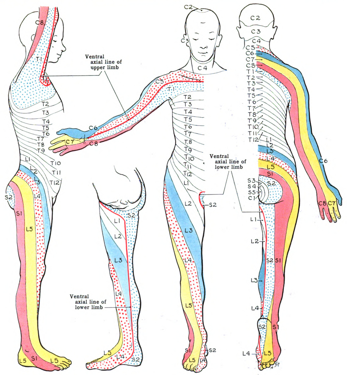
Dermatome, myotome, and reflex screens help identify which spinal nerve root may be involved. Review the dermatome map and myotome chart below.
How each screen is performed: dermatomes are tested with light touch or pinprick, comparing side to side for symmetry. Myotomes are tested with a resisted isometric hold of about 5 seconds against each muscle group. Reflexes are tested by tapping the tendon with a reflex hammer and observing the response.
Key dermatome landmarks to remember:
- C5: lateral arm / deltoid
- C6: thumb
- C7: middle finger
- C8: little finger
- T4: nipple line
- T10: umbilicus
- L1: inguinal region
- L3: anterior/medial thigh to knee
- L4: medial leg / medial malleolus
- L5: dorsum of foot / great toe
- S1: lateral foot / heel
Myotomes
- Upper extremity
- C1-C2: neck flexion
- C2-C4: shoulder elevation
- C5: shoulder abduction/elbow flexion
- C6: wrist extension
- C7: elbow extension/wrist flexion
- C8: digit flexion/thumb extension
- T1: finger adduction
- Lower extremity
- L1-L2: hip flexion
- L3: knee extension
- L4: knee extension/ankle dorsiflexion
- L5: ankle dorsiflexion/great toe extension
- S1: ankle eversion/ankle plantar flexion
- S2: ankle plantar flexion
Spinal reflexes
Reflexes are graded on a 0 to 4+ scale:
| Grade | Finding |
|---|---|
| 0 | Absent |
| 1+ | Diminished |
| 2+ | Normal |
| 3+ | Brisk |
| 4+ | Very brisk, with clonus |
- C5: biceps reflex
- C6: brachioradialis reflex
- C7: triceps reflex
- L3-L4: patellar reflex
- L5: semitendinosus reflex
- S1-S2: Achilles reflex
A diminished or absent reflex often points to a lower motor neuron (LMN) or nerve root lesion, while a hyperactive reflex points to an upper motor neuron (UMN) lesion:
| Finding | UMN lesion | LMN lesion |
|---|---|---|
| Tone | Spastic (increased) | Flaccid (decreased) |
| Reflexes | Hyperreflexia | Hyporeflexia |
| Babinski sign | Present | Absent |
| Atrophy | Minimal | Significant |
| Fasciculations | Absent | Present |
A root lesion follows a dermatome/myotome pattern, while a peripheral nerve lesion follows that nerve’s own sensory field and muscles. For example, an L3 root lesion can weaken both knee extension (femoral nerve) and hip adduction (obturator nerve), because both nerves carry L3 fibers, and its numbness stays in the thigh and over the knee. A femoral nerve lesion also weakens knee extension and diminishes the patellar reflex, but it spares hip adduction, and its numbness can reach the medial leg through the saphenous branch.
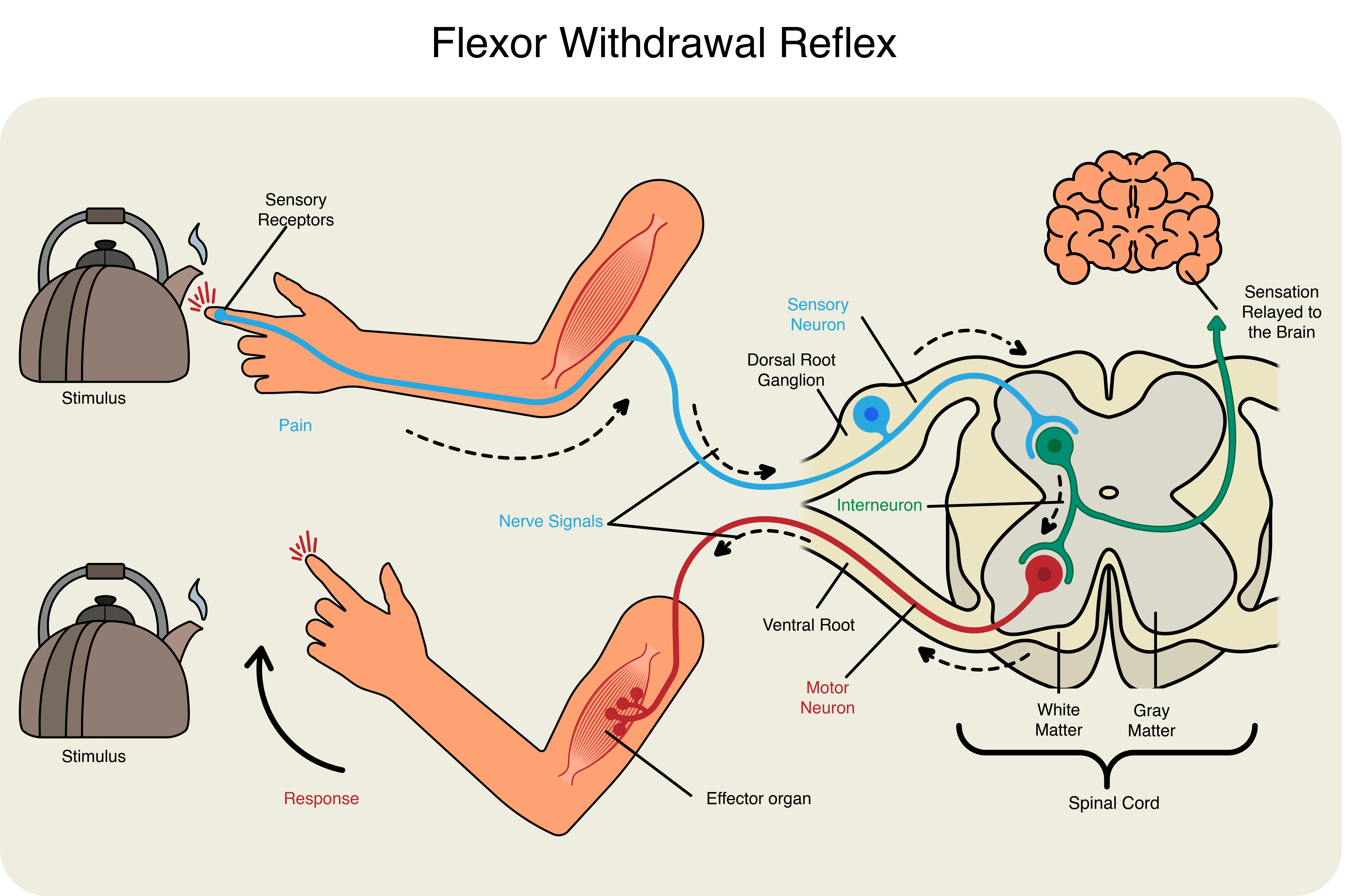
Example of reflexes working in the spinal cord below:
Example: Localizing a nerve root lesion
A patient reports weakness with knee extension and mild weakness with hip adduction, decreased sensation over the anterior/medial thigh, and a diminished patellar reflex. Which nerve root does this pattern point to?
- Myotome: knee extension is primarily controlled by L3.
- Reflex: the patellar reflex tests L3-L4.
- Dermatome: the anterior/medial thigh corresponds to the L2-L3 dermatomes.
- Hip adduction: the adductors are supplied by the obturator nerve, not the femoral nerve, but share the L3 root, so adduction weakness points past the femoral nerve to the root.
All four findings overlap at the L3 level, so L3 is the most likely nerve root involved. This is the same reasoning you’ll use clinically: a single finding can have several causes, but when the sensory, motor, and reflex findings all point to the same level, that level is the most likely one involved.
Answer: L3 nerve root
Other PTA-administered screens
- Light touch and sharp/dull sensation: confirms dermatome findings and flags sensory loss that affects safety with hot, cold, or sharp objects.
- Finger-to-nose and heel-to-shin: screens cerebellar coordination; dysmetria signals fall risk during gait training.
- Romberg test: screens proprioceptive/vestibular control; loss of balance with eyes closed signals a need for closer guarding.
- Berg balance scale and timed up and go (TUG): quantify fall risk and help set the level of assistance needed.
- ASIA scale: the PT uses it to classify the completeness of a spinal cord injury and predict recovery; the PTA can collect the sensory and strength findings it is built on and reports changes to the PT.
Brachial, lumbar, and sacral plexuses
Most nerves to the limbs come from these three plexuses. The upper limb is supplied by the brachial plexus, and the lower limb by the lumbar and sacral plexuses (together called the lumbosacral plexus).
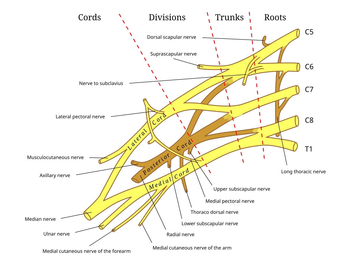
Brachial plexus
Muscles innervated by the brachial plexus
The brachial plexus gives off a few proximal branches before ending in five terminal nerves. The tested skill is recognizing each terminal nerve and its headline muscle action, not memorizing every muscle it supplies.
Proximal branches
- Dorsal scapular nerve (C5): rhomboids and levator scapulae
- Long thoracic nerve (C5-C7): serratus anterior
- Suprascapular nerve (C5-C6): supraspinatus and infraspinatus
Terminal nerves
- Musculocutaneous nerve (C5-C7): biceps brachii, brachialis, and coracobrachialis (elbow flexion)
- Axillary nerve (C5-C6): deltoid and teres minor (shoulder abduction)
- Radial nerve (C5-T1): triceps brachii and the wrist/finger extensors, recalled using the BEAST acronym:
- B: Brachioradialis
- E: Extensor muscles of the wrist and fingers
- A: Abductor pollicis longus, anconeus
- S: Supinator
- T: Triceps brachii
- Median nerve (C5-T1): most anterior forearm flexors (e.g., pronator teres, flexor carpi radialis, flexor digitorum superficialis), the thenar muscles, and the 1st and 2nd lumbricals
- Ulnar nerve (C8-T1): flexor carpi ulnaris, the medial half of flexor digitorum profundus, and most of the intrinsic hand muscles
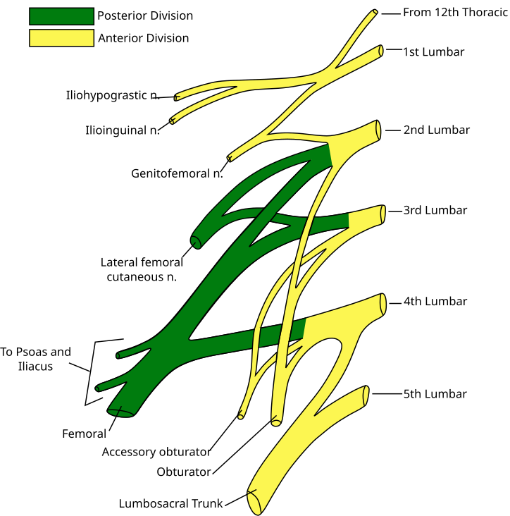
Lumbar plexus
Muscles innervated by the lumbar plexus (L1-L4)
The major muscles and sensory territory supplied by each nerve are listed below.
- Iliohypogastric nerve (L1): internal oblique and transversus abdominis
- Femoral nerve (L2-L4): iliacus, sartorius, and the quadriceps femoris group (knee extension)
- Obturator nerve (L2-L4): the hip adductor group and gracilis (hip adduction)
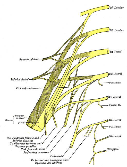
Sacral plexus
Muscles innervated by the sacral plexus (L4-S4)
- Superior gluteal nerve (L4-S1): gluteus medius, gluteus minimus, and tensor fasciae latae (hip abduction)
- Inferior gluteal nerve (L5-S2): gluteus maximus (hip extension)
- Sciatic nerve (L4-S3): the largest peripheral nerve in the body, splitting above the knee into two terminal divisions:
- Tibial nerve: the hamstrings (except the short head of biceps femoris), the posterior leg muscles (e.g., gastrocnemius, soleus), and the intrinsic foot muscles (ankle plantarflexion)
- Common peroneal (fibular) nerve: the short head of biceps femoris, then splits into the deep peroneal nerve (anterior compartment, ankle dorsiflexion) and the superficial peroneal nerve (lateral compartment, ankle eversion)