



Anatomy, examination, and disorder classification
Vestibular disorders
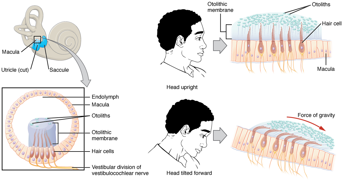
Hair cells, whose stereocilia bend with head movement, are the mechanoreceptors within the ear that sense head position, head movement, and when the body is in motion. Head position is sensed by the maculae of the utricle and saccule. Head movement is sensed via the semicircular canals. The movement of fluid within the semicircular canals generates nerve impulses that travel to cranial nerve VIII (vestibulocochlear nerve) and then to the brainstem and cerebellum. The vestibular nuclei receive input from the vestibular apparatus and are essential in maintaining equilibrium, posture, head position, and clear vision with movement.
Vestibular dysfunction can develop at any age. Factors precipitating vestibular disorders can be external or internal. External factors could be whiplash, a fall, an airplane ride, or pollutants. Internal factors may be infections, use of medications, stroke, brain injury, or migraine. Vestibular dysfunctions can further be divided into central and peripheral disorders - management of symptoms will be based on the classification of the vestibular disorder.
Generalized symptoms of vestibular disorders
- Dizziness
- Balance deficits
- Vertigo
- Blurred vision
- Recurrent falls
- Nausea and vomiting
The examination for vestibular dysfunction is critical in determining the type of disorder and the appropriate treatment options.
Determining the type of disorder
- Symptom history and duration
- Vestibular ocular reflex testing - assessing nystagmus, clarity of vision with head movements, and body movements
- Head impulse (head-thrust) test: the examiner suddenly rotates the patient’s head about 10 degrees while the patient looks at the examiner’s nose; a quick corrective saccade back to the target afterward is a positive result, suggesting weakness of the vestibular organ on the side the head was turned toward
- Vertebral artery testing
- Sensory testing
- Clinical test of sensory interaction on balance (CTSIB)
- Functional testing - transfers, gait, dynamic sitting, and dynamic balance
- Vestibulospinal reflex testing - examining posture and balance in all positions
- Positional changes and their impact on symptoms
- Cervical range of motion
- Outcome measures as appropriate, such as the dynamic gait index
The CTSIB is conducted with the patient standing with their hands at their sides and feet together, performing the following six sensory conditions:
- Stand on a firm surface with eyes open - baseline condition, with vision, somatosensory, and vestibular input all available
- Stand on a firm surface with eyes closed - removes visual input, so balance relies on the somatosensory and vestibular systems
- Stand on a firm surface with a visual conflict dome - vision is inaccurate, challenging the patient to rely on somatosensory and vestibular input instead
- Stand on a foam surface with eyes open - somatosensory input is degraded, so balance relies on vision and the vestibular system
- Stand on a foam surface with eyes closed - somatosensory input is degraded and vision is removed, leaving balance mostly dependent on the vestibular system
- Stand on a foam surface with a visual conflict dome - somatosensory input is degraded and vision is inaccurate, leaving balance mostly dependent on the vestibular system
Central vestibular disorders
Treatment for central vestibular dysfunction
- Identify triggers, if any, that are noted to be external
- Gaze stabilization exercises to assist with improving the vestibular ocular reflex
- Coordination activities to address cerebellar symptoms
- Adaptation exercises to decrease dizziness to allow for retraining of the brain
- Substitution of visual and somatosensory systems in the presence of vestibular dysfunction
Peripheral vestibular dysfunction
Most peripheral vestibular dysfunction is unilateral. Two mechanisms are introduced here - endolymph dysfunction and BPPV; labyrinthitis, Meniere’s disease, and vestibular neuritis are covered in the next chapter, Peripheral vestibular disorders and intervention strategies.
The table below compares the hallmark presentations of central and peripheral vestibular disorders - this is the distinction the exam most often tests:
| Feature | Central | Peripheral |
|---|---|---|
| Nystagmus direction | Pure vertical (especially downbeating) or pure torsional, or changes direction with gaze without any change in head position | Horizontal-torsional, beating in one direction; on positional testing, posterior-canal BPPV produces upbeating torsional nystagmus |
| Fatigability | Positional nystagmus does not fatigue and persists while the position is held | Positional nystagmus of BPPV fatigues with repeated testing and resolves in under a minute; the spontaneous nystagmus of vestibular neuritis does not fatigue and lasts through the acute episode (a day or more), but is suppressed by fixation |
| Effect of visual fixation | No change | Suppressed |
| Hearing/tinnitus | Absent | May be present (e.g., Meniere’s disease, labyrinthitis); usually absent in BPPV and vestibular neuritis |
| Vertigo | Sometimes present; imbalance is often the main complaint | Usually present and often intense; may be absent in bilateral or uncompensated hypofunction (e.g., medication ototoxicity), which causes imbalance and oscillopsia instead |
| Disequilibrium | Severe; often unable to stand or walk | Mild to moderate; able to walk |
| Other neuro signs | May accompany other cranial nerve or cerebellar signs | None |