


This browser is too old to run Achievable.

Update it, or open this page in a current version of Chrome, Safari, Firefox or Edge. On an iPhone or iPad, update iOS: every browser there runs on the version of Safari that comes with it.

Achievable logoAchievable logo
NPTE-PTA
Sign in
Sign up
Purchase
Textbook
Practice exams
Support
How it works
Resources
Exam catalog
Mountain with a flag at the peak
Textbook
Introduction
1. Cardiovascular system
2. Pulmonary system
3. Neuromuscular system
3.1 Central nervous system
3.2 Anatomy and function of spinal cord
3.3 Peripheral nervous system
3.4 Stroke deficits and recovery
3.5 Traumatic brain injury
3.6 Spinal cord injury
3.7 Neurodegenerative disorders and epilepsy
3.8 Peripheral nervous system conditions
3.9 Other neurological conditions
3.10 Interventions for neurological conditions
3.11 Vestibular system
3.11.1 Anatomy, examination, and disorder classification
3.11.2 Peripheral vestibular disorders and intervention strategies
4. Pediatrics
5. Musculoskeletal system
6. Other systems
7. Non-systems
Wrapping up
Achievable logoAchievable logo
3.11.2 Peripheral vestibular disorders and intervention strategies
Achievable NPTE-PTA
3. Neuromuscular system
3.11. Vestibular system
Our NPTE-PTA course is now in "early access" - get 50% off for a limited time.

Peripheral vestibular disorders and intervention strategies

7 min read
Font
Discuss
Share
Feedback

Identifying and managing peripheral vestibular disorders

Benign paroxysmal positional vertigo (BPPV)

Peripheral vestibular disorders
US Gov
/
Wikimedia Commons
/
Public domain

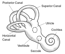
A mechanical inner ear disorder causing brief, intense vertigo with a change in head position: otoconia (calcium carbonate crystals) dislodge from the utricle and migrate into a semicircular canal, where they disturb endolymph flow during head movement.

Confirmation of BPPV

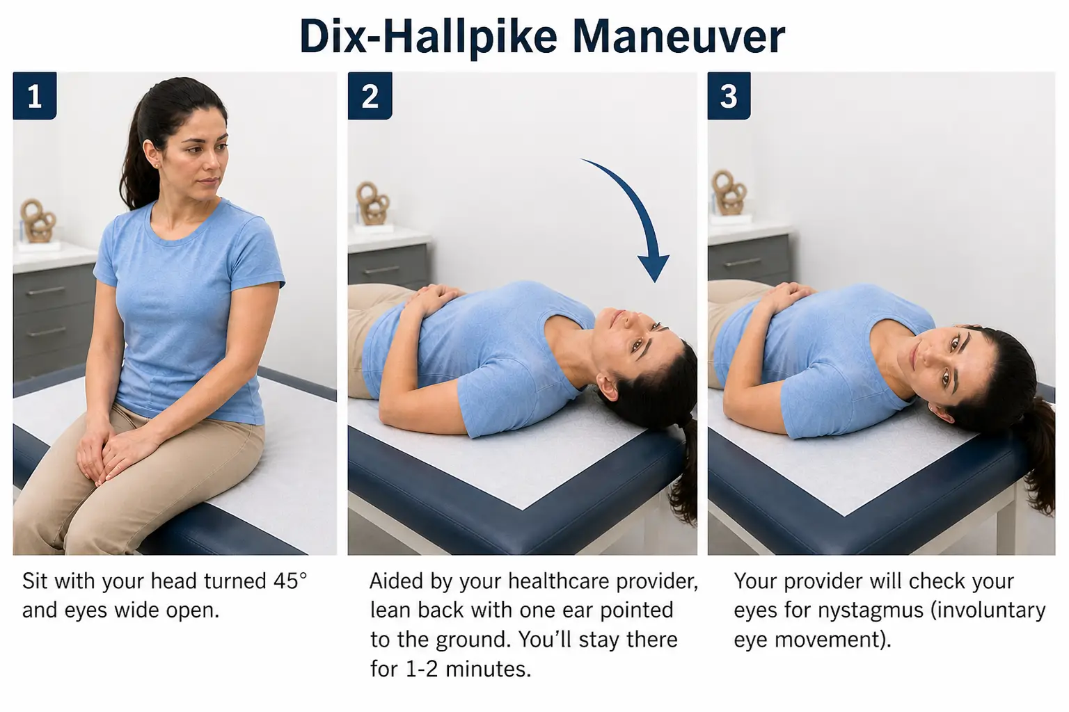
  • Confirmed by performing the Dix-Hallpike test - before completing the test, the therapist must perform a vertebral artery test and ensure appropriate cervical range of motion is present
    • Patient positioned in long sitting, with the head turned 45 degrees toward the affected side
    • Patient is quickly lowered to supine with the head remaining at 45 degrees and hanging over the edge of the mat, while the therapist observes for nystagmus
      • Nystagmus will resolve within 30-60 seconds
      • A positive test occurs when the patient experiences nystagmus and vertigo with a head position change
    • Repeat the test on the contralateral side
Dix-Hallpike maneuver showing the patient lying supine with the head turned 45 degrees and extended off the table to assess BPPV.
Dix Hallpike
Achievable

PTA role and stop signs: the Dix-Hallpike test and repositioning maneuvers are carried out within the supervising PT’s plan of care - the PTA collects data and performs the established maneuvers rather than setting or changing the diagnosis or plan. Stop positioning and notify the PT immediately if you see neurological symptoms (diplopia, dysarthria, dysphagia, facial numbness, drop attacks), nystagmus that doesn’t fatigue or lasts longer than 60 seconds, or pure downbeat nystagmus - these suggest a central rather than peripheral cause.

Understanding what nystagmus means in relation to BPPV

  • Ageotropic (apogeotropic)
    • Fast beating phase of the nystagmus is moving away from the ground (towards the upper ear) - this indicates lateral canal BPPV
  • Geotropic
    • Fast beating phase of nystagmus is moving towards the ground (towards the lower ear) - this also indicates lateral canal BPPV
  • Torsional (rotary), upbeating
    • Indicates posterior canal BPPV

Differentiation of BPPV and canals impacted

Lateral canal BPPV

  • Tested with the supine roll test (not the Dix-Hallpike): with the patient supine and the head flexed about 30 degrees, the head is turned quickly to each side while the clinician watches for horizontal nystagmus
  • Can manifest as ageotropic or geotropic horizontal nystagmus
  • Barbeque (log/Lempert) roll: with the patient supine and the head turned 90 degrees toward the affected ear, the clinician rolls the patient in 90-degree increments away from the affected side - supine → unaffected side-lying → prone (nose down) → sitting - pausing 30-60 seconds at each position to guide the otoconia out of the lateral canal.

Posterior canal BPPV (most common BPPV)

  • Typically presents with torsional (rotary), upbeating nystagmus
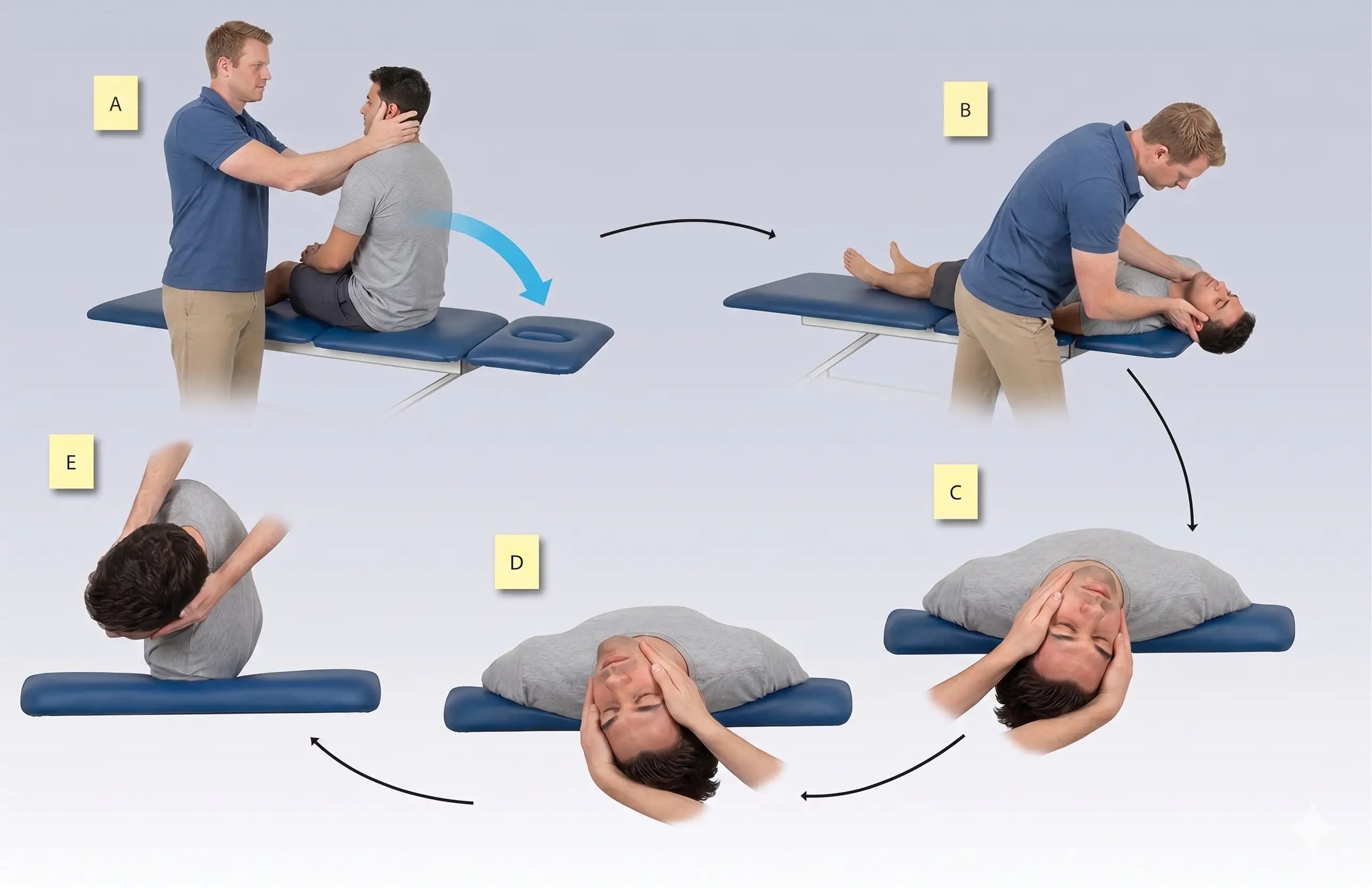
  • The Epley maneuver is performed to reposition the otoconia: the patient moves from sitting to supine with the head turned toward the affected side, then rotates the head and body through a series of positions to guide the otoconia out of the posterior canal, finishing in sitting.

Anterior canal BPPV (least common BPPV)

  • Typically presents with downbeat nystagmus (often with a small torsional component) that is brief and fatigues with repetition; downbeat nystagmus that persists or does not fatigue is a central red flag
  • The Yacovino maneuver is performed to reposition the otoconia: the patient moves from sitting to supine with the head extended below horizontal, then to a chin-to-chest position, and back to sitting, guiding the otoconia out of the anterior canal.
Epley maneuver illustrating the sequential head and body positions used to reposition displaced otoconia in the inner ear.
Epley maneuver
Achievable

Meniere’s disease

Definitions
Meniere’s disease
A chronic inner ear disorder with common symptoms of fullness in the ear, ringing in the ear, loss of hearing, and vertigo due to abnormal fluid buildup in the ear.

Episodes of vertigo typically last 20 minutes to 12 hours and recur episodically. Due to the chronicity of the disease, management focuses on reducing the frequency of attacks and managing symptoms.

Management of symptoms

  • Low sodium diet - reduces fluid levels
  • Regular exercise - improves balance and reduces stress
  • Management of stress - stress can exacerbate an episode of vertigo
  • Medical management - diuretics reduce fluid within the ear; surgery (fluid drainage or removal of the damaged inner ear) is reserved for refractory cases

Physical therapy management of individuals with Meniere’s disease is symptom-based, and episodes of care are short. The focus will primarily be to assist with any balance deficits or muscle weakness, provide an individualized home exercise program, and reinforce patient education.

Vestibular neuritis

Definitions
Vestibular neuritis
Inflammation of the vestibular nerve, most often presumed viral (post-viral).

Symptoms include sudden onset of severe vertigo (spinning sensation), nausea and vomiting, dizziness and lightheadedness, difficulty walking, and nystagmus. Symptoms are typically sudden and improve with medical intervention over a few days.

Treatment options

  • Vestibular suppressants and anti-nausea medication - for short-term relief of acute symptoms; prolonged use can delay central compensation
  • Corticosteroids - for management of inflammation

Acute vertigo typically subsides within days, but vestibular rehabilitation is recommended to address the lingering imbalance and dizziness from unilateral vestibular hypofunction:

  • Gaze stabilization (VOR x1) - the patient keeps the eyes fixed on a stationary target while moving the head, progressing speed and visual background to retrain the vestibulo-ocular reflex
  • Habituation - repeated exposure to the movements or positions that provoke symptoms (e.g., Brandt-Daroff exercises) to reduce the brain’s response over time
  • Balance and substitution training - progressing base of support and surface compliance while using visual and somatosensory cues to compensate for the vestibular loss

Labyrinthitis

Definitions
Labyrinthitis
Inflammation of the inner ear caused by viral infections, bacterial infections, autoimmune disorders, or head trauma.

Symptoms include vertigo, nausea and vomiting, balance problems, hearing loss, tinnitus (ringing in the ears), and ear pain. Most symptoms resolve within a few weeks, but some permanent hearing loss may be present.

Treatment interventions

  • Antihistamines and anti-nausea drugs - for symptom control of vertigo, nausea, and vomiting
  • Corticosteroids - for management of inflammation
  • Antibiotics - for a confirmed or suspected bacterial cause

Once the acute phase settles, PT uses the same vestibular rehabilitation (gaze stabilization, habituation, and balance training) described above for vestibular neuritis; long-term use of vestibular suppressants can delay central compensation.

Comparing the peripheral vestibular disorders

Condition Vertigo duration / trigger Hearing loss / tinnitus PT role
BPPV Seconds, triggered by a position change None Repositioning maneuvers (Epley, Yacovino, barbeque roll)
Meniere’s disease Minutes to hours, episodic Fluctuating hearing loss, tinnitus, aural fullness Balance training and HEP between attacks
Vestibular neuritis Constant for days None Gaze stabilization, habituation, balance training
Labyrinthitis Constant for days Hearing loss and tinnitus Same vestibular rehabilitation as neuritis

BPPV overview and diagnosis

  • Otoconia dislodge from utricle, migrate into semicircular canal, disrupt endolymph flow during head movement
  • Confirmed with Dix-Hallpike test: patient moved from long sitting to supine with head turned 45° and extended off table; observe for nystagmus (resolves in 30-60 sec); repeat on both sides
  • Must check vertebral artery status and cervical ROM before testing
  • PTA performs established maneuvers within PT’s plan of care; does not diagnose
  • Stop and notify PT if: neurological signs (diplopia, dysarthria, dysphagia, facial numbness, drop attacks), nystagmus lasting >60 sec or not fatiguing, or pure downbeat nystagmus (central red flags)

Nystagmus patterns and canal involvement

  • Ageotropic: fast phase beats away from ground → lateral canal BPPV
  • Geotropic: fast phase beats toward ground → lateral canal BPPV
  • Torsional/rotary, upbeating: posterior canal BPPV (most common type)
  • Downbeat (brief, fatigues): anterior canal BPPV (least common); persistent/non-fatiguing = central cause

BPPV treatment maneuvers by canal

  • Lateral canal: diagnosed via supine roll test (not Dix-Hallpike); treated with Barbeque (Lempert) roll — 90° increments supine → unaffected side → prone → sitting, pausing 30-60 sec each position
  • Posterior canal: treated with Epley maneuver — series of head/body turns from supine to sitting
  • Anterior canal: treated with Yacovino maneuver — supine with head extended, to chin-tuck, to sitting

Meniere’s disease

  • Chronic inner ear disorder: ear fullness, tinnitus, hearing loss, vertigo from fluid buildup
  • Vertigo episodes last 20 min–12 hrs, recurrent/episodic
  • Management: low-sodium diet, exercise, stress management, diuretics; surgery only for refractory cases
  • PT role: symptom-based, short episodes of care, address balance/weakness, HEP, patient education

Vestibular neuritis vs. labyrinthitis

  • Vestibular neuritis: vestibular nerve inflammation (often post-viral); sudden severe vertigo, nausea, gait difficulty, nystagmus; no hearing loss
  • Labyrinthitis: inner ear inflammation (viral, bacterial, autoimmune, trauma); same symptoms plus hearing loss and tinnitus
  • Acute treatment: vestibular suppressants/anti-nausea meds (short-term only — prolonged use delays compensation), corticosteroids; antibiotics if bacterial (labyrinthitis)
  • Vestibular rehab (both conditions) once acute phase resolves:
    • Gaze stabilization (VOR x1): fixate on target while moving head, progress speed/background
    • Habituation: repeated exposure to provoking movements (e.g., Brandt-Daroff) to reduce response
    • Balance/substitution training: progress base of support/surface compliance using visual/somatosensory cues

Comparing disorders

  • Key differentiators: duration/trigger of vertigo, presence of hearing loss/tinnitus, and PT intervention focus
  • BPPV = brief, positional, no hearing changes, treated with repositioning maneuvers
  • Meniere’s = episodic (min-hrs), fluctuating hearing loss/tinnitus, symptom management between attacks
  • Neuritis/labyrinthitis = constant for days; labyrinthitis includes hearing loss/tinnitus; both use same rehab approach

Sign up for free to take 13 quiz questions on this topic

Previous
Next  | 4.1 Pediatric foundations
All rights reserved ©2016 - 2026 Achievable, Inc.

Peripheral vestibular disorders and intervention strategies

Identifying and managing peripheral vestibular disorders

Benign paroxysmal positional vertigo (BPPV)

A mechanical inner ear disorder causing brief, intense vertigo with a change in head position: otoconia (calcium carbonate crystals) dislodge from the utricle and migrate into a semicircular canal, where they disturb endolymph flow during head movement.

Confirmation of BPPV

  • Confirmed by performing the Dix-Hallpike test - before completing the test, the therapist must perform a vertebral artery test and ensure appropriate cervical range of motion is present
    • Patient positioned in long sitting, with the head turned 45 degrees toward the affected side
    • Patient is quickly lowered to supine with the head remaining at 45 degrees and hanging over the edge of the mat, while the therapist observes for nystagmus
      • Nystagmus will resolve within 30-60 seconds
      • A positive test occurs when the patient experiences nystagmus and vertigo with a head position change
    • Repeat the test on the contralateral side

PTA role and stop signs: the Dix-Hallpike test and repositioning maneuvers are carried out within the supervising PT’s plan of care - the PTA collects data and performs the established maneuvers rather than setting or changing the diagnosis or plan. Stop positioning and notify the PT immediately if you see neurological symptoms (diplopia, dysarthria, dysphagia, facial numbness, drop attacks), nystagmus that doesn’t fatigue or lasts longer than 60 seconds, or pure downbeat nystagmus - these suggest a central rather than peripheral cause.

Understanding what nystagmus means in relation to BPPV

  • Ageotropic (apogeotropic)
    • Fast beating phase of the nystagmus is moving away from the ground (towards the upper ear) - this indicates lateral canal BPPV
  • Geotropic
    • Fast beating phase of nystagmus is moving towards the ground (towards the lower ear) - this also indicates lateral canal BPPV
  • Torsional (rotary), upbeating
    • Indicates posterior canal BPPV

Differentiation of BPPV and canals impacted

Lateral canal BPPV

  • Tested with the supine roll test (not the Dix-Hallpike): with the patient supine and the head flexed about 30 degrees, the head is turned quickly to each side while the clinician watches for horizontal nystagmus
  • Can manifest as ageotropic or geotropic horizontal nystagmus
  • Barbeque (log/Lempert) roll: with the patient supine and the head turned 90 degrees toward the affected ear, the clinician rolls the patient in 90-degree increments away from the affected side - supine → unaffected side-lying → prone (nose down) → sitting - pausing 30-60 seconds at each position to guide the otoconia out of the lateral canal.

Posterior canal BPPV (most common BPPV)

  • Typically presents with torsional (rotary), upbeating nystagmus
  • The Epley maneuver is performed to reposition the otoconia: the patient moves from sitting to supine with the head turned toward the affected side, then rotates the head and body through a series of positions to guide the otoconia out of the posterior canal, finishing in sitting.

Anterior canal BPPV (least common BPPV)

  • Typically presents with downbeat nystagmus (often with a small torsional component) that is brief and fatigues with repetition; downbeat nystagmus that persists or does not fatigue is a central red flag
  • The Yacovino maneuver is performed to reposition the otoconia: the patient moves from sitting to supine with the head extended below horizontal, then to a chin-to-chest position, and back to sitting, guiding the otoconia out of the anterior canal.

Meniere’s disease

Definitions
Meniere’s disease
A chronic inner ear disorder with common symptoms of fullness in the ear, ringing in the ear, loss of hearing, and vertigo due to abnormal fluid buildup in the ear.

Episodes of vertigo typically last 20 minutes to 12 hours and recur episodically. Due to the chronicity of the disease, management focuses on reducing the frequency of attacks and managing symptoms.

Management of symptoms

  • Low sodium diet - reduces fluid levels
  • Regular exercise - improves balance and reduces stress
  • Management of stress - stress can exacerbate an episode of vertigo
  • Medical management - diuretics reduce fluid within the ear; surgery (fluid drainage or removal of the damaged inner ear) is reserved for refractory cases

Physical therapy management of individuals with Meniere’s disease is symptom-based, and episodes of care are short. The focus will primarily be to assist with any balance deficits or muscle weakness, provide an individualized home exercise program, and reinforce patient education.

Vestibular neuritis

Definitions
Vestibular neuritis
Inflammation of the vestibular nerve, most often presumed viral (post-viral).

Symptoms include sudden onset of severe vertigo (spinning sensation), nausea and vomiting, dizziness and lightheadedness, difficulty walking, and nystagmus. Symptoms are typically sudden and improve with medical intervention over a few days.

Treatment options

  • Vestibular suppressants and anti-nausea medication - for short-term relief of acute symptoms; prolonged use can delay central compensation
  • Corticosteroids - for management of inflammation

Acute vertigo typically subsides within days, but vestibular rehabilitation is recommended to address the lingering imbalance and dizziness from unilateral vestibular hypofunction:

  • Gaze stabilization (VOR x1) - the patient keeps the eyes fixed on a stationary target while moving the head, progressing speed and visual background to retrain the vestibulo-ocular reflex
  • Habituation - repeated exposure to the movements or positions that provoke symptoms (e.g., Brandt-Daroff exercises) to reduce the brain’s response over time
  • Balance and substitution training - progressing base of support and surface compliance while using visual and somatosensory cues to compensate for the vestibular loss

Labyrinthitis

Definitions
Labyrinthitis
Inflammation of the inner ear caused by viral infections, bacterial infections, autoimmune disorders, or head trauma.

Symptoms include vertigo, nausea and vomiting, balance problems, hearing loss, tinnitus (ringing in the ears), and ear pain. Most symptoms resolve within a few weeks, but some permanent hearing loss may be present.

Treatment interventions

  • Antihistamines and anti-nausea drugs - for symptom control of vertigo, nausea, and vomiting
  • Corticosteroids - for management of inflammation
  • Antibiotics - for a confirmed or suspected bacterial cause

Once the acute phase settles, PT uses the same vestibular rehabilitation (gaze stabilization, habituation, and balance training) described above for vestibular neuritis; long-term use of vestibular suppressants can delay central compensation.

Comparing the peripheral vestibular disorders

Condition Vertigo duration / trigger Hearing loss / tinnitus PT role
BPPV Seconds, triggered by a position change None Repositioning maneuvers (Epley, Yacovino, barbeque roll)
Meniere’s disease Minutes to hours, episodic Fluctuating hearing loss, tinnitus, aural fullness Balance training and HEP between attacks
Vestibular neuritis Constant for days None Gaze stabilization, habituation, balance training
Labyrinthitis Constant for days Hearing loss and tinnitus Same vestibular rehabilitation as neuritis
Key points

BPPV overview and diagnosis

  • Otoconia dislodge from utricle, migrate into semicircular canal, disrupt endolymph flow during head movement
  • Confirmed with Dix-Hallpike test: patient moved from long sitting to supine with head turned 45° and extended off table; observe for nystagmus (resolves in 30-60 sec); repeat on both sides
  • Must check vertebral artery status and cervical ROM before testing
  • PTA performs established maneuvers within PT’s plan of care; does not diagnose
  • Stop and notify PT if: neurological signs (diplopia, dysarthria, dysphagia, facial numbness, drop attacks), nystagmus lasting >60 sec or not fatiguing, or pure downbeat nystagmus (central red flags)

Nystagmus patterns and canal involvement

  • Ageotropic: fast phase beats away from ground → lateral canal BPPV
  • Geotropic: fast phase beats toward ground → lateral canal BPPV
  • Torsional/rotary, upbeating: posterior canal BPPV (most common type)
  • Downbeat (brief, fatigues): anterior canal BPPV (least common); persistent/non-fatiguing = central cause

BPPV treatment maneuvers by canal

  • Lateral canal: diagnosed via supine roll test (not Dix-Hallpike); treated with Barbeque (Lempert) roll — 90° increments supine → unaffected side → prone → sitting, pausing 30-60 sec each position
  • Posterior canal: treated with Epley maneuver — series of head/body turns from supine to sitting
  • Anterior canal: treated with Yacovino maneuver — supine with head extended, to chin-tuck, to sitting

Meniere’s disease

  • Chronic inner ear disorder: ear fullness, tinnitus, hearing loss, vertigo from fluid buildup
  • Vertigo episodes last 20 min–12 hrs, recurrent/episodic
  • Management: low-sodium diet, exercise, stress management, diuretics; surgery only for refractory cases
  • PT role: symptom-based, short episodes of care, address balance/weakness, HEP, patient education

Vestibular neuritis vs. labyrinthitis

  • Vestibular neuritis: vestibular nerve inflammation (often post-viral); sudden severe vertigo, nausea, gait difficulty, nystagmus; no hearing loss
  • Labyrinthitis: inner ear inflammation (viral, bacterial, autoimmune, trauma); same symptoms plus hearing loss and tinnitus
  • Acute treatment: vestibular suppressants/anti-nausea meds (short-term only — prolonged use delays compensation), corticosteroids; antibiotics if bacterial (labyrinthitis)
  • Vestibular rehab (both conditions) once acute phase resolves:
    • Gaze stabilization (VOR x1): fixate on target while moving head, progress speed/background
    • Habituation: repeated exposure to provoking movements (e.g., Brandt-Daroff) to reduce response
    • Balance/substitution training: progress base of support/surface compliance using visual/somatosensory cues

Comparing disorders

  • Key differentiators: duration/trigger of vertigo, presence of hearing loss/tinnitus, and PT intervention focus
  • BPPV = brief, positional, no hearing changes, treated with repositioning maneuvers
  • Meniere’s = episodic (min-hrs), fluctuating hearing loss/tinnitus, symptom management between attacks
  • Neuritis/labyrinthitis = constant for days; labyrinthitis includes hearing loss/tinnitus; both use same rehab approach

More from Vestibular system

  • Anatomy, examination, and disorder classification