Disorders of the gallbladder and bile ducts
-
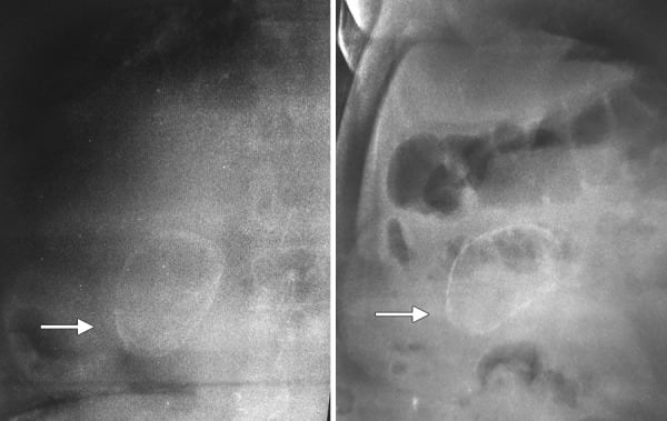
Cholelithiasis or gallstones: This is the presence of solid calculi in the gallbladder. Calculi form when bile becomes supersaturated and precipitates. Gallstones may be:
- Cholesterol stones (most common)
- Pigmented stones (seen in hemolytic anemias), composed of calcium bilirubinate
Risk factors include high-calorie diet, obesity, female sex, pregnancy, rapid weight loss, estrogen therapy, cholestasis, low physical activity, diabetes mellitus, dyslipidemias, total parenteral nutrition, low fibre diets, hyperinsulinism, and metabolic syndrome.
Many people are asymptomatic. Others present with biliary colic, which is acute abdominal pain in the RUQ or epigastrium. It may be aggravated by fatty foods and typically resolves gradually over 1-5 hours. Upper abdominal tenderness is present. Some patients progress to complications (e.g., acute cholecystitis).
Gallstones can be visualized on ultrasound.
Management
- Asymptomatic patients with incidentally detected gallstones are managed expectantly.
- Exceptions (treat with cholecystectomy): hemolytic anemias, porcelain gallbladder, gallstones >3 cm, and patients planning liver transplant or bariatric surgery.
- Symptomatic patients are treated with cholecystectomy, preferably laparoscopic.
- Pain control: NSAIDs are preferred. Meperidine is used in pregnant women for pain relief.
- Medical dissolution: Ursodeoxycholic acid and chenodeoxycholic acid help dissolve cholesterol gallstones <5 mm.
- Non-surgical candidates: extracorporeal shock wave lithotripsy (ESWL) may help.
Complications of gallstones: As follows
Acute cholecystitis: This is acute inflammation of the gallbladder caused by obstruction of the cystic duct by a gallstone. There is often a past history of recurrent biliary colic. It presents with RUQ pain, fever, and chills. An RUQ mass may be palpable. A positive Murphy sign is present (inspiratory arrest on deep palpation in the RUQ).
Laboratory findings include leukocytosis and slightly elevated bilirubin.
Diagnosis
- Ultrasound can diagnose gallstones and may show:
- Pericholecystic fluid
- Gallbladder wall thickening >4 mm
- Gallstones
- Sonographic Murphy sign (RUQ pain when pressing down the probe)
- HIDA scan can be used if needed. Failure to visualize the gallbladder in acute cholecystitis suggests cystic duct obstruction.
- Failure of HIDA (hepatobiliary iminodiacetic acid) to fill the gallbladder at two hours after injection is indicative of cystic duct obstruction.
- Ultrasound is preferred to diagnose gallstones, while HIDA scan is more sensitive and specific for acute cholecystitis.
- A cholecystokinin HIDA (CCK-HIDA) scan evaluates gallbladder contractility and can be done if clinical suspicion of gallstones or cholecystitis is high but ultrasound is negative.
- CCK-HIDA will also be positive in acalculous cholecystitis.
- CT scan and MRI can evaluate complications such as empyema and gangrene.
Treatment
- Early laparoscopic cholecystectomy (within 24-72 h of symptom onset)
- Antibiotics and supportive management
- In critically ill or older patients who are poor surgical candidates, percutaneous cholecystostomy may help.
- Uncomplicated cases with no underlying conditions and easy access to medical care can be treated outpatient with antibiotics and analgesics, as long as they are afebrile and do not have common bile duct stones.
Gangrenous cholecystitis: This is necrosis and perforation of the gallbladder wall due to ischemia from progressive vascular insufficiency, occurring as a complication of gallstones. It is seen in diabetics, older age, delayed surgery for acute cholecystitis, and cardiovascular disease. It carries a very high mortality rate.
Imaging
- Ultrasound: thickened gallbladder wall, pericholecystic fluid
- CT: air in the gallbladder wall or lumen, irregular or absent gallbladder wall, intraluminal membranes, pericholecystic abscess, and lack of gallbladder wall enhancement
Treatment is laparoscopic or open cholecystectomy plus supportive management including antibiotics.
Emphysematous cholecystitis: This is seen with infections due to Clostridia, E.coli, and other gas-forming organisms. It is rapidly progressive and can lead to gangrene. It is more common in older patients and diabetics.
- Imaging shows air within the gallbladder wall or lumen.
- Perforation may occur; in that case, crepitus may be present with generalised peritonitis.
- Clinically, it presents like gangrenous cholecystitis but may be insidious.
Treatment is antibiotics plus open cholecystectomy or percutaneous cholecystostomy (in some cases).
Acalculous cholecystitis: This is inflammation of the gallbladder caused by hypo- or dyskinesia of the gallbladder with reduced emptying, in the absence of stones. It can follow long periods of fasting or starvation, TPN, rapid weight loss, critical illness (ICU patients), severe burns, and surgery. It has a high mortality rate because gangrene and perforation are common.
Patients present with typical symptoms of acute cholecystitis plus sepsis and shock.
Diagnosis
- CCK-HIDA scan shows <35% gallbladder ejection fraction.
- Ultrasound shows a thickened gallbladder wall and pericholecystic fluid.
- CT and regular HIDA scan can also be done.
Management
- Percutaneous cholecystostomy or stent via an ERCP to aid drainage of the gallbladder
- Antibiotics
- Cholecystectomy when the patient is stable
Choledocholithiasis and ascending cholangitis: Gallstones may migrate into the common bile duct.
- Choledocholithiasis is the presence of a stone in the common bile duct.
- This can cause obstruction and infection of the bile duct, called cholangitis.
Cholangitis presents with Charcot’s triad:
- Fever
- Obstructive jaundice
- Abdominal pain
If severe with septicaemia, Reynolds pentad develops:
- Hypotension
- Altered mental status
This is a medical emergency and should be treated with intravenous antibiotics and ERCP to retrieve the stone. ERCP is both diagnostic and therapeutic.
- Ultrasound shows a dilated common bile duct.
- Other imaging studies include MRCP and endoscopic ultrasound, but these are not therapeutic.
- Laboratory findings include leukocytosis and elevated liver enzymes.
Treatment is antibiotics and ERCP with or without stenting.
Gallstone pancreatitis: A stone may migrate further and block the opening of the pancreatic duct at the level of the Sphincter of Oddi, causing pancreatitis.
- Lipase and amylase are elevated.
- ERCP is done for diagnosis and treatment.
Gallstone ileus: This is mechanical obstruction of the intestine caused by an impacted gallstone. It typically follows acute cholecystitis. Inflammation leads to pressure and ischemic necrosis of the gallbladder wall, adhesions, and formation of a cholecysto-enteric fistula. The gallstone enters the intestine through the fistula and obstructs the intestine, most commonly the terminal ileum.
Patients present with nausea, vomiting, crampy abdominal pain, and abdominal distension. Jaundice may be present. Perforation peritonitis may occur.
Imaging studies show:
- Partial or complete intestinal obstruction with dilated bowel loops and air-fluid levels
- Pneumobilia or contrast material in the biliary tree
- An aberrant gallstone with shifting positions on serial films
Treatment is supportive, with stone removal and closure of the fistula. Some cases may require bowel resection.