Cerebrovascular disorders
The brain receives about 20% of the cardiac output. When systolic blood pressure drops below 50 mmHg, cerebral perfusion pressure falls, which increases the risk of ischemia.
Ischemia lasting 4-5 minutes can cause irreversible injury to:
- Hippocampal and neocortical pyramidal cells
- Striatal neurons
- Purkinje cells
With more prolonged ischemia, thalamic and brainstem neurons can also be damaged. In the neonatal brain, injury to the periventricular white matter is common, in addition to the cortical damage described above.
Section A: Stroke
Stroke is classically defined as a neurological deficit caused by an acute focal injury of the central nervous system (CNS) due to a vascular cause. This includes cerebral infarction, intracerebral hemorrhage (ICH), and subarachnoid hemorrhage (SAH).
(I) Cerebral infarction: Cerebral ischemia can result from:
- Atherosclerosis with or without thrombosis of major vessels (e.g., MCA)
- Embolization from the heart or from clots in blood vessels
- Vasculitis
- Hypercoagulable states (e.g., Factor V Leiden)
- Polycythemia
- Small vessel disease (more common in women)
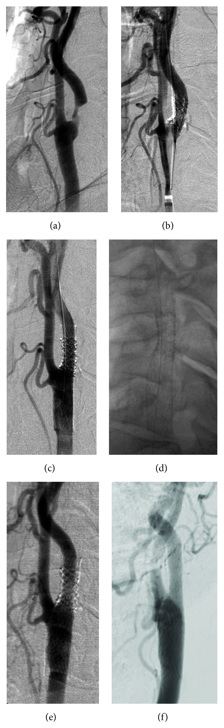
In older patients, atherosclerosis with thrombosis is the most common cause. The MCA is the most commonly involved artery in both atherosclerotic and embolic strokes.
In younger patients, you should consider non-atherosclerotic causes, especially embolization and hypercoagulable states. Venous thrombosis is associated with estrogen therapy and oral contraceptive pill use. Vasospasm accompanying SAH can also cause cerebral infarcts.
Clinical features: Clinical features depend on the area infarcted. Symptoms are typically acute to subacute and evolve over hours to days. Embolic strokes tend to present more abruptly than arterial thrombosis.
Features of strokes according to arterial distribution
| Artery involved | Presenting features |
| MCA | Hemiparesis and hemianesthesia of the contralateral face and body; contralateral homonymous hemianopia; deviation of eye toward the affected hemisphere.
Left hemisphere involvement presents, in addition, with Broca’s, Wernicke’s, conduction or global aphasia. Right hemisphere lesions present, in addition, with contralateral hemineglect or speech alterations (e.g., “computer generated” speech or a foreign accent; dysprosody). |
| ACA | Hemiparesis and hemianesthesia of the contralateral lower limb; gait apraxia, urinary incontinence, primitive reflexes like grasping and sucking, frontal lobe involvement presenting as disinhibition, lack of motivation, impaired judgement. |
| PCA | Contralateral homonymous hemianopia typically with macular sparing; left PCA lesions will also have alexia without agraphia (can write but cannot read) |
| Vertebral artery or anterior spinal artery | Medial medullary syndrome presenting with contralateral spastic hemiparesis, loss of touch, vibration and pressure sensations in the limbs and trunk. Dysarthria (improper articulation of speech), paralysis and atrophy of tongue ipsilateral to the lesion, deviation of the tongue toward the side of lesion. |
| Vertebral artery or PICA* (posterior inferior cerebellar artery) | Lateral medullary or Wallenberg’s syndrome presenting with contralateral loss of pain and temperature sensations in the body and limbs, ipsilateral Horner’s syndrome (miosis, anhidrosis, ptosis), vertigo, nausea, vomiting, vestibular nystagmus (fast phase away from the side of the lesion), ipsilateral sensory hearing loss, bulbar signs like dysphagia, hoarseness, deviation of uvula away from the side of lesion, absent gag reflex; loss of pain and temperature from ipsilateral hemi face and scalp, loss of taste from ipsilateral tongue |
| AICA or circumferential branches of the basilar artery | Lateral pontine syndrome presenting with vertigo, nausea, vomiting, vestibular nystagmus (fast phase away from the side of the lesion), ipsilateral sensory hearing loss, loss of pain and temperature from ipsilateral hemi face and scalp, weakness of muscles of mastication. |
| Paramedian branches of basilar artery | Medial pontine syndrome presenting with spastic hemiparesis of contralateral loss of pain and temperature sensations in the body and limbs, internal strabismus (eye moves medially due to paralysis of lateral rectus), diplopia, if medial lemniscus is involved then contralateral loss of touch, vibration, pressure sensations; is facial nerve is involved then ipslateral facial weakness, dry eyes, loss of taste from anterior ⅔ of the tongue. |
| Basilar artery (main stem) | Locked in syndrome presenting with the inability to move the body and limbs except III cranial nerve innervated eye muscles; hearing and sensations are preserved, patients are fully awake and aware. |
| Thalamoperforating branches of the PCA | Weber’s or medial midbrain syndrome presenting with contralateral spastic paresis, contralateral lower face weakness**, deviation of the tongue away from the lesion, oculomotor nerve palsy with dilated pupil, ptosis, external strabismus, ipsilateral loss of light reflex and accommodation reflex. |
*Differentiate AICA from PICA stroke by the presence of bulbar signs only in PICA.
** An intact blink reflex means it is an UMN facial nerve lesion as it spares the upper half of the face.
#A fully awake and aware person suffering from locked - in syndrome can legally give consent.
Lacunar infarcts: These are typically seen in hypertension and diabetes due to severe atherosclerosis of the deep penetrating branches of the major cerebral arteries. Lipohyalinosis, hyaline arteriosclerosis of small vessels, and aneurysm formation can occur. Lacunar infarcts cause small infarcts in the basal ganglia, thalamus, internal capsule, and brainstem. Multiple or recurrent lacunar infarctions can lead to dementia. They may present as pure motor or pure sensory (thalamic pain syndrome, etc.) strokes.
Watershed infarcts: Watershed infarcts occur at the junction of the distal fields of two non-anastomosing arterial systems.
- They can occur between the cortical territories of the ACA, MCA, and PCA (cortical watershed areas).
- They can also occur in the white matter along and slightly above the lateral ventricle, between the deep and superficial arterial systems of the MCA, or between the superficial systems of the MCA and ACA (deep watershed areas).
Microemboli or hypotension (from any cause) can reduce perfusion pressure in cerebral arteries, leading to watershed zone ischemia and sometimes infarction. This risk is increased by stenosis in the cerebral arteries.
Watershed infarcts can present with syncope, episodic or progressive weakness of the hand, partial seizures, proximal upper and lower limb paresis (“man in barrel sign”), mood changes, and visual deficits. Bilateral, symmetrical watershed infarcts are seen in global ischemia and HIE.
**Hypoxic ischemic encephalopathy or HIE or global ischemia: **Global ischemia can result from hypovolemic shock, cardiac arrest, CO poisoning, hypoglycemia, severe anemia, and status epilepticus, etc.
Neurons require a steady supply of glucose and ATP (via oxidative phosphorylation) to meet their high metabolic demands. In global ischemia:
- ATP production falls, leading to failure of the Na+K+ATPase pump.
- Na+ influx causes cellular edema and cell death.
- Membrane depolarization triggers glutamate release into the synaptic cleft.
- Activation of NMDA and AMPA glutamate receptors causes Ca++ influx.
- Increased intracellular Ca++ activates lytic enzymes and promotes free radical formation.
Severe HIE is accompanied by cerebral edema, and its effects are compounded by increased intracranial pressure. Cytotoxic (intracellular) edema develops early in the insult. Interstitial cerebral edema follows due to vascular injury. Cerebral atrophy can occur due to neuronal loss from apoptosis and necrosis.