


Achievable logoAchievable logo
NCLEX
Sign in
Sign up
Purchase
Textbook
Practice exams
Support
How it works
Exam catalog
Mountain with a flag at the peak
Textbook
Introduction
1. Safe and effective care environment
2. Health promotion and maintenance
3. Psychosocial Integrity
4. Physiological Integrity
4.1 Basic care and comfort
4.2 Pharmacological and parenteral therapies
4.3 Reduction of risk potential
4.3.1 Diagnostic tests and lab values
4.3.2 Monitoring for complications of procedures
4.3.3 Changes in vital signs and neurological status
4.3.4 Electrolyte imbalances and fluid status
4.3.5 Medical equipment use and safety
4.3.6 Potential complications of devices and procedures
4.4 Physiological adaptation
Wrapping up
Achievable logoAchievable logo
4.3.6 Potential complications of devices and procedures
Achievable NCLEX
4. Physiological Integrity
4.3. Reduction of risk potential
Our NCLEX course is currently in development and is a work-in-progress.

Potential complications of devices and procedures

7 min read
Font
Discuss
Share
Feedback

Introduction

Medical devices and invasive procedures improve outcomes, but they also introduce predictable risks. On the Next Generation NCLEX (NGN), learners are expected to recognize early warning signs, prioritize nursing actions, and prevent harm through vigilant monitoring and timely escalation.

This section integrates prior content on post-procedure monitoring, vital signs, electrolytes, and equipment safety, focusing on what can go wrong and how nurses respond first.

Learning objectives

By the end of this section, the learner should be able to:

  • Identify common complications associated with medical devices and procedures
  • Recognize early vs late clinical indicators
  • Prioritize immediate nursing actions
  • Apply NGN-style clinical judgment to device-related scenarios

Core concept: Devices bypass natural defenses

Any device that enters the body increases the risk of infection, bleeding, obstruction, or malfunction.

The nurse’s role is anticipation, not reaction.

Early vs late complications

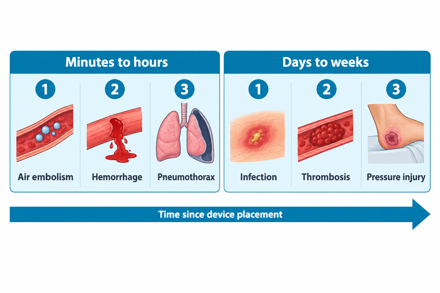
Complications often follow predictable timelines. NGN questions frequently test whether the nurse recognizes when something is abnormal.

Early complications (minutes to hours)

  • Air embolism
  • Hemorrhage
  • Pneumothorax
  • Acute allergic reaction
  • Sedation-related respiratory depression

Late complications (days to weeks)

  • Device-associated infection
  • Thrombosis
  • Pressure injury
  • Device occlusion
  • Fistula formation
alt_text
//////Caption: Timeline of Device-Related Complications
Illustration type: Timeline
Illustration note: Illustrate common early (minutes to hours) and late (days to weeks) complications of medical devices and invasive procedures, highlighting examples in each category///////

Infection-related complications

Common devices involved

  • Central venous catheters
  • Urinary catheters
  • Surgical drains
  • Endotracheal tubes

Early warning signs

  • Local redness, warmth, or drainage
  • Fever or hypothermia
  • Rising white blood cell count
  • Altered mental status in older adults

High-Risk Device-Associated Infections

Certain devices are strongly linked to specific infections that are frequently tested.

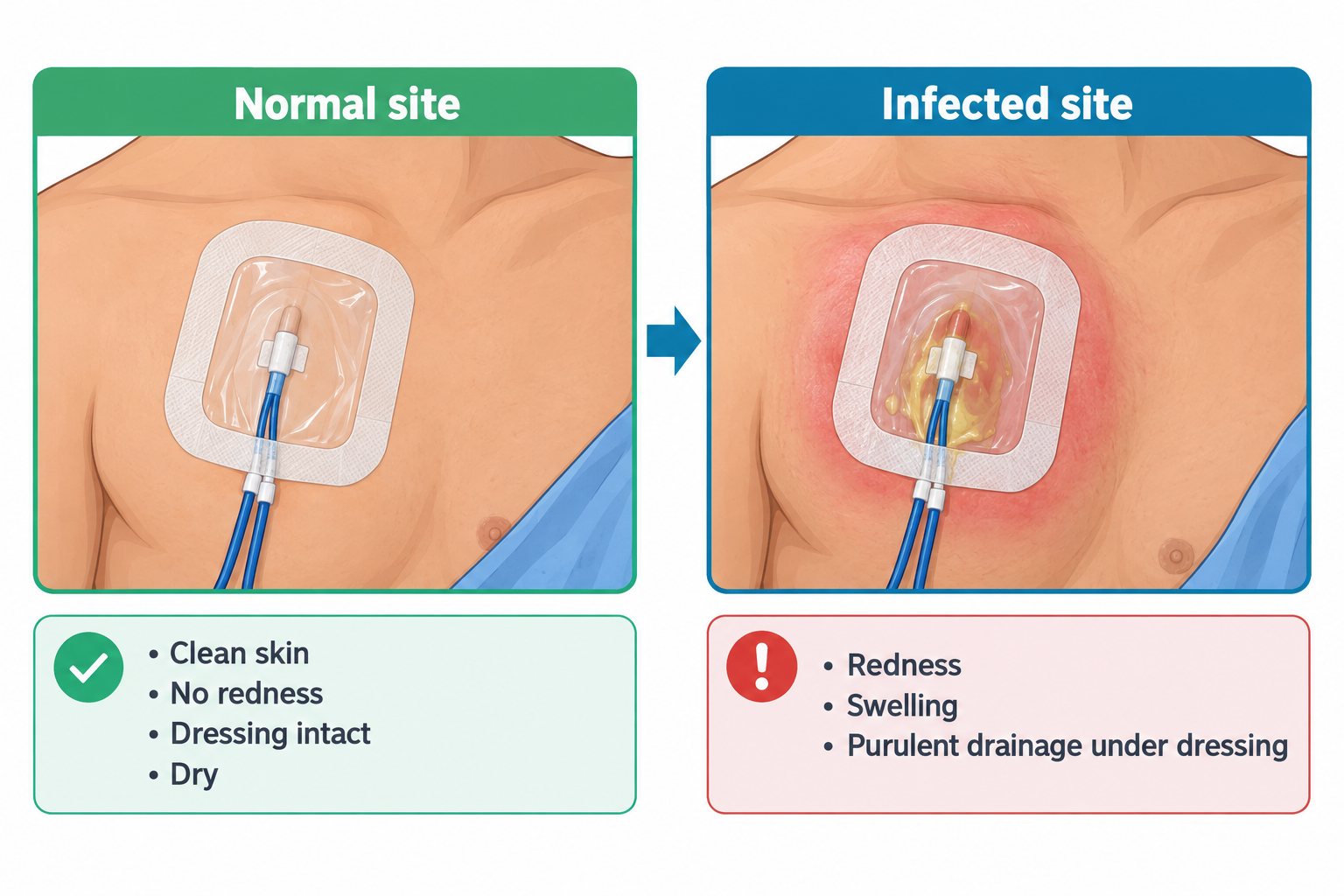
Central venous catheters → CLABSI risk

  • Monitor insertion site
  • Assess for unexplained fever
  • Maintain sterile dressing integrity
alt_text
//////Caption: Central Line Dressing: Normal vs. Infected Insertion Site
Illustration type: Side-by-side comparison photo/diagram
Illustration note: Compare a normal central line insertion site with a clean, dry, intact occlusive dressing versus an infected insertion site showing erythema, swelling, and purulent drainage to reinforce early recognition of CLABSI////////

Urinary catheters → CAUTI risk

  • Maintain closed system
  • Keep bag below bladder
  • Avoid dependent loops

Endotracheal tubes → Ventilator-associated pneumonia (VAP)

  • Elevate HOB 30–45°
  • Perform oral care
  • Monitor secretions

Surgical drains → Localized wound infection

  • Sudden change in drainage color
  • Foul odor
  • Increased pain at site

Complications during device removal

Insertion risks are commonly discussed. Removal risks are often overlooked.

Central line removal

  • Risk: Air embolism
  • Nursing action: Apply an occlusive dressing immediately after removal and position the client supine during removal to reduce the risk of air embolism (left lateral Trendelenburg is used if an air embolism occurs)

Chest tube removal

  • Risk: Tension pneumothorax
  • Nursing action: Apply occlusive dressing immediately

Foley catheter removal

  • Risk: Urinary retention
  • Nursing action: Monitor first void within 6–8 hours

Epidural catheter removal

  • Risk: Epidural hematoma (neurological deficit)
  • Nursing action: Assess sensation and motor function
Definitions
Device-associated infection
Infection occurring due to the presence of an indwelling device
Hematoma
Localized collection of blood outside blood vessels
Device dislodgement
Partial or complete movement of a device from its intended position
NGN insight:
Confusion may be the first sign of infection in older adults, even before fever or other classic symptoms develop.

Bleeding and vascular complications

High-risk procedures

  • Central line insertion
  • Arterial line placement
  • Surgical drain removal
  • Cardiac catheterization

Key assessment findings

  • Oozing or frank bleeding
  • Expanding hematoma
  • Hypotension
  • Tachycardia
alt_text
//////Caption: Expanding Hematoma at an Arterial Line Site
Illustration type: Annotated clinical photo/diagram
Illustration note: Illustrate an expanding hematoma at an arterial line insertion site with labels identifying increasing size, firmness, and the need to assess distal pulses and circulation.///////

Air embolism

Risk factors

  • Central line disconnection
  • Improper priming of IV tubing
  • Line removal without occlusion

Clinical signs

  • Sudden dyspnea
  • Chest pain
  • Hypotension
  • Anxiety or confusion

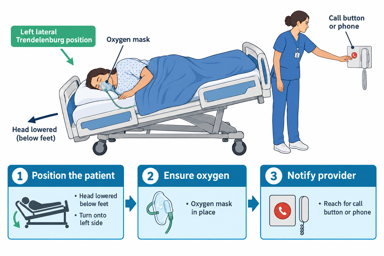
Medical emergency: Air embolism can cause rapid cardiovascular collapse.

Immediate nursing action: Place the client in the left lateral Trendelenburg position, administer oxygen as indicated, and notify the provider immediately.

alt_text
//////Caption: Recognizing and Responding to an Air Embolism
Illustration type: Clinical flowchart
Illustration note: Illustrate the progression from central line manipulation to sudden dyspnea, chest pain, hypotension, and oxygen desaturation, followed by immediate positioning in the left lateral Trendelenburg position, oxygen administration as indicated, and provider notification.///////

Obstruction and dislodgement

Affected devices

  • Feeding tubes
  • Chest tubes
  • Urinary catheters
  • Surgical drains

Warning signs

  • Sudden change in output
  • Increased pressure or pain
  • Alarms sounding
  • Absent expected drainage
alt_text
//////Caption: Normal vs. Obstructed Chest Tube Drainage System
Illustration type: Side-by-side comparison diagram
Illustration note: Compare a normal chest tube drainage system with expected tidaling and drainage versus an obstructed system showing absent tidaling and little or no expected drainage to reinforce early recognition of chest tube obstruction.///////

Tissue injury and pressure-related complications

Contributing factors

  • Prolonged device contact
  • Edema
  • Poor circulation
  • Reduced sensation

Assessment findings

  • Skin redness or breakdown
  • Pain or numbness
  • Swelling
NGN tip:
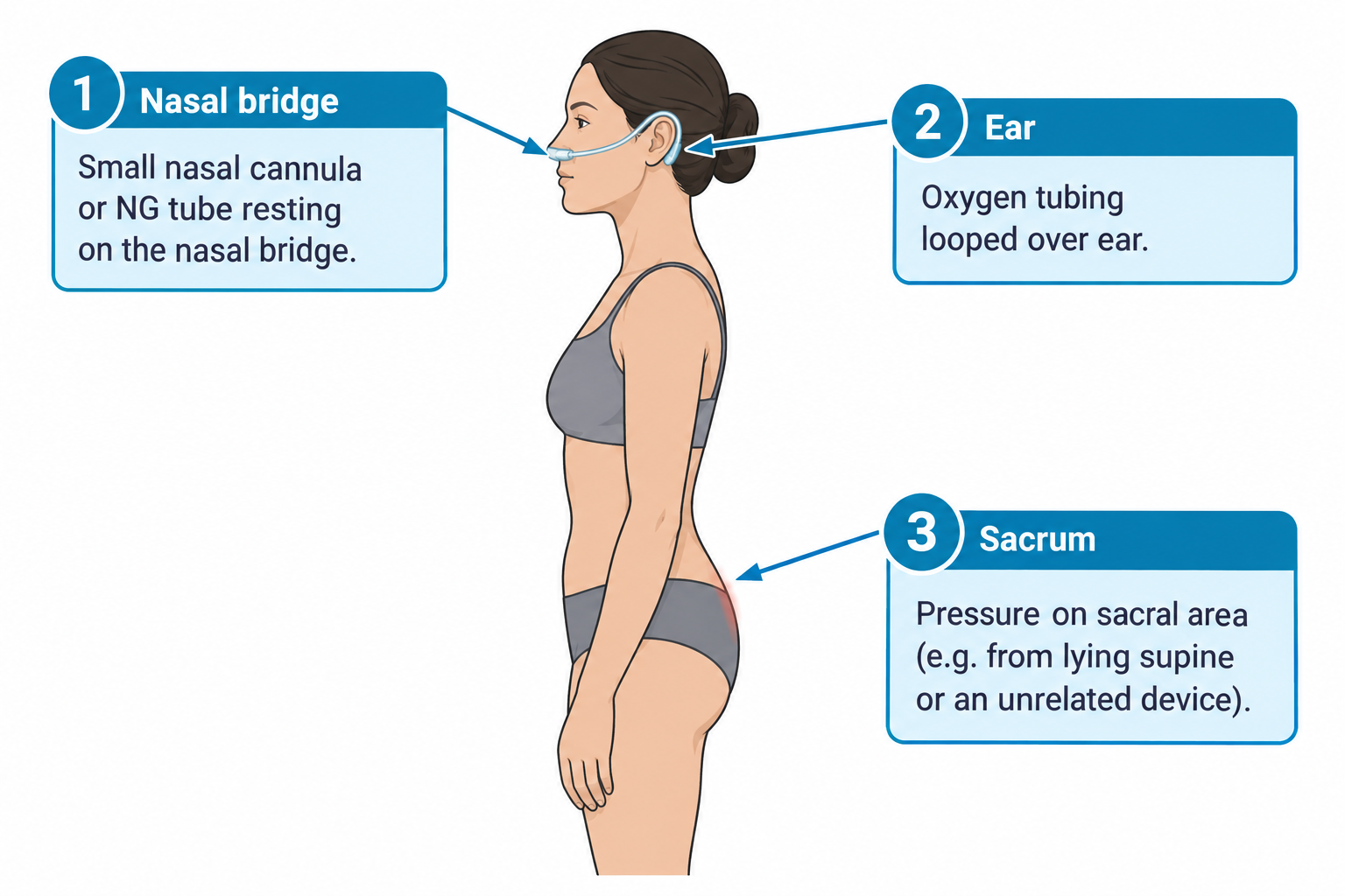
Medical devices are a common cause of hospital-acquired pressure injuries.
alt_text
//////Caption: Common Device-Related Pressure Injury Sites
Illustration type: Annotated anatomical diagram
Illustration note: Illustrate common locations of device-related pressure injuries, including the nasal bridge (nasal cannula/NG tube), ears (oxygen tubing), and sacrum (prolonged immobility with medical devices), highlighting areas at highest risk for skin breakdown.///////

Procedure-specific complications

Anterior axillary line (blue) Mid-axillary line (red)

Examples

  • Lumbar puncture: headache, CSF leak
  • Thoracentesis: pneumothorax
  • Endoscopy: bleeding, perforation
  • Biopsy: infection, hemorrhage
NGN insight:
Focus on recognizing the most life-threatening complication, not every possible complication.
alt_text
//////Caption: High-Risk Procedure Complications
Illustration type: Comparison table
Illustration note: Compare lumbar puncture, thoracentesis, endoscopy, and biopsy by identifying their most serious complications and the priority nursing assessments following each procedure.///////

OR

alt_text ////OPTIONAL “Anterior axillary line (blue) Mid-axillary line (red)” 📷 Image suggestion: “Thoracentesis landmark anatomy” — diagram showing anterior axillary and mid-axillary lines for procedure site landmarking, if this fragment is meant to anchor an actual image here./////

Putting it together: NGN prioritization

NGN integration

  • Sudden change = assess immediately
  • Device complication + abnormal vital signs = escalate immediately
  • Airway and circulation override device concerns

Clinical vignette 1

A client with a central line becomes acutely short of breath during a tubing change. Blood pressure drops and oxygen saturation decreases.

(spoiler)

Nursing action: Place the client in the left lateral Trendelenburg position, administer oxygen as indicated, notify the provider immediately, and prepare for further intervention.

Rationale: Sudden dyspnea, hypotension, and oxygen desaturation during central line manipulation suggest an air embolism, a medical emergency requiring immediate intervention.

Clinical vignette 2

A client with a surgical drain reports increasing pressure at the incision site. Drain output has stopped abruptly.

(spoiler)

Nursing action: Assess the surgical site and drain, notify the provider promptly, and continue close monitoring.

Rationale: Abrupt cessation of drainage with increasing pressure at the incision site may indicate drain obstruction or hematoma formation requiring prompt evaluation.

Common pitfalls on the NCLEX

  • Ignoring subtle early signs
  • Assuming devices function correctly
  • Treating alarms without client assessment
  • Delaying escalation of care
  • Devices increase risk while providing benefit
  • Early recognition prevents severe complications
  • Older adults present atypically
  • NGN rewards prioritization and trend analysis
  • The nurse is the final safety checkpoint

Sign up for free to take 5 quiz questions on this topic

Previous
Next  | 4.4.1 Medical emergencies
All rights reserved ©2016 - 2026 Achievable, Inc.

Potential complications of devices and procedures

Introduction

Medical devices and invasive procedures improve outcomes, but they also introduce predictable risks. On the Next Generation NCLEX (NGN), learners are expected to recognize early warning signs, prioritize nursing actions, and prevent harm through vigilant monitoring and timely escalation.

This section integrates prior content on post-procedure monitoring, vital signs, electrolytes, and equipment safety, focusing on what can go wrong and how nurses respond first.

Learning objectives

By the end of this section, the learner should be able to:

  • Identify common complications associated with medical devices and procedures
  • Recognize early vs late clinical indicators
  • Prioritize immediate nursing actions
  • Apply NGN-style clinical judgment to device-related scenarios

Core concept: Devices bypass natural defenses

Any device that enters the body increases the risk of infection, bleeding, obstruction, or malfunction.

The nurse’s role is anticipation, not reaction.

Early vs late complications

Complications often follow predictable timelines. NGN questions frequently test whether the nurse recognizes when something is abnormal.

Early complications (minutes to hours)

  • Air embolism
  • Hemorrhage
  • Pneumothorax
  • Acute allergic reaction
  • Sedation-related respiratory depression

Late complications (days to weeks)

  • Device-associated infection
  • Thrombosis
  • Pressure injury
  • Device occlusion
  • Fistula formation
alt_text
//////Caption: Timeline of Device-Related Complications
Illustration type: Timeline
Illustration note: Illustrate common early (minutes to hours) and late (days to weeks) complications of medical devices and invasive procedures, highlighting examples in each category///////

Infection-related complications

Common devices involved

  • Central venous catheters
  • Urinary catheters
  • Surgical drains
  • Endotracheal tubes

Early warning signs

  • Local redness, warmth, or drainage
  • Fever or hypothermia
  • Rising white blood cell count
  • Altered mental status in older adults

High-Risk Device-Associated Infections

Certain devices are strongly linked to specific infections that are frequently tested.

Central venous catheters → CLABSI risk

  • Monitor insertion site
  • Assess for unexplained fever
  • Maintain sterile dressing integrity
alt_text
//////Caption: Central Line Dressing: Normal vs. Infected Insertion Site
Illustration type: Side-by-side comparison photo/diagram
Illustration note: Compare a normal central line insertion site with a clean, dry, intact occlusive dressing versus an infected insertion site showing erythema, swelling, and purulent drainage to reinforce early recognition of CLABSI////////

Urinary catheters → CAUTI risk

  • Maintain closed system
  • Keep bag below bladder
  • Avoid dependent loops

Endotracheal tubes → Ventilator-associated pneumonia (VAP)

  • Elevate HOB 30–45°
  • Perform oral care
  • Monitor secretions

Surgical drains → Localized wound infection

  • Sudden change in drainage color
  • Foul odor
  • Increased pain at site

Complications during device removal

Insertion risks are commonly discussed. Removal risks are often overlooked.

Central line removal

  • Risk: Air embolism
  • Nursing action: Apply an occlusive dressing immediately after removal and position the client supine during removal to reduce the risk of air embolism (left lateral Trendelenburg is used if an air embolism occurs)

Chest tube removal

  • Risk: Tension pneumothorax
  • Nursing action: Apply occlusive dressing immediately

Foley catheter removal

  • Risk: Urinary retention
  • Nursing action: Monitor first void within 6–8 hours

Epidural catheter removal

  • Risk: Epidural hematoma (neurological deficit)
  • Nursing action: Assess sensation and motor function
Definitions
Device-associated infection
Infection occurring due to the presence of an indwelling device
Hematoma
Localized collection of blood outside blood vessels
Device dislodgement
Partial or complete movement of a device from its intended position
NGN insight:
Confusion may be the first sign of infection in older adults, even before fever or other classic symptoms develop.

Bleeding and vascular complications

High-risk procedures

  • Central line insertion
  • Arterial line placement
  • Surgical drain removal
  • Cardiac catheterization

Key assessment findings

  • Oozing or frank bleeding
  • Expanding hematoma
  • Hypotension
  • Tachycardia
alt_text
//////Caption: Expanding Hematoma at an Arterial Line Site
Illustration type: Annotated clinical photo/diagram
Illustration note: Illustrate an expanding hematoma at an arterial line insertion site with labels identifying increasing size, firmness, and the need to assess distal pulses and circulation.///////

Air embolism

Risk factors

  • Central line disconnection
  • Improper priming of IV tubing
  • Line removal without occlusion

Clinical signs

  • Sudden dyspnea
  • Chest pain
  • Hypotension
  • Anxiety or confusion

Medical emergency: Air embolism can cause rapid cardiovascular collapse.

Immediate nursing action: Place the client in the left lateral Trendelenburg position, administer oxygen as indicated, and notify the provider immediately.

alt_text
//////Caption: Recognizing and Responding to an Air Embolism
Illustration type: Clinical flowchart
Illustration note: Illustrate the progression from central line manipulation to sudden dyspnea, chest pain, hypotension, and oxygen desaturation, followed by immediate positioning in the left lateral Trendelenburg position, oxygen administration as indicated, and provider notification.///////

Obstruction and dislodgement

Affected devices

  • Feeding tubes
  • Chest tubes
  • Urinary catheters
  • Surgical drains

Warning signs

  • Sudden change in output
  • Increased pressure or pain
  • Alarms sounding
  • Absent expected drainage
alt_text
//////Caption: Normal vs. Obstructed Chest Tube Drainage System
Illustration type: Side-by-side comparison diagram
Illustration note: Compare a normal chest tube drainage system with expected tidaling and drainage versus an obstructed system showing absent tidaling and little or no expected drainage to reinforce early recognition of chest tube obstruction.///////

Tissue injury and pressure-related complications

Contributing factors

  • Prolonged device contact
  • Edema
  • Poor circulation
  • Reduced sensation

Assessment findings

  • Skin redness or breakdown
  • Pain or numbness
  • Swelling
NGN tip:
Medical devices are a common cause of hospital-acquired pressure injuries.
alt_text
//////Caption: Common Device-Related Pressure Injury Sites
Illustration type: Annotated anatomical diagram
Illustration note: Illustrate common locations of device-related pressure injuries, including the nasal bridge (nasal cannula/NG tube), ears (oxygen tubing), and sacrum (prolonged immobility with medical devices), highlighting areas at highest risk for skin breakdown.///////

Procedure-specific complications

Anterior axillary line (blue) Mid-axillary line (red)

Examples

  • Lumbar puncture: headache, CSF leak
  • Thoracentesis: pneumothorax
  • Endoscopy: bleeding, perforation
  • Biopsy: infection, hemorrhage
NGN insight:
Focus on recognizing the most life-threatening complication, not every possible complication.
alt_text
//////Caption: High-Risk Procedure Complications
Illustration type: Comparison table
Illustration note: Compare lumbar puncture, thoracentesis, endoscopy, and biopsy by identifying their most serious complications and the priority nursing assessments following each procedure.///////

OR

alt_text ////OPTIONAL “Anterior axillary line (blue) Mid-axillary line (red)” 📷 Image suggestion: “Thoracentesis landmark anatomy” — diagram showing anterior axillary and mid-axillary lines for procedure site landmarking, if this fragment is meant to anchor an actual image here./////

Putting it together: NGN prioritization

NGN integration

  • Sudden change = assess immediately
  • Device complication + abnormal vital signs = escalate immediately
  • Airway and circulation override device concerns

Clinical vignette 1

A client with a central line becomes acutely short of breath during a tubing change. Blood pressure drops and oxygen saturation decreases.

(spoiler)

Nursing action: Place the client in the left lateral Trendelenburg position, administer oxygen as indicated, notify the provider immediately, and prepare for further intervention.

Rationale: Sudden dyspnea, hypotension, and oxygen desaturation during central line manipulation suggest an air embolism, a medical emergency requiring immediate intervention.

Clinical vignette 2

A client with a surgical drain reports increasing pressure at the incision site. Drain output has stopped abruptly.

(spoiler)

Nursing action: Assess the surgical site and drain, notify the provider promptly, and continue close monitoring.

Rationale: Abrupt cessation of drainage with increasing pressure at the incision site may indicate drain obstruction or hematoma formation requiring prompt evaluation.

Common pitfalls on the NCLEX

  • Ignoring subtle early signs
  • Assuming devices function correctly
  • Treating alarms without client assessment
  • Delaying escalation of care
Key points
  • Devices increase risk while providing benefit
  • Early recognition prevents severe complications
  • Older adults present atypically
  • NGN rewards prioritization and trend analysis
  • The nurse is the final safety checkpoint

More from Reduction of risk potential

  • Diagnostic tests and lab values
  • Monitoring for complications of procedures
  • Changes in vital signs and neurological status
  • Electrolyte imbalances and fluid status
  • Medical equipment use and safety